. 2001 Mar 1;20(5):1123-33.
doi: 10.1093/emboj/20.5.1123.

Regulation of the Sko1 transcriptional repressor by the Hog1 MAP kinase in response to osmotic stress

Affiliations

Regulation of the Sko1 transcriptional repressor by the Hog1 MAP kinase in response to osmotic stress

M Proft et al. EMBO J. .

Abstract

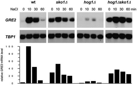
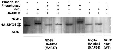
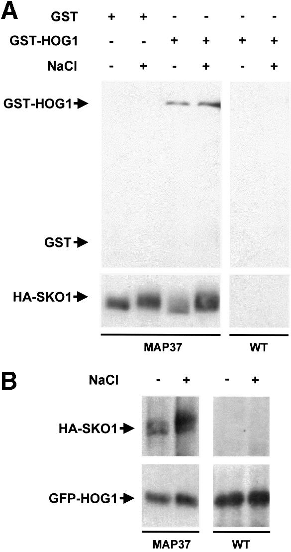
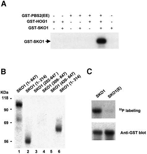
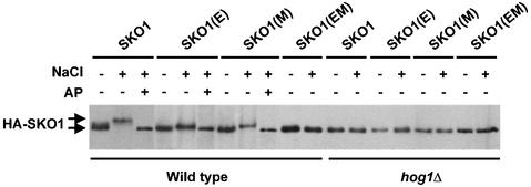
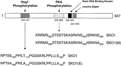
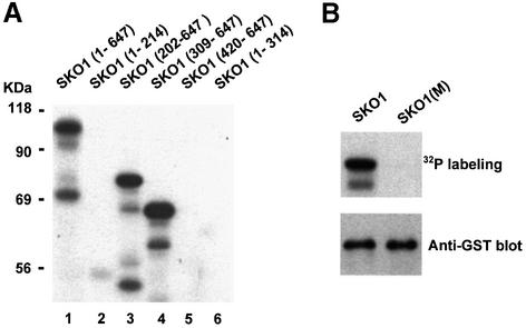
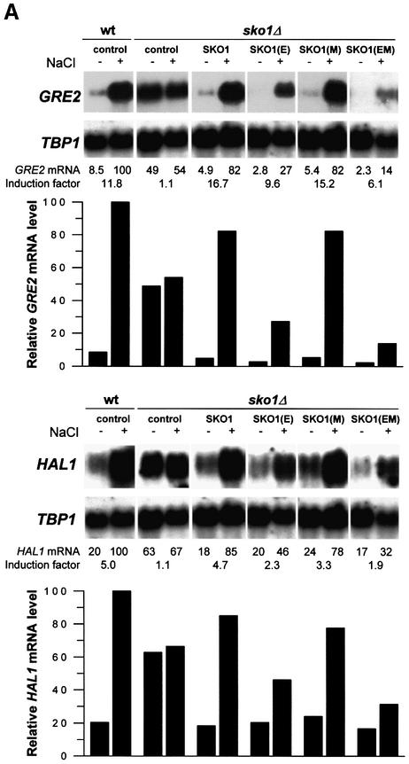
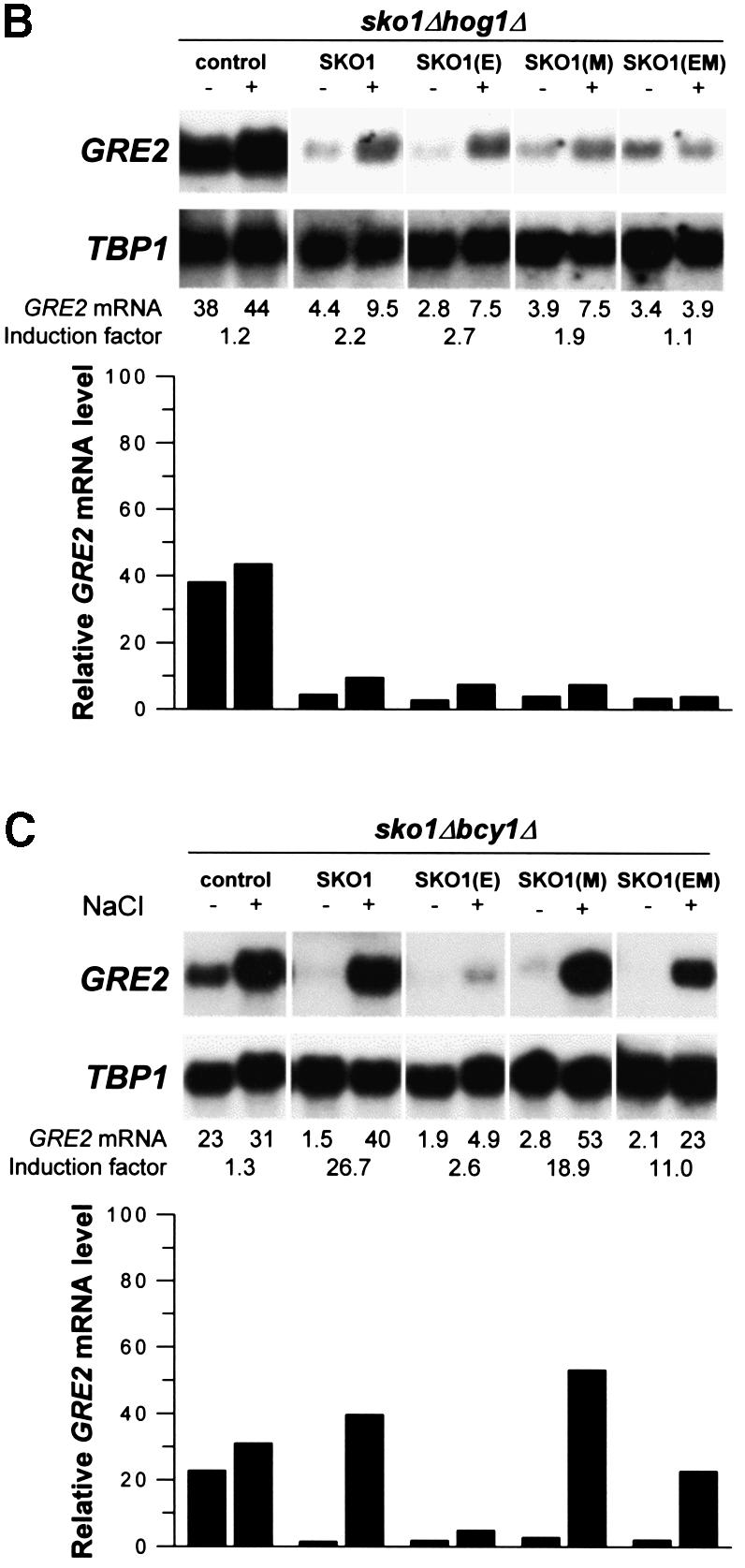
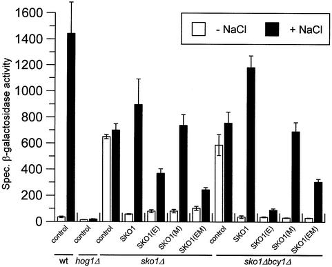
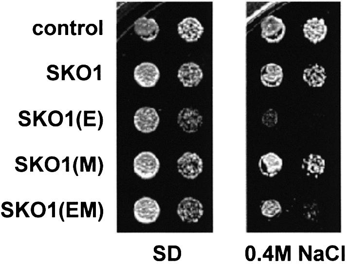
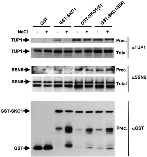
Exposure of yeast to increases in extracellular osmolarity activates the Hog1 mitogen-activated protein kinase (MAPK), which is essential for the induction of gene expression required for cell survival upon osmotic stress. Several genes are regulated in response to osmotic stress by Sko1, a transcriptional repressor of the ATF/CREB family. We show by in vivo coprecipitation and phosphorylation studies that Sko1 and Hog1 interact and that Sko1 is phosphorylated upon osmotic stress in a Hog1-dependent manner. Hog1 phosphorylates Sko1 in vitro at multiple sites within the N-terminal region. Phosphorylation of Sko1 disrupts the Sko1-Ssn6-Tup1 repressor complex, and consistently, a mutant allele of Sko1, unphosphorylatable by Hog1, exhibits less derepression than the wild type. Interestingly, Sko1 repressor activity is further enhanced in strains with high protein kinase A (PKA) activity. PKA phosphorylates Sko1 near the bZIP domain and mutation of these sites eliminates modulation of Sko1 responses to high PKA activity. Thus, Sko1 transcriptional repression is controlled directly by the Hog1 MAPK in response to stress, and this effect is further modulated by an independent signaling mechanism through the PKA pathway.


Figures

None
Fig. 1. Roles of Sko1 and Hog1 in osmotic stress-induced transcription of GRE2. Transcript levels of GRE2 were monitored by northern analysis before and during osmotic stress caused by 0.4 M NaCl. GRE2 mRNA levels were quantified, normalized for the TBP1 internal loading control and depicted in the lower panel. The different small blots presented as well as the quantification data come from the same original blot for each mutant. The isogenic yeast strains W303-1A (wt), MAP19 (sko1Δ), MAP32 (hog1Δ) and MAP33 (hog1Δ sko1Δ) were used.
None
Fig. 2. In vivo phosphorylation of Sko1 upon osmotic stress depends on Hog1. HA–SKO1 was expressed from its chromosomal locus in wild-type (MAP37) and hog1Δ (MAP36) cells. Approximately 50 µg of yeast total protein extracts were separated by SDS–PAGE, and the fusion protein was detected by monoclonal HA-specific antibodies. Cells were subjected or not to brief osmotic shock (0.4 M NaCl, 10 min) and the extracts were treated or not with 10 U of alkaline phosphatase in the absence or presence of phosphatase inhibitors as indicated. Control strain (without HA–SKO1) was wild-type strain W303-1A.
None
Fig. 3. In vivo binding of Hog1 to Sko1. (A) MAP37 strain (which expresses HA-tagged Sko1 from the wild-type locus) was transformed with a plasmid expressing GST or GST–HOG1 under the PGAL1 promoter. Cells were grown in the presence of galactose and samples were taken before (–) or 5 min after (+) the addition of NaCl to a final concentration of 0.4 M. HA–SKO1 was precipitated using anti-HA monoclonal antibody (12CA5) and protein A–Sepharose beads, and the presence of GST proteins in precipitates was probed by immunoblotting using anti-GST (upper panel). Antibody against HA was used to detect the HA–SKO1 fusion protein (lower panel). (B) Yeast MAP37 cells were transformed with a single copy plasmid containing GFP-tagged Hog1, expressed under its own promoter. Cells were grown in the presence of glucose and treated as in (A). HA–SKO1 and GFP–HOG1 in precipitates were probed with anti-HA and anti-GFP antibodies.
None
Fig. 4. In vitro phosphorylation of Sko1 by Hog1. (AIn vitro activated Hog1 phosphorylates Sko1. Recombinant tagged proteins were purified from E.coli as described in Materials and methods. Hog1 and the constitutively activated Pbs2 allele [PBS2(EE)] were incubated in presence of kinase buffer and ATP. Sko1 was then added (when indicated) in the presence of radioactive ATP. Phosphorylated proteins were resolved by SDS–PAGE and detected by autoradiography. The position of tagged Sko1 is indicated on the left. (B) Hog1 phosphoryl ates the region between residues 1 and 202 of Sko1. Various Sko1 fragments were tested for their ability to be phosphorylated by an in vitro activated Hog1 (as described in Materials and methods). After in vitro kinase assay, phosphorylated proteins were resolved by SDS–PAGE and detected by autoradiography. The positions of the Sko1 fragments included in the constructs are indicated in parentheses. (C) Mutation of Sko1 Ser108, Thr113 and Ser126 to Ala abolishes Hog1 phosphorylation. Full-length Sko1 and the triple mutant SKO1(E) protein (mutant in the Ser108, Thr113 and Ser126) were tested for Hog1 phosphorylation as in (A). After phosphorylation, proteins were resolved by SDS–PAGE and transferred to a nylon membrane. Phosphorylated proteins were detected by autoradiography (upper panel). GST-tagged Sko1 proteins were detected by immunoblot using the anti-GST monoclonal antibody (lower panel).
None
Fig. 5. In vivo phosphorylation of Sko1 mutant proteins. Wild-type or hog1Δ cells were transformed with HA-tagged wild-type Sko1 (SKO1), the Sko1 mutant in the Hog1 phosphorylation sites [SKO1(E)], the Sko1 mutant in the PKA phosphorylation sites [SKO1(M)] or the Sko1 mutant in both Hog1 and PKA phosphorylation sites [SKO1(EM)]. Cells were grown in the presence of galactose for 4 h and samples were taken before (–) or 5 min after (+) the addition of NaCl to a final concentration of 0.4 M, and the extracts were treated (+) or not (–) with 10 U of alkaline phosphatase (AP). HA–SKO1 was detected by immunoblotting using anti-HA monoclonal antibody (12CA5).
None
Fig. 6. Schematic overview of the phosphorylation sites in the Sko1 protein. Three Hog1 phosphorylation sites are present in the N-terminus of Sko1, whereas three PKA phosphorylation sites are located near the bZIP structural domain. (E) and (M) triple mutations, which correspond to the Hog1 and PKA phosphorylation sites, are indicated.
None
Fig. 7. Sko1 is phosphorylated by PKA in vitro. (A) PKA phosphorylates the region between residues 309 and 420 of Sko1. Full-length or various Sko1 fragments were tested by their ability to be phosphorylated by PKA. Sko1 recombinant tagged proteins were purified from E.coli as described in Materials and methods, and incubated with PKA in the presence of kinase buffer and radioactive ATP. Phosphorylated proteins were resolved by SDS–PAGE and detected by autoradiography. The positions of the Sko1 fragments included in the constructs are indicated in parentheses. (B) Mutation of Sko1 Ser380, Ser393 and Ser399 to Ala abolishes PKA phosphorylation. Full-length Sko1 and the triple mutant SKO1(M) protein (mutant in Ser380, Thr393 and Ser399) were tested for PKA phosphorylation as in (A). After phosphorylation, proteins were resolved by SDS–PAGE and transferred to a nylon membrane. Phosphorylated proteins were detected by autoradiography (upper panel). GST-tagged Sko1 proteins were detected by immunoblot using the anti-GST monoclonal antibody (lower panel).
None
Fig. 8. Hog1 and PKA phosphorylations modulate Sko1-mediated repression in vivo. Yeast strains were transformed with the empty vector pYEX-4T (control) or the wild-type GST–Sko1 fusion (SKO1), the GST–Sko1 mutant allele in the Hog1 phosphorylation sites [SKO1(E)], the GST–Sko1 mutant allele in the PKA phosphorylation sites [SKO1(M)] or the GST–Sko1 mutant allele in both Hog1 and PKA phosphorylation sites [SKO1(EM)]. Cells were grown selectively to mid-log phase and then subjected (+) or not (–) to brief osmotic shock (0.4 M NaCl, 10 min). Total RNA was assayed by northern blot analysis for transcript levels of GRE2, HAL1 and TBP1. The induction factor represents the degree of induction plus or minus stress. The different small blots presented as well as the quantification data come from the same original blot for each mutant. (A) Hog1 phosphorylation sites are important for release from Sko1-mediated repression. GST–Sko1 fusions were expressed in wild type (W303-1A) and Δsko1 (MAP19). (B) Sko1-mediated derepression is controlled by Hog1. GST–Sko1 fusions were expressed in Δsko1Δhog1 (MAP33) cells. (C) PKA phosphorylation sites modulate Sko1 function in cells with high PKA activity. GST–Sko1 fusions were expressed in Δsko1Δbcy1 (MAP44) cells. The amount of Sko1 protein was similar for the different alleles and was not altered when cells were bcy1Δ (data not shown).
None
Fig. 9. Roles of Hog1 and PKA phosphorylation sites in the regulation of a CRE-driven reporter gene. The yeast strains used in Figure 8 were co-transformed with the CYC1–(2×CRE)–lacZ reporter pMP253 and subjected (+ NaCl) or not (– NaCl) to brief osmotic shock (0.4 M NaCl, 15 min). Specific β-galactosidase activity is given in nmol/min/mg and is the result of the measurement in duplicate of three independent transformants. The amount of Sko1 protein was similar for the different alleles and was not altered when cells were bcy1Δ (data not shown).
None
Fig. 10. Hog1 phosphorylation of Sko1 is critical for salt stress resistance. Yeast Δsko1 mutant cells (MAP19) were transformed with vector control pYEX-4T, GST–SKO1 (wild type), GST–SKO1(E) (mutant in the Hog1 phosphorylation sites), GST–SKO1(M) (mutant in the PKA phosphorylation sites) or GST–SKO1(EM) (mutant in both Hog1 and PKA phosphorylation sites) alleles. Overexpression of the GST fusions was achieved by addition of CuSO4 to a final concentration of 0.3 mM to the plates. Serial dilutions of the transformed yeast strains were spotted onto SD plates lacking uracil containing or not 0.4 M NaCl as indicated.
None
Fig. 11. In vivo binding of Sko1 to the Ssn6–Tup1 co-repressor is affected by phosphorylation upon stress. MAP19 strain (sko1Δ) was transformed with a plasmid expressing GST, GST–SKO1, GST–SKO(E) or GST–SKO1(EM). Cells were grown and subjected (+) or not (–) to a brief osmotic shock (5 min, 0.4 M NaCl). GST proteins were pulled down with glutathione–Sepharose 4B and the presence of wild-type Tup1 or Ssn6 proteins was probed by immunoblotting using specific antibodies against Tup1 (upper panel) or Ssn6 (middle panel), respectively. Total extract represents <10% of the input protein and Prec. is the total amount of Ssn6 or Tup1 coprecipitated. The amount of precipitated GST proteins was detected using anti-GST (lower panel).

Similar articles

Cited by

References

    1. Bilsland-Marchesan E., Ariño,J., Saito,H., Sunnerhagen,P. and Posas,F. (2000) Rck2 kinase is a substrate for the osmotic stress-activated mitogen-activated protein kinase Hog1. Mol. Cell. Biol., 20, 3887–3895. - PMC - PubMed
    1. Boguslawski G. (1992) PBS2, a yeast gene encoding a putative protein kinase, interacts with the RAS2 pathway and affects osmotic sensitivity of Saccharomyces cerevisiae. J. Gen. Microbiol., 138, 2425–2432. - PubMed
    1. Brewster J.L., de Valoir,T., Dwyer,N.D., Winter,E. and Gustin,M.C. (1993) An osmosensing signal transduction pathway in yeast. Science, 259, 1760–1763. - PubMed
    1. Carlson M. and Botstein,D. (1982) Two differentially regulated mRNAs with different 5′-ends encode secreted and intracellular forms of yeast invertase. Cell, 28, 145–154. - PubMed
    1. DeVit M.J. and Johnston,M. (1999) The nuclear exportin Msn5 is required for nuclear export of the Mig1 glucose repressor of Saccharomyces cerevisiae. Curr. Biol., 9, 1231–1241. - PubMed

Publication types

MeSH terms