. 2001 Mar 1;20(5):1173-83.
doi: 10.1093/emboj/20.5.1173.

Ten1 functions in telomere end protection and length regulation in association with Stn1 and Cdc13

Affiliations

Ten1 functions in telomere end protection and length regulation in association with Stn1 and Cdc13

N Grandin et al. EMBO J. .

Abstract

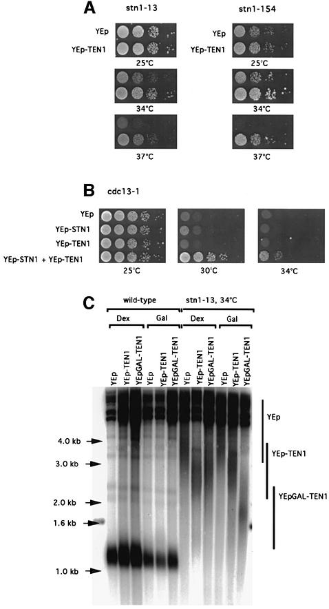
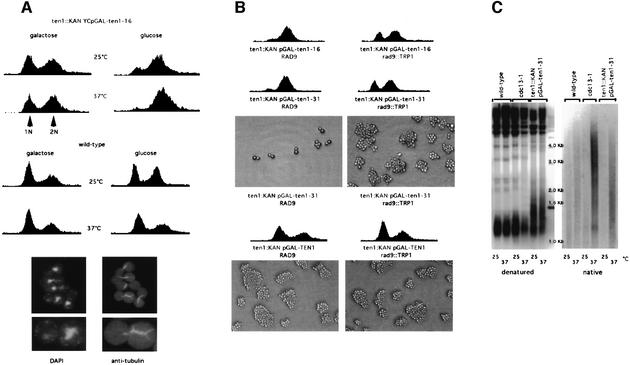
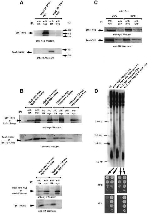
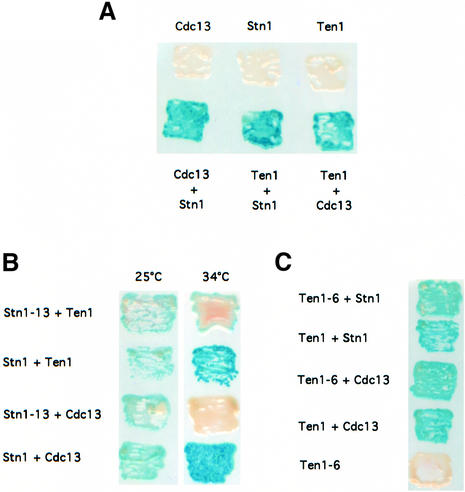
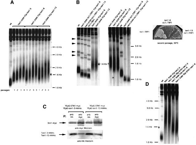
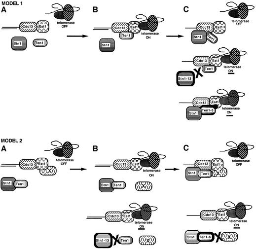
In Saccharomyces cerevisiae, Cdc13 has been proposed to mediate telomerase recruitment at telomere ends. Stn1, which associates with Cdc13 by the two-hybrid interaction, has been implicated in telomere maintenance. Ten1, a previously uncharacterized protein, was found to associate physically with both Stn1 and Cdc13. A binding defect between Stn1-13 and Ten1 was responsible for the long telomere phenotype of stn1-13 mutant cells. Moreover, rescue of the cdc13-1 mutation by STN1 was much improved when TEN1 was simultaneously overexpressed. Several ten1 mutations were found to confer telomerase-dependent telomere lengthening. Other, temperature-sensitive, mutants of TEN1 arrested at G(2)/M via activation of the Rad9-dependent DNA damage checkpoint. These ten1 mutant cells were found to accumulate single-stranded DNA in telomeric regions of the chromosomes. We propose that Ten1 is required to regulate telomere length, as well as to prevent lethal damage to telomeric DNA.


Figures

None
Fig. 1. Suppression of the temperature-sensitive growth and telomere length defects of stn1 and cdc13 mutations following TEN1 overexpression. (Astn1-13 (left) and stn1::TRP1 YCp111-stn1-154 (right) strains were transformed with a multicopy (episomal, 2µ) plasmid expressing TEN1 under the control of its own promoter (YEp-TEN1) or vector alone (YEp) and 10-fold serial dilutions (from left to right in each row) of transformants grown for 2 days at the indicated temperatures and photographed. (B) Co-overexpression with TEN1 improves the rescue of cdc13-1 by STN1. Both STN1 and TEN1 were overexpressed from a 2µ plasmid under the control of their respective promoter. Temperature-sensitive cdc13-1 cells were transformed with a plasmid expressing STN1 (YEp-STN1), with a plasmid expressing TEN1 (YEp-TEN1), with both plasmids (YEp-STN1 + YEp-TEN1) or with vector alone (YEp), spotted as in (A) and incubated for 2 days at the temperatures indicated. (C) Telomere length in wild-type cells (left) and stn1-13 mutant cells (right) harbouring either YEp-TEN1, YEp-GAL-TEN1 or YEp alone, grown for ∼60 generations either on glucose- (Dex) or galactose-based medium (Gal) to induce expression of the GAL1 promoter. The relevant genotypes are indicated above the lanes. A Y′ 32P-labelled probe was used to detect telomeric sequences (see Materials and methods). Note that the telomere length of stn1-13 cells grown at 34°C was much larger than that at 30 or 32°C (see Figures 5D and 3D, respectively). The existence of such extremely heterogeneous-length telomeres is not unprecedented (see, for instance, rif1 rif2 double mutants; Wotton and Shore, 1997). Vertical bars schematically outline the upper and lower limits of the average telomere lengths in each category of strains considered.
None
Fig. 2. The arrest conferred by the temperature-sensitive ten1-16 and ten1-31 mutant alleles occurs at G2/M and depends on the Rad9-mediated DNA damage checkpoint. (A) Top: FACS analysis of ten1 arrested cells. Cells disrupted for TEN1 (ten1::kanMX4) and surviving owing to the presence of the temperature-sensitive ten1-16 allele on a single-copy plasmid, under the control of the GAL1 promoter (ten1::KAN YCpGAL-ten1-16), were grown to mid-log phase at 25°C and shifted up to 37°C for 4 h or left at 25°C, either on galactose-based medium, a condition that induced expression of the ten1 allele, or on glucose-based medium, and prepared for FACS analysis, together with wild-type control cells. The ten1 mutant cells arrested in G2/M on glucose medium at 37°C. When grown on glucose medium at 25°C or on galactose medium at 37°C, these mutant strains became enriched in dumb-bell-shaped cells but still continued to grow. ten1-31 cells behaved similarly (not shown). Bottom: cells from the ten1::kanMX4 YCp111-GAL1-ten1-31 strain were grown on galactose-based medium at 25°C, then on glucose-based medium at 37°C (repressive conditions), fixed 4 h later and processed for tubulin immunofluorescence and staining of the DNA with DAPI. Arrested ten1 mutant cells contained a single nucleus located close to the neck joining the mother and daughter. They also exhibited a short tubulin spindle, typical of a G2/M arrest, extending through the nucleus between the two duplicated spindle bodies. (B) Cell cycle profiles of ten1 mutant cells bearing (rad9::TRP1, right) or not (RAD9, left) a disruption of RAD9, a DNA damage checkpoint gene, were fixed for FACS analysis 4 h after transfer to glucose medium at 37°C, a condition that inactivated these temperature-sensitive ten1 alleles. In the presence of the rad9 mutation, both the ten1-16 and ten1-31 mutant cells failed to mark the G2/M arrest exhibited by the RAD9+ ten1 cells. As a consequence, rad9 ten1 double mutants continued to proliferate for a few cell divisions, as attested by their ability to form numerous microcolonies (shown only for ten1-31), a phenotype indicative of a checkpoint defect. In contrast, RAD9+ ten1-31 cells, which are checkpoint proficient, stopped progression through the cell cycle. Meanwhile, ten1 disruptants bearing wild-type TEN1 (ten1::KAN pGAL-TEN1) bearing (rad9::TRP1) or not (RAD9) a disruption of RAD9, and growing on galactose medium, continued to proliferate indefinitely. (C) Non-denaturing Southern hybridization to a Y′ 32P-labelled probe (native, right) revealed the presence of abnormally high levels of single-stranded DNA in the telomeric regions of ten1-31 mutant cells (ten1::TRP1 pGAL-ten1-31) at 37°C. Cells were grown on galactose-based medium at 25°C, then for 4 h on glucose-based medium at 37°C (repressive conditions) or 25°C (permissive conditions) and harvested for preparation of genomic DNA. cdc13-1 mutant cells grown at 37°C were used as a positive control, while cdc13-1 cells grown at the permissive temperature of 25°C and wild-type cells grown at 25 or 37°C served as negative controls. Genomic DNAs from these same strains were run in parallel and processed for hybridization with the same probe under denaturing conditions (denatured, left) to serve as additional controls.
None
Fig. 3. Ten1 associates with Stn1 in vivo, an interaction that is defective in stn1 mutants. (A) Control experiments were performed to reveal the position of Stn1–myc and Ten1–HAHis on western blots. Extracts from wild-type cells overexpressing STN1–myc alone under GAL1 promoter control (lanes 1 and 2 in both top and bottom panels) or TEN1–HAHis alone under GAL1 promoter control (lanes 3 and 4 in both top and bottom panels), at 30°C, were subjected to immunoprecipitation with monoclonal anti-HA antibody (IP: anti-HA, lanes 1 and 3 in both panels) or monoclonal anti-myc antibody (IP: anti-myc, lanes 2 and 4 in both panels). The band migrating below Stn1–myc in this and subsequent blots presumably represents an Stn1 degradation product. (B) Co-immunoprecipitation of Stn1–myc and Ten1–HAHis. Protein extracts were prepared from cells of the indicated genotype. All experiments were performed at 30°C. (C) Stn1 and Ten1 still co-immunoprecipitate in cdc13-1 mutant cells co-expressing YEp181-GAL1-STN1–myc and YEp195-GAL1-TEN1–GFP and shifted up for 4 h at the restrictive temperature of 37°C; same methods and nomenclature as in (A) and (B). (D) Suppression of stn1-associated telomere elongation following expression of fusions between wild-type Ten1 and mutant Stn1 proteins. An stn1::TRP1 haploid strain kept alive on galactose owing to the presence of YCp33-GAL1-STN1 was transformed with either one of the indicated centromeric plasmids. The plasmid containing wild-type STN1 was then shuffled out following counterselection on 5-FOA medium. The resulting strains were then grown for ∼60 generations at 32°C and the length of their telomeres measured using a Y′ probe. Expression of either stn1-13 or stn1-154 alone produced dramatic telomere elongation (lanes 2 and 4, respectively) compared with wild-type cells (wt, lane 1). In contrast, expression of the Ten1–Stn1-13 (lane 3) or Ten1–Stn1-154 (lane 5) fusions totally suppressed the stn1-induced telomere elongation. In addition, the Ten1–Stn1 hybrid proteins rescued not only the inviability due to the stn1 disruption, but also the temperature-sensitive growth defect conferred by the Stn1-13 or Stn1-154 mutant protein. The four patches of cells in each row represent 10-fold dilutions of the same culture.
None
Fig. 4. Two-hybrid interactions between Ten1, Stn1 and Cdc13. (A) Ten1 associates with Cdc13, as well as with Stn1, in a yeast two-hybrid system. Strains simultaneously expressing pAS2-TEN1 and pACT2-STN1 (bottom row, Ten1 + Stn1) or pAS2-TEN1 and pACT2-CDC13 (bottom row, Ten1 + Cdc13) were positive for β-galactosidase activity, measured as described in Materials and methods, like cells co-expressing pAS2-CDC13 and pACT2-STN1 (bottom row, Cdc13 + Stn1) used here as a positive control. On the other hand, cells simultaneously expressing pAS2-TEN1 and pACT2 alone (top row, Ten1), pAS2 alone and pACT2-CDC13 (top row, Cdc13) or pAS2 alone and pACT2-STN1 (top row, Stn1) scored negative in the X-gal assay. Patches of cells replica-plated on nitrocellulose membrane were incubated for 2 h at 30°C and then photographed. For each one of the six pAS2/pACT2 pairs shown here, three different strains were monitored (four transformants for each). (B) The Stn1-13 mutant protein fails to interact with either wild-type Ten1 or wild-type Cdc13 at restrictive temperature. The stn1-13 allele was expressed from pACT2 together with pAS2-TEN1 or pAS2-CDC13, as explained above. Both pairs scored negative for β-galactosidase activity (white patches) at 34°C, but not at 25°C (permissive temperature), while the corresponding controls expressing wild-type STN1 were positive at both temperatures (blue patches). (C) The Ten1-6 mutant protein still interacts with both wild-type Stn1 and wild-type Cdc13. The ten1-6 allele was expressed from pAS2 together with pACT2-STN1 or pACT2-CDC13, as explained in (A). Both pairs scored positive in the X-gal assay (blue patches) at 30°C (ten1-6 is not a ts allele), similarly to the corresponding controls expressing wild-type TEN1.
None
Fig. 5. Telomerase-dependent telomere elongation associated with ten1 mutations is not due to a defect in Stn1 binding. (A) All three ten1 mutant strains selected for telomere length deregulation (ten1::KAN YCp-ten1-3, lanes 2–5; ten1::KAN YCp-ten1-6, lanes 6–9; ten1::KAN YCp-ten1-13, lanes 10–13) exhibited dramatic telomere elongation compared with wild-type cells (wt, lane 1). All strains were grown at 30°C for various numbers of generations (one passage or restreak is typically performed after ∼25 generations). (B) Disruption of the RNA subunit of telomerase, TLC1, in ten1-6 and ten1-13 resulted in total suppression of telomere elongation (left panel, compare lanes 2 and 3 with lanes 5 and 6, respectively). Lanes 1 (wt cells) and 4 (tlc1::TRP1 cells) served as controls. The ten1 tlc1Δ mutant cells shown in the left panel are at a pre-survival stage, as attested by the presence of non-Y′ bands (indicated by arrowheads near lane 1 of the left panel), contrary to those shown in the middle panel, which have already entered senescence (same strains as in the left panel), attested by the disappearance of non-Y′ bands. Accelerated senescence in the ten1-6 tlc1::TRP1 and ten1-13 tlc1::TRP1 double mutants, compared with tlc1::TRP1 singles, is illustrated in the right panel. (C) The Ten1-3–HAHis and Ten1-13–HAHis mutant proteins still interact physically with wild-type Stn1. Protein extracts were prepared from cells of the indicated genotype and IP–western blots performed as described in the legend to Figure 3. (D) Expression of Stn1–Ten1-3 or Stn1–Ten1-6 fusion protein (under the control of STN1 natural promoter) does not abolish the telomere elongation phenotype conferred by the ten1 mutation. ten1Δ YEp195-TEN1 was transformed with either one of the plasmids indicated. The plasmid containing wild-type TEN1 was then shuffled out on 5-FOA medium. The resulting strains were then grown for ∼60 generations at 30°C and the length of their telomeres measured. Expression of either ten1-3 or ten1-6 alone produced dramatic telomere elongation (lanes 2 and 4, respectively) compared with wild-type cells (wt, lane 1). Expression of the Stn1–Ten1-3 (lane 3) or Stn1–Ten1-6 (lane 5) fusion had no effect on ten1-3- or ten1-6-induced telomere elongation.
None
Fig. 6. Speculative models for the functions of the Cdc13–Stn1–Ten1 complex at telomere ends. Two models are proposed to account for the data described here, on the basis of previously published data (see text). The principal actors in both models are represented in (A). Stn1 and Ten1, which physically associate together as well as with Cdc13, a single-stranded telomeric DNA binding protein, act as inhibitors of Cdc13-mediated telomerase recruitment (telomerase contains the catalytic subunit Est2, and TLC1, the RNA template; Est3, also shown here, is a regulator of telomerase). Est1, another regulator of telomerase, which also binds single-stranded telomeric DNA, associates physically with TLC1 and Cdc13. In model 1, Ten1 binding to Cdc13–Est1 might represent the signal allowing interactions between Cdc13–Est1 and telomerase (telomerase ON, B). Binding of Stn1 to Cdc13 and Ten1 might then result in termination of telomere elongation (telomerase OFF, top panel in C). Ten1 could possibly be required to recruit Stn1 in this latter step. Subsequently, Stn1 and Ten1 release from the Cdc13–Stn1–Ten1 complex followed by Ten1 rebinding, or, alternatively, release of Stn1 alone from this complex, might represent the signal allowing interactions between Cdc13, Est1 and telomerase for a new round of elongation of telomeric DNA (not shown). Abolition of the physical interaction between Stn1-13 and both Cdc13 and Ten1 is proposed to explain telomere lengthening in stn1-13 mutant cells (middle panel in C), while the Ten1-6 mutant protein, which is still capable of binding Cdc13 and Stn1, might confer a situation favourable to an increased association between itself and Cdc13–Est1 (bottom panel in C). In model 2, Stn1 and Ten1 are always in complex together and Ten1 is viewed basically as a cargo to convey Stn1 to Cdc13 or, alternatively, as a regulator of Stn1 function; protein X is an as yet unidentified telomeric protein that associates with Est1 (A). Release of protein X from Est1 allows interactions between Cdc13–Est1 and telomerase (top panel in B). Binding of Stn1–Ten1 to Cdc13 and of protein X to both Est1 and Ten1 might terminate the reaction of telomerase recruitment (telomerase OFF, top panel in C). Subsequently, Stn1 and Ten1 release from the Cdc13–Stn1–Ten1 complex accompanied by release of protein X from Est1 and Ten1 might represent the signal allowing interactions between Cdc13, Est1 and telomerase for a new round of telomeric DNA elongation (not shown). Failure of the Stn1-13 mutant protein to bind Ten1 would prevent termination of telomerase recruitment (telomerase ON, bottom panel in B), while a defect in the interaction between the Ten1-6 mutant protein and protein X would prevent re-association with Cdc13–Est1 and inhibition of the interaction between Cdc13–Est1 and telomerase (telomerase ON, bottom panel in C).

Similar articles

Cited by

References

    1. Ausubel F.A., Brent,R., Kingston,R.E., Moore,D.D., Seidman,J.G., Smith,J.A. and Struhl,K. (eds) (1998) Current Protocols in Molecular Biology. John Wiley and Sons, New York, NY.
    1. Bertuch A. and Lundblad,V. (1998) Telomeres and double-strand breaks: trying to make the ends meet. Trends Cell Biol., 8, 339–342. - PubMed
    1. Bourns B.D., Alexander,M.K., Smith,A.M. and Zakian,V.A. (1998) Sir proteins, Rif proteins, and Cdc13p bind Saccharomyces telomeres in vivo. Mol. Cell. Biol., 18, 5600–5608. - PMC - PubMed
    1. Diede S.J. and Gottschling,D.E. (1999) Telomerase-mediated telomere addition in vivo requires DNA primase and DNA polymerases α and δ. Cell, 99, 723–733. - PubMed
    1. Dionne I. and Wellinger,R.J. (1996) Cell cycle-regulated generation of single-stranded G-rich DNA in the absence of telomerase. Proc. Natl Acad. Sci. USA, 93, 13902–13907. - PMC - PubMed

Publication types

MeSH terms