. 2001 Mar 1;20(5):1203-12.
doi: 10.1093/emboj/20.5.1203.

The small DNA binding domain of lambda integrase is a context-sensitive modulator of recombinase functions

Affiliations

The small DNA binding domain of lambda integrase is a context-sensitive modulator of recombinase functions

D Sarkar et al. EMBO J. .

Abstract

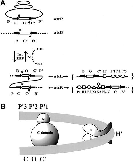
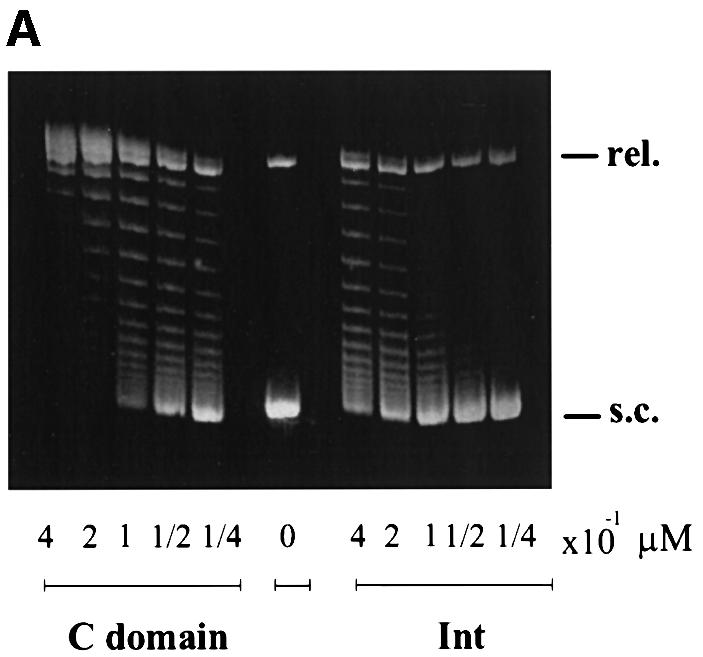
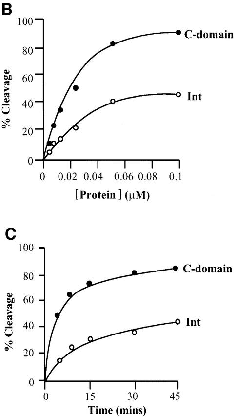
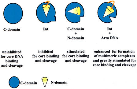
lambda Integrase (Int) has the distinctive ability to bridge two different and well separated DNA sequences. This heterobivalent DNA binding is facilitated by accessory DNA bending proteins that bring flanking Int sites into proximity. The regulation of lambda recombination has long been perceived as a structural phenomenon based upon the accessory protein-dependent Int bridges between high-affinity arm-type (bound by the small N-terminal domain) and low-affinity core-type DNA sites (bound by the large C-terminal domain). We show here that the N-terminal domain is not merely a guide for the proper positioning of Int protomers, but is also a context-sensitive modulator of recombinase functions. In full-length Int, it inhibits C-terminal domain binding and cleavage at the core sites. Surprisingly, its presence as a separate molecule stimulates the C-terminal domain functions. The inhibition in full-length Int is reversed or overcome in the presence of arm-type oligonucleotides, which form specific complexes with Int and core-type DNA. We consider how these results might influence models and experiments pertaining to the large family of heterobivalent recombinases.


Figures

None
Fig. 1. Reaction scheme, protein binding sites and basic architectural motif of λ site-specific recombination. (A) Integrative recombination between the phage and bacterial att sites, attP and attB, respectively, requires the phage-encoded integrase (Int) and the bacteria-encoded IHF. The products of this recombination, attL and attR, form the junctions between bacterial DNA (broken line) and the phage DNA (solid line). Excisive recombination between attL and attR to regenerate attP and attB requires, in addition to Int and IHF, the phage-encode Xis (excisionase) protein. Exicision is stimulated by the host-encoded FIS protein and is inhibited by excess IHF. In both directions, the reaction involves an initial top-strand cleavage (vertical downward arrow) followed by strand swapping and ligation of the new junctions. The resulting Holliday junction intermediate (not shown) is resolved to products by a second round of DNA cleavages, strand swapping and ligation on the bottom strands (vertical upward arrow). The top (vertical downward arrow) and the bottom (vertical upward arrow) strand cleavages are staggered by 7 bp and define the overlap region O (rectangle), which is flanked by inverted core-type Int binding sites (horizontal arrows), C, C′, B and B′. The enlarged diagram of attL and attR (right panel) shows the protein binding sites of the P and P′ arms: five arm-type Int binding sites P1, P2, P′1, P′2 and P′3 (ovals); three IHF sites H1, H2 and H′ (squares); two Xis sites, X1 and X2 (triangles); and one Fis F, (diamond). (B) A model for the IHF-induced bending of attL DNA. This segment of DNA binds one protomer of IHF and one protomer of λ Int. IHF binds at the H′ site and bends the DNA by >140°. Int recognizes two segments of attL DNA, namely the arm- and core-type sites (indicated by P′1 and C′) with its N- and C-domain, respectively.
None
Fig. 2. Comparison of the cleavage activities by Int and its C-domain. (A) Topoisomerase activities of λ Int and the C-domain. Supercoiled pBR322 plasmid DNA (0.36 µg/assay) was incubated with the indicated amounts of protein at 25°C for 1 h (see Materials and methods). The reactions were quenched in 0.2% SDS and analyzed by agarose gel electrophoresis. The positions of supercoiled (s.c.) and relaxed (rel.) plasmid DNA comprise the two ends of the ladder of topoisomers. (B) DNA cleavage as a function of protein concentration. 32P-labeled top-strand nicked full att-site suicide substrate (0.02 µM) was incubated with the indicated amounts of Int or C-domain for 30 min at 25°C and reactions were quenched with 0.2% SDS. (C) DNA cleavage as a function of time. Int or C-domain (0.1 µM) was incubated for the indicated times and reactions were quenched in 0.2% SDS. In both (B) and (C), the reaction conditions were as described in Materials and methods and the amount of covalent complex formed was analyzed by SDS–PAGE and quantitated using a phosphorimager (Fuji).
None
Fig. 3. Gel-shift assays of Int and the C-domain. The indicated amounts of protein were mixed with either 0.1 µM 32P-labeled synthetic 35 bp COC′ core-type DNA (left panel) or 36 bp P′1,2 arm-type DNA (right panel), as described in Materials and methods. ‘Zero’ lanes indicate the substrate DNA alone. The reaction was analyzed by electrophoresis in 8% polyacrylamide gel.
None
Fig. 4. Effect of N-domain in trans on cleavage activity and binding of the C-domain. (A) DNA cleavage as a function of C-domain concentration in the presence of different amounts of N-domain. Labeled suicide substrate (0.02 µM) (see legends to Figure 2B) was incubated at 20°C for 30 min with different concentrations of C-domain in either the absence (open circles) or presence of 0.5 µM (closed circles), 0.75 µM (triangles) or 1.0 µM (squares) N-domain (see Materials and methods). The reactions were quenched in 0.2% SDS. (B) DNA cleavage by a fixed concentration of C-domain as a function of time, in the presence of different amounts of N-domain. As described in (A) and Materials and methods, 0.1 µM C-domain in either the absence (open circles) or presence of 0.5 µM (closed circles), 0.75 µM (triangles) or 1.0 µM (squares) N-domain. The reactions were quenched in 0.2% SDS and analyzed by electrophoresis through 12% (w/v) SDS–PAGE (see Materials and methods). (C) Effect of N-domain in trans on binding of C-domain to core-type DNA. The indicated amounts of C-domain were incubated with 0.1 µM 32P-labeled 35 bp core-type DNA at room temperature for 20 min in the absence or presence of different concentrations of N-domain, as described in Materials and methods. The reaction was analyzed by electrophoresis in 8% polyacrylamide gel and the amount of protein–DNA complex formed was quantitated by scanning the gels on a phosphorimager (Fuji). Retarded complexes with electrophoretic mobilities corresponding to one or two bound C-domains (open or closed symbols, respectively) are plotted as a function of C-domain concentration. Reactions were either in the absence of N-domain (circles) or in the presence of 0.5 µM (triangles) or 1.0 µM (squares) N-domain.
None
Fig. 5. The effect of arm-type oligonucleotide on Int binding to core-type DNA. (A) Int binding to core-type DNA in the presence and absence of arm-type oligonucleotide. Left panel: 32P-labeled 26 bp arm-type DNA (P′1,2*) at 0.28 µM was incubated in the absence or presence of Int (0.55 µM) and in the absence or presence of 0.1 µM unlabeled core-type DNA (COC′). Right panel: 32P-labeled core-type DNA (COC*) at 0.1 µM was incubated in the absence or presence of Int (0.55 µM) and in the absence or presence of 0.28 µM unlabeled arm-type DNA (P′1,2) for 25 min at room temperature. The reactions were analyzed by electrophoresis on native 7% polyacrylamide gels and visualized by autoradiography (see Materials and methods). One complex dependent on all three components appears to be labeled by either [32P]COC′ or [32P]P′1,2. (B) The mobility of a COC′-labeled ternary complex of Int depends on the size of the P′1,2 oligonucleo tides. Using the same conditions described in (A), Int complexes were formed with 32P-labeled COC′ in the presence of either a 26 or 36 bp P′1,2 oligonucleotide. The reactions were analyzed on a 7% polyacrylamide gel and visualized by autoradiography.
None
Fig. 6. Arm–Int–core complex formation requires specific arm- and core-type DNA sequences. (A32P-labeled COC′ (0.1 µM) was incubated with 0.55 µM Int and 0.28 µM unlabeled P′1,2 in the presence of increasing amounts of unlabeled COC′ (specific competitor) or unlabeled non-specific competitor DNA for COC′ (see Materials and methods) and the percentage of labeled ternary complex was determined by gel electrophoresis as described in the legends to Figure 5A and quantitated with a phosphorimager (Fuji). The amount of ternary complex formed in the absence of any competitor was 94% and was normalized to 100% in the figure. (B32P-labeled P′1,2 (0.28 µM) was incubated with 0.55 µM Int and 0.1 µM unlabeled COC′ in the presence of increasing amounts of unlabeled P′1,2 (specific competitor) or unlabeled non-specific competitor DNA for P′1,2 (see Materials and methods). The percentage of labeled ternary complex was determined as in (A). The amount of ternary complex formed in the absence of any competitor was 70% and was normalized to 100% in the figure.
None
Fig. 7. Arm-type DNA stimulates Int cleavage and binding at COC′. (A) Time course of Int cleavage of COC′ as assayed by the formation of covalent complexes on a top-strand nicked suicide substrate. 32P-labeled COC′ covalent complexes with Int were resolved on a 7% SDS–polyacrylamide gel and quantitated on a phosphorimager (see Materials and methods). (B) Time course of ternary complexes formed with COC′, Int and P′1,2. Ternary complexes of Int, P′1,2 and 32P-labeled COC′ were resolved on 7% native polyacrylamide gels and quantitated on a phosphorimager as described in Materials and methods. In both experiments, reaction mixes contained 0.25 µM Int, 0.05 µM COC′, 0.05 mg/ml herring sperm DNA. Arm-type DNA (P′1,2) was added to a final concentration of 0.025 µM (diamonds) or 0.06 µM (triangles) or omitted (circles). Reactions were incubated at 19°C and 10 µl aliquots were taken at the indicated time points.
None
Fig. 8. Schematic summary of the context-sensitive modulation of C-domain recombinase functions at COC′ by the N-domain and arm-type oligonucleotides.

Similar articles

Cited by

References

    1. Azaro M.A. and Landy,A. (2001) Integrase and the λ Int family. In Craig,N.L., Craigie,R., Gellert,M. and Lambowitz,A. et al. (eds), Mobile DNA II. ASM Press, Washington, DC.
    1. Ball C.A. and Johnson,R.C. (1991a) Efficient excision of phage λ from the Escherichia coli chromosome requires the Fis protein. J. Bacteriol., 173, 4027–4031. - PMC - PubMed
    1. Ball C.A. and Johnson,R.C. (1991b) Multiple effects of Fis on integration and the control of lysogeny in phage λ. J. Bacteriol., 173, 4032–4038. - PMC - PubMed
    1. Better M., Lu,C., Williams,R.C. and Echols,H. (1982) Site-specific DNA condensation and pairing mediated by the Int protein of bacteriophage λ. Proc. Natl Acad. Sci. USA, 79, 5837–5841. - PMC - PubMed
    1. Bushman W., Thompson,J.F., Vargas,L. and Landy,A. (1985) Control of directionality in λ site-specific recombination. Science, 230, 906–911. - PMC - PubMed

Publication types

MeSH terms