Comparative Study
. 2001 Feb 15;15(4):428-43.
doi: 10.1101/gad.869501.

Targeting histone deacetylase complexes via KRAB-zinc finger proteins: the PHD and bromodomains of KAP-1 form a cooperative unit that recruits a novel isoform of the Mi-2alpha subunit of NuRD

Affiliations
Comparative Study

Targeting histone deacetylase complexes via KRAB-zinc finger proteins: the PHD and bromodomains of KAP-1 form a cooperative unit that recruits a novel isoform of the Mi-2alpha subunit of NuRD

D C Schultz et al. Genes Dev. .

Abstract

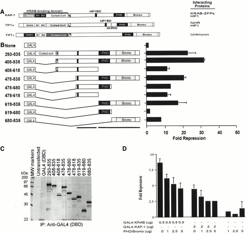
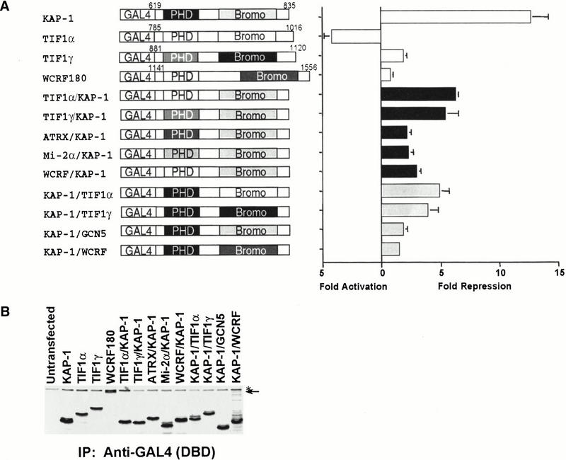
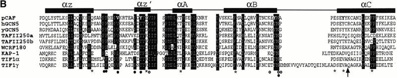
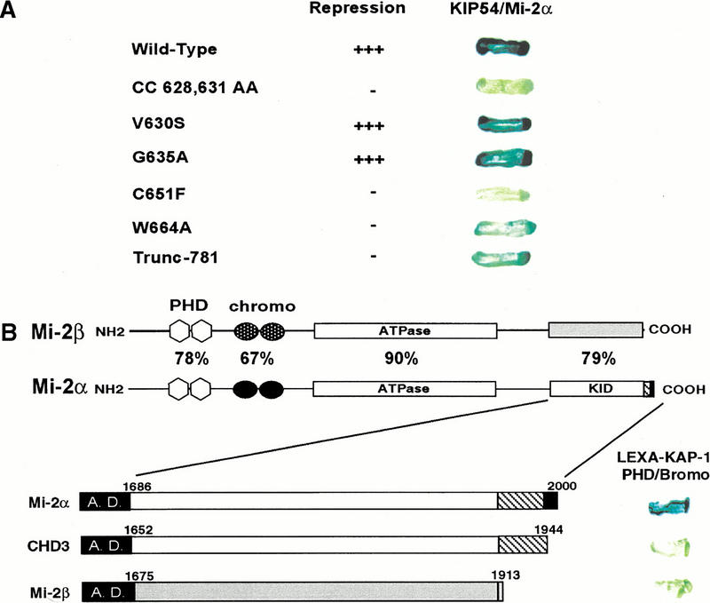
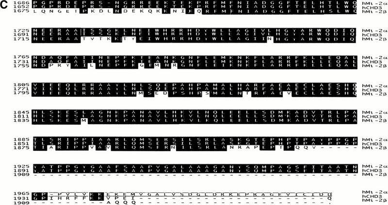
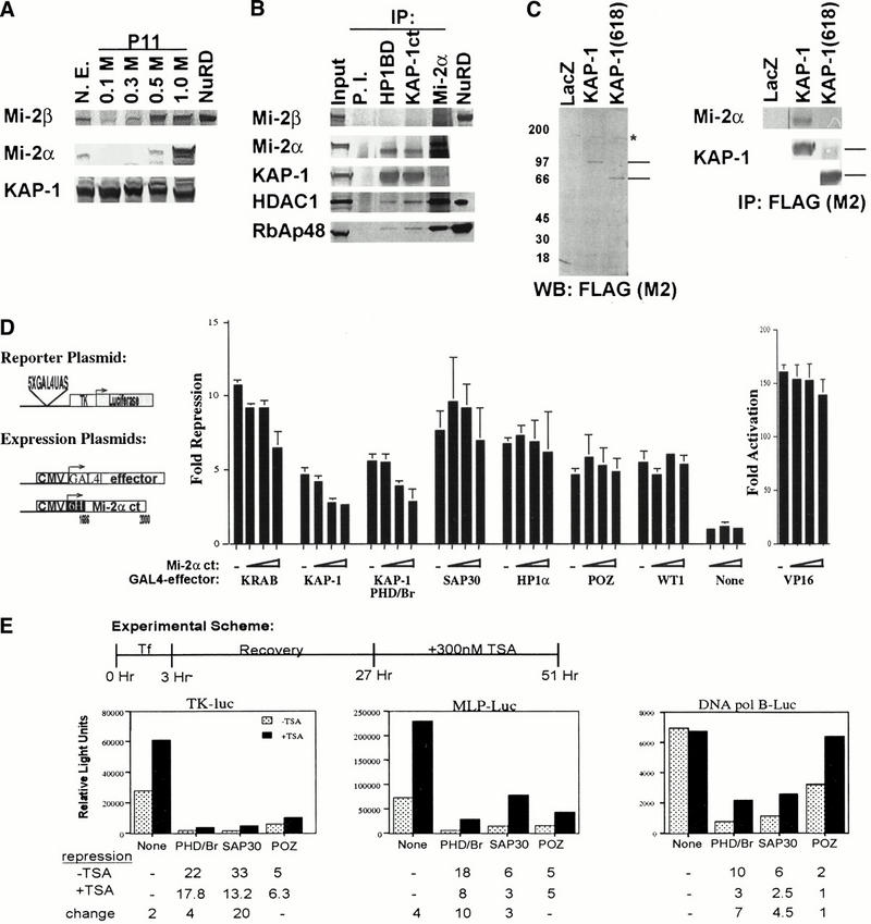
Macromolecular complexes containing histone deacetylase and ATPase activities regulate chromatin dynamics and are vitally responsible for transcriptional gene silencing in eukaryotes. The mechanisms that target these assemblies to specific loci are not as well understood. We show that the corepressor KAP-1, via its PHD (plant homeodomain) and bromodomain, links the superfamily of Krüppel associated box (KRAB) zinc finger proteins (ZFP) to the NuRD complex. We demonstrate that the tandem PHD finger and bromodomain of KAP-1, an arrangement often found in cofactor proteins but functionally ill-defined, form a cooperative unit that is required for transcriptional repression. Substitution of highly related PHD fingers or bromodomains failed to restore repression activity, suggesting high specificity in their cooperative function. Moreover, single amino acid substitutions in either the bromodomain or PHD finger, including ones that mimic disease-causing mutations in the hATRX PHD finger, abolish repression. A search for effectors of this repression function yielded a novel isoform of the Mi-2alpha protein, an integral component of the NuRD complex. Endogenous KAP-1 is associated with Mi-2alpha and other components of NuRD, and KAP-1-mediated silencing requires association with NuRD and HDAC activity. These data suggest the KRAB-ZFP superfamily of repressors functions to target the histone deacetylase and chromatin remodeling activities of the NuRD complex to specific gene promoters in vivo.


Figures

Figure 1
Figure 1
Analysis of the intrinsic repression activity of KAP-1 indicates that the PHD finger and bromodomain are required and sufficient to repress transcription. (A) Schematic illustration of the KAP-1/TIF1 family of transcriptional regulators. The KRAB binding domain in KAP-1 is indicated. (Striped box) TIF signature sequence (TSS), (black box) HP1 binding domain (HP1BD), (thin black line) nuclear hormone receptor interaction domain. (B) Schematic illustration of the heterologous GAL4–KAP-1 constructs, as defined by the amino acid numbers at the left. The intrinsic repressor activity of KAP-1 was measured in a transient assay, using a minimal TK-luciferase reporter template regulated by five consensus GAL4 UAS. All experiments were done in NIH/3T3 cells with 0.5 μg of reporter plasmid and 5 μg of the indicated heterologous GAL4–KAP-1 construct. Fold repression represents the ratio of luciferase activity measured for the reporter alone to the activity measured in the presence of the indicated effector proteins after normalization for transfection efficiency. Error bars represent the standard deviation for three independent transfections. Absence of error bars indicates a standard deviation too small to physically illustrate. (C) Each plasmid was tested for stable protein expression via transfection into COS1 cells followed by immunoprecipitation of [35S]methionine-labeled whole cell extracts with anti-GAL4 (DBD) IgG (1 μg). (D) Overexpression of the KAP-1 PHD finger and bromodomain (amino acids 619 to 835) demonstrated a dose-dependent dominant effect on GAL4–KRAB- and GAL4–KAP-1-mediated transcriptional repression. Transcriptional effects were monitored in a transient assay in which 0.5 μg of the 5x-GAL4 UAS-SV40 luciferase reporter and the indicated amounts of expression plasmids transfected into NIH/3T3 cells. Fold repression was calculated as described. Expression of the dominant negative protein was confirmed in COS1 cells (data not shown).
Figure 2
Figure 2
Heterologous chimeric fusions of related PHD fingers or bromodomains to KAP-1. (A) Schematic illustration of chimeric fusion proteins engineered. The chimeric fusion proteins possess the following amino acid sequences of the individual proteins, respectively: TIF1α/KAP-1 (815–876/680–835), TIF1γ/KAP-1 (880–941/680–835), ATRX/KAP-1 (210–283/680–835), Mi-2α/KAP-1 (372–423/680–835), WCRF180/KAP-1 (1141–1205/680–835), KAP-1/TIF1α (619–679/877–1016), KAP-1/TIF1γ (619–679/942–1120), KAP-1/GCN5 (619–679/714–837), and KAP-1/WCRF180 (619–679/1389–1556). All experiments were done in NIH/3T3 cells, as described in Fig. 1. (White bars) Natural PHD finger/bromodomain fusions, (black bars) chimeras with the KAP-1 bromodomain, (gray bars) chimeras with the KAP-1 PHD finger. (B) Each plasmid was tested for stable protein expression via transfection into COS1 cells followed by immunoprecipitation of [35S]methionine-labeled whole cell extracts with anti-GAL4 (DBD) IgG (1 μg). (Arrow) the migration of the heterologous GAL4–WCRF180 PHD finger/bromodomain, (asterisk) a nonspecific band retained during the immunoprecipitation.
Figure 3
Figure 3
Mutations in the PHD finger and bromodomain significantly impair the intrinsic repression activity of this integrated transcriptional repression domain. (A) Amino acid sequence alignment of the KAP-1 PHD finger with related sequences from 15 independent proteins. The numbers in brackets indicate the corresponding amino acids in each protein. Strictly, conserved amino acids are shaded in black. Boxed amino acids are those in which the chemical nature of the side chain has been conserved. Asterisks indicate amino acids that were mutated to match naturally occurring mutations in the hATRX protein (Gibbons et al. 1997). Each mutation was made in the context of the GAL4–KAP-1 (619–835) expression construct. (B) Amino acid sequence alignment of the KAP-1 bromodomain with related sequences from TIF1α, TIF1γ, WCRF180, hGCN5, yGCN5, hTAFII250, and pCAF. Schematic illustration of the secondary structure and relevant position of each structural element in the bromodomain sequence are indicated above. Amino acid residues that have been conserved or where the chemical nature of the side chain has been maintained are shaded in black. Asterisks mark the amino acids that were mutated in this study. The arrow identifies the position of the 781trunc mutation. Each mutation was made in the context of the GAL4–KAP-1 (619–835) expression construct. Bolded amino acids in the pCAF sequence are those mutated in the study by Dhalliun et al. (1999). (Filled circles) Amino acids in the crystal structure that contact the acetyl moiety of acetylated histones (Owen et al. 2000). (Open circles) Amino acids in the crystal structure that contact the amide backbone of histone H4 (Owen et al. 2000). (C) Schematic diagram of the luciferase reporter and the heterologous GAL4-effector plasmid. Stable expression of each protein was determined via transfection into COS1 cells followed by immunoprecipitation of [35S]methionine-labeled whole cell extracts with anti-GAL4 (DBD) IgG (1 μg). (D) All experiments were done in NIH/3T3 cells, as described in Fig. 1. (Black bars) PHD finger mutations, (gray bars) bromodomain mutations.
Figure 3
Figure 3
Mutations in the PHD finger and bromodomain significantly impair the intrinsic repression activity of this integrated transcriptional repression domain. (A) Amino acid sequence alignment of the KAP-1 PHD finger with related sequences from 15 independent proteins. The numbers in brackets indicate the corresponding amino acids in each protein. Strictly, conserved amino acids are shaded in black. Boxed amino acids are those in which the chemical nature of the side chain has been conserved. Asterisks indicate amino acids that were mutated to match naturally occurring mutations in the hATRX protein (Gibbons et al. 1997). Each mutation was made in the context of the GAL4–KAP-1 (619–835) expression construct. (B) Amino acid sequence alignment of the KAP-1 bromodomain with related sequences from TIF1α, TIF1γ, WCRF180, hGCN5, yGCN5, hTAFII250, and pCAF. Schematic illustration of the secondary structure and relevant position of each structural element in the bromodomain sequence are indicated above. Amino acid residues that have been conserved or where the chemical nature of the side chain has been maintained are shaded in black. Asterisks mark the amino acids that were mutated in this study. The arrow identifies the position of the 781trunc mutation. Each mutation was made in the context of the GAL4–KAP-1 (619–835) expression construct. Bolded amino acids in the pCAF sequence are those mutated in the study by Dhalliun et al. (1999). (Filled circles) Amino acids in the crystal structure that contact the acetyl moiety of acetylated histones (Owen et al. 2000). (Open circles) Amino acids in the crystal structure that contact the amide backbone of histone H4 (Owen et al. 2000). (C) Schematic diagram of the luciferase reporter and the heterologous GAL4-effector plasmid. Stable expression of each protein was determined via transfection into COS1 cells followed by immunoprecipitation of [35S]methionine-labeled whole cell extracts with anti-GAL4 (DBD) IgG (1 μg). (D) All experiments were done in NIH/3T3 cells, as described in Fig. 1. (Black bars) PHD finger mutations, (gray bars) bromodomain mutations.
Figure 4
Figure 4
The PHD finger and bromodomain of KAP-1 specifically interact with the C-terminal amino acids of Mi-2α/CHD3. (A) The yeast strain L40 cotransformed with the indicated KAP-1 mutants as LEXA DNA binding domain fusions and the rescued Mi-2α–ct: GAL4 activation domain clone (KIP54). Positive interaction is inferred by blue yeast due to the activation of the integrated LEXA responsive LacZ reporter gene and subsequent hydrolysis of the synthetic substrate X-gal. (B) The interaction between KAP-1 and Mi-2 is specific for the C terminus of a unique isoform of Mi-2α. Schematic illustration of Mi-2α and Mi-2β. Mi-2α/CHD3 is a member of the CHD family of proteins which are characterized by the seven signature motifs of a “DEAH” box ATPase/Helicase. Other signature motifs include two PHD fingers (light gray hexagons), two chromodomains (black circles). Percentages represent percent identity between the two proteins at the indicated motifs. The putative KAP-1 interaction domain (KID) is indicated. In-frame fusion between the C-terminal sequences of hMi-2α (accession no. 3298562), hCHD3 (accession no. 2645433), hMi-2β/CHD4 (accession no. 1107696) was designed with the GAL4 activation domain. Numbers represent the corresponding amino acid in the respective sequences. L40 yeast cotransformed with the indicated GAL4 activation domain fusion proteins and the wild-type LEXA KAP-1 PHD finger and bromodomain illustrated the specificity of this interaction for Mi-2α. (C) Amino acid sequence alignment of the C-terminal residues schematically depicted in B. Identical residues indicated in black. Boxed amino acids are unique to Mi-2α.
Figure 4
Figure 4
The PHD finger and bromodomain of KAP-1 specifically interact with the C-terminal amino acids of Mi-2α/CHD3. (A) The yeast strain L40 cotransformed with the indicated KAP-1 mutants as LEXA DNA binding domain fusions and the rescued Mi-2α–ct: GAL4 activation domain clone (KIP54). Positive interaction is inferred by blue yeast due to the activation of the integrated LEXA responsive LacZ reporter gene and subsequent hydrolysis of the synthetic substrate X-gal. (B) The interaction between KAP-1 and Mi-2 is specific for the C terminus of a unique isoform of Mi-2α. Schematic illustration of Mi-2α and Mi-2β. Mi-2α/CHD3 is a member of the CHD family of proteins which are characterized by the seven signature motifs of a “DEAH” box ATPase/Helicase. Other signature motifs include two PHD fingers (light gray hexagons), two chromodomains (black circles). Percentages represent percent identity between the two proteins at the indicated motifs. The putative KAP-1 interaction domain (KID) is indicated. In-frame fusion between the C-terminal sequences of hMi-2α (accession no. 3298562), hCHD3 (accession no. 2645433), hMi-2β/CHD4 (accession no. 1107696) was designed with the GAL4 activation domain. Numbers represent the corresponding amino acid in the respective sequences. L40 yeast cotransformed with the indicated GAL4 activation domain fusion proteins and the wild-type LEXA KAP-1 PHD finger and bromodomain illustrated the specificity of this interaction for Mi-2α. (C) Amino acid sequence alignment of the C-terminal residues schematically depicted in B. Identical residues indicated in black. Boxed amino acids are unique to Mi-2α.
Figure 5
Figure 5
Role of Mi-2α and histone deacetylase in KAP-1 repression. (A) Western blot analysis of 10 μg of phosphocellulose (P11) fractionated HeLa nuclear extracts. NuRD, conventionally purified complex as described previously (Zhang et al. 1998a). (B) In vivo association between endogenous KAP-1 and Mi-2α. Three hundred μg of 1.0 M P11-DEAE-bound HeLa nuclear extract was immunoprecipitated by rabbit IgG (P.I.) (lane 2), anti-KAP-1 (HP1BD/423–589) antibodies (lane 3), or anti-KAP-1 (Ct/619–835) antibodies (lane 4), or anti-Mi2α antibodies (lane 5). Immunoprecipitated proteins were eluted by boiling in SDS sample buffer and then separated on a 4–12% NuPAGE gradient gel (Invitrogen), followed by immunoblot analysis with antibodies against Mi-2β, Mi-2α, KAP-1 (RBCC), HDAC1, and RbAp48. (Input) 10 μg of 1.0 M P11-DEAE bound extract, (NuRD) conventionally purified complex as described previously (Zhang et al. 1998a). (C) The in vivo association between KAP-1 and Mi-2α requires the PHD finger and bromodomain of KAP-1. Expression plasmids for LacZ, FLAG-KAP-1, or FLAG-KAP-1 (618) were transfected into 293 cells. Nuclear extracts (3–5 mg) were immunoprecipitated with anti-FLAG mAb M2 (lanes 1–3). The immune complexes were separated on a 4–12% NuPAGE gradient gel (Invitrogen) followed by immunoblot analysis with anti-Mi-2α pAb or anti-KAP-1 (RBCC/20–418) pAb. (Left panel) Anti-FLAG (M2) Western blot of transfected 293 cell nuclear extracts. Lines on right indicate the migration of full-length KAP-1 and KAP-1 (618), respectively. (Asterisk) An anti-FLAG cross-reacting species. (Right panel) The western blot analysis of anti-FLAG (M2) immunoprecipitates for Mi-2α and KAP-1 (RBCC). Lines on the right of the KAP-1 immunoblot represent the migrations of full-length KAP-1 and KAP-1 (618), respectively. (D) Schematic diagram of the luciferase reporter and the heterologous GAL4-effector plasmid. Expression of the KAP-1 interaction domain of Mi-2α (amino acids 1686 to 2000) dominantly inhibits heterologous KRAB- and KAP-1-mediated repression with minimal effects on heterologous repression observed for SAP30, HP1α, BCL6-POZ, WT1 or the activation potential of VP16. (Dashes) Repression observed in the absence of transfected Mi-2α dominant negative plasmid DNA. All experiments were done in NIH/3T3 cells with 0.1 μg of GAL4–VP16, 0.5 μg of GAL4–KRAB, 1 μg of each remaining GAL4-repressor, and 0.5 μg of the reporter plasmid. (Open triangles) Titrating amounts (1 μg, 2 μg, and 4 μg) of the dominant negative Mi-2α expression plasmid transfected. Expression of all proteins was confirmed in COS1 cells (data not shown). Fold repression was calculated as described in Fig. 1. (E) Addition of the histone deacetylase inhibitor, TSA, partially reverses the repression activity of the KAP-1 PHD finger/bromodomain. NIH/3T3 cells were transiently cotransfected with 0.5 μg of the indicated reporter plasmid and 5 μg of the heterologous GAL4 expression plasmids KAP-1 (619 to 835), SAP30, and BCL6-POZ. Twenty-four hours posttransfection, the cells were treated with 300 nM TSA (Wako) for an additional 24 h prior to harvesting. Fold repression was calculated as described in Fig. 1. Fold repression in the absence of TSA (stippled bars); fold repression in the presence of TSA (black bars).

Similar articles

Cited by

References

    1. Aasland R, Gibson TJ, Stewart AF. The PHD finger: Implications for chromatin-mediated transcriptional regulation. Trends Biochem Sci. 1995;20:56–59. - PubMed
    1. Agata Y, Matsuda E, Shimizu A. Two novel Kruppel-associated box-containing zinc-finger proteins, KRAZ1 and KRAZ2, repress transcription through functional interaction with the corepressor KAP-1 (TIF1beta/KRIP-1) J Biol Chem. 1999;274:16412–16422. - PubMed
    1. Bellefroid EJ, Poncelet DA, Lecocq PJ, Revelant O, Martial JA. The evolutionarily conserved Kruppel-associated box domain defines a subfamily of eukaryotic multifingered proteins. Proc Natl Acad Sci. 1991;88:3608–3612. - PMC - PubMed
    1. Bjorses P, Halonen M, Palvimo JJ, Kolmer M, Aaltonen J, Ellonen P, Perheentupa J, Ulmanen I, Peltonen L. Mutations in the AIRE gene: Effects on subcellular location and transactivation function of the autoimmune polyendocrinopathy–candidiasis–ectodermal dystrophy protein. Am J Hum Genet. 2000;66:378–392. - PMC - PubMed
    1. Bochar DA, Savard J, Wang W, Lafleur DW, Moore P, Cote J, Shiekhattar R. A family of chromatin remodeling factors related to Williams syndrome transcription factor. Proc Natl Acad Sci. 2000;97:1038–1043. - PMC - PubMed

Publication types

MeSH terms

Substances

LinkOut - more resources