

Children with macrocrania: clinical and imaging predictors of disorders requiring surgery
- PMID: 11237985
- PMCID: PMC7976845
Children with macrocrania: clinical and imaging predictors of disorders requiring surgery
Abstract
Background and purpose: Macrocrania is a common pediatric clinical condition affecting up to 5% of the population. The purpose of this study was to determine clinical and imaging predictors that are useful in the differentiation of disorders requiring surgical treatment from those that can be treated medically in children with macrocrania.
Methods: In a 3-year 7-month retrospective study, 88 patients (median age, 8 months; interquartile range, 5--13 months) with macrocrania and no known underlying neurologic disorder underwent imaging of the brain (sonography, n = 36; CT, n = 31; MR imaging = 21). The study was conducted in a pediatric tertiary care referral center. Clinical and imaging data were correlated to final diagnosis by means of logistic regression and receiver operating characteristic curves.
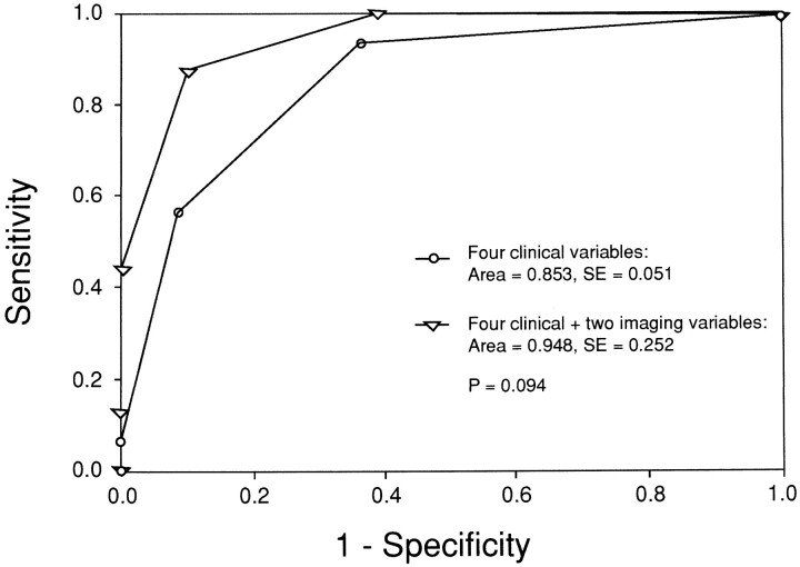
Results: Sixteen (18%) of the patients had disorders requiring surgery: communicating hydrocephalus, n = 7; noncommunicating hydrocephalus, n = 3; hemorrhagic subdural collections, n = 3; neoplasm, n = 1; encysted cavum septi pellucidi, n = 1; and vein of Galen malformation, n = 1. Clinical predictors of disorders requiring surgery included vomiting (P =.007), labor instrumentation (P =.026), developmental delay (P =.008), and abnormal neurologic findings (P =.028). Imaging predictors of disorders requiring surgery included a focal space-occupying lesion (P <.0001) and moderate-to-severe ventriculomegaly (P <.0001). The diagnostic sensitivity of the combination of independent clinical and imaging predictors was higher than that of independent clinical predictors alone, being 100% (95% confidence interval = 96.9%, 100%) and 93.8% (95% confidence interval = 88.7%, 98.8%), respectively. A trend indicated that the area under the receiver operating characteristic curve for clinical plus imaging findings (0.95) was greater than that for clinical findings alone (0.85) (P =.09). An increase in the number of clinical and imaging predictors was highly correlated with an increased risk of a disorder requiring surgery (P <.0001).
Conclusion: Baseline neuroimaging is indicated for children with macrocrania because the combination of clinical and imaging predictors has the best diagnostic performance in determining the need for surgical versus nonsurgical management.
Figures
References
-
- Hamill PV, Drizd TA, Johnson CL, Reed RB, Roche AF, Moore WM. Physical growth: National Center for Health Statistics percentiles. Am J Clin Nutr 1979;32:607-629 - PubMed
-
- Alvarez LA, Maytal J, Shinnar S. Idiopathic external hydrocephalus: natural history and relationship to benign familial macrocrania. Pediatrics 1986;77:901-907 - PubMed
-
- Gooskens RH, Gielen CC, Hanlo PW, Faber JA, Willemse J. Intracranial spaces in childhood macrocrania: comparison of length measurements and volume calculations. Dev Med Child Neurol 1988;30:509-519 - PubMed
-
- Nickel RE, Gallenstein JS. Developmental prognosis for infants with benign enlargement of the subarachnoid spaces. Dev Med Child Neurol 1987;29:181-186 - PubMed
-
- Babcock DS, Han BK, Dine MS. Sonographic findings in infants with macrocrania. AJR Am J Roentgenol 1988;150:1359-1365 - PubMed
MeSH terms
LinkOut - more resources
Full Text Sources