. 2001 Mar;158(3):855-66.
doi: 10.1016/S0002-9440(10)64034-5.

The transmembrane form of the CX3CL1 chemokine fractalkine is expressed predominantly by epithelial cells in vivo

Affiliations

The transmembrane form of the CX3CL1 chemokine fractalkine is expressed predominantly by epithelial cells in vivo

A D Lucas et al. Am J Pathol. 2001 Mar.

Abstract

Fractalkine (CX3CL1) is synthesized as a type I transmembrane protein. Its unique CX(3)C chemokine domain is attached to a 241-amino acid mucin stalk, a 19-amino acid transmembrane domain, and a 37-amino acid intracellular domain of unknown function. A soluble form of fractalkine can be generated by proteolytic cleavage at the base of the mucin stalk. Novel monoclonal and polyclonal antibodies that specifically recognize only the amino- or carboxyl-terminal ends of the human fractalkine molecule have revealed that epithelial cells are the predominant cell type expressing transmembrane forms of fractalkine in human skin, the tonsil, and the large intestine. Using these specific anti-fractalkine reagents we do not detect high-level expression of fractalkine on endothelial cells in normal or inflamed colon samples obtained from patients with Crohn's disease or ulcerative colitis. In contrast to previous reports we do not detect fractalkine expression by Langerhans cells or immature dendritic cells in mucosal-associated lymphoid tissues in vivo. We show that the reagent used in previous studies, an anti-fractalkine N-terminal peptide antisera, cross-reacts with human CD84. Finally we discuss potential roles for fractalkine in constitutive leukocyte trafficking based on its observed pattern of expression in epithelia.


Figures

Figure 1.
Figure 1.
Distinguishing between cleaved and membrane-tethered fractalkine, generation of specific reagents. A: Samples of Western lysates from WT CHO-K1 and CHO-K1 cells transfected with a human fractalkine expression vector along with samples of supernatant taken from fractalkine-transfected CHO-K1 cells, were run on 7.5% acrylamide gels under standard reducing conditions. Samples were transferred to nitrocellulose membranes and identical membranes probed using goat anti-fractalkine polyclonal reagent (goat α-Fkn, R&D Systems) (lanes 1–3) or chicken anti-C-peptide polyclonal reagent (chicken α-C-pep, lanes 4–6). The goat α-Fkn reagent is reactive against the chemokine domain of the molecule and specifically detects twobands at the predicted size of 95 kd (lane 2, asterisk). These two bands are also detected by the chicken α-C-pep reagent (lane 5, asterisk). In addition, these reagents discriminate between cleaved and intact forms of the molecule as the goat α-Fkn detects the cleaved form of fractalkine within transfected cell supernatant (lane 3, 85 to 90 kd), whereas the chicken α-C-pep does not (lane 6). Furthermore, the goat α-Fkn detects one larger (lane 2, 100 kd) and two smaller bands (lane 2, 75 and 66 kd) within transfected CHO-K1 samples that are not detected by the chicken α-C-pep (lane 5). The larger band may be nonspecific because it has no counterpart detected by the chicken α-C-pep. The two smaller bands may indicate partially degraded forms of fractalkine, still containing the N-terminus chemokine domain. B–I: The specificity of a range of anti-fractalkine antibodies was evaluated by immunohistochemistry. Cytospins were prepared from NIH/3T3 cells transiently transfected as above, with fractalkine (3T3-Fkn) and were stained as follows. B: 3T3-Fkn stained with mouse IgG1 control as a control for C. C: 3T3-Fkn stained with mouse anti-fractalkine chemokine domain (mouse α-Fkn, clone 51636.11; R&D Systems) mAb. D: 3T3-Fkn stained with no primary antibody as a control for E and F. E: 3T3-Fkn stained with goat α-Fkn. F: 3T3-Fkn stained with chicken α-C-pep. G: 3T3-Fkn stained with rabbit IgG as a control for H and I. H: 3T3-Fkn stained with rabbit α-C-peptide. I: 3T3-Fkn stained with rabbit α-N-pep polyclonal reagent. Note that although there is light nonspecific staining of the nucleus within the control sections (B, D, and F) this is in marked contrast to the strong cell surface staining in sections stained with the specific reagents. Similar results were obtained using transfected CHO-K1 cells and via immunofluorescence. Original magnification, ×400.
Figure 2.
Figure 2.
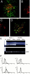
The transmembrane form of fractalkine is expressed by the human colorectal adenocarcinoma cell line, DLD-1. A: DLD-1, cells were grown to confluence on glass coverslips and stained using indirect immunofluorescence for transmembrane-expressed fractalkine using the anti-fractalkine chemokine domain (mouse α-Fkn, clone 51636.11; green) mAb and rabbit anti-C-peptide reagent (α-C-pep; red). Strong double labeling (orange) occurred on a subset of cells where the intracellular epitope was most strongly expressed. Lower levels of anti-chemokine domain staining could be detected on most cells. B: Anti-chemokine domain reagent specificity was demonstrated by double labeling using an isotype control antibody for the anti-chemokine mAb (green) and α-C-pep (red). α-C-pep staining was also competed out by addition of 10× molar excess of the immunizing peptide (data not shown). C: The α-Fkn (green) but not α-C-pep staining (red) couldbe competed totally by pre-incubation with a 10× molar excess of recombinant human fractalkine chemokine domain (rhFkn; 362-CX-025; R&D Systems). D: Cells were double-labeled with α-cytokeratin (clone AE1/AE3, DAKO; green). Original magnifications, ×400 (A–D). E: Total RNA was prepared from DLD-1 and HUVECs cultured with or without 10 U/ml TNF-α. RNA was reverse-transcribed and triplicate 25 ng cDNA samples subjected to PCR reactions using primers specific for fractalkine (Fkn) or HPRT. There was no fractalkine or HPRT signal amplified in reverse transcriptase samples (data not shown). F: DLD-1 cells were permeabilized and stained using i) mouse α-Fkn (clone 51636.11) mAb or control mouse IgG1 mAb (Serotech), ii) goat α-Fkn polyclonal or 10% goat serum, iii) α-C-pep or rabbit IgG, iv) α-N-pep polyclonal or rabbit IgG, and fractalkine expression analyzed by FACS. The bold trace shows the fluorescence of cells stained with the specific antibody, whereas the normal trace shows the background fluorescence of cells stained with the control reagent.
Figure 3.
Figure 3.
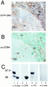
Rabbit α-N-pep polyclonal reagent cross-reacts with human CD84. Human tonsils were collected after routine tonsillectomy. Frozen sections (8 to 10 μm) were cut and examined for fractalkine expression by immunohistochemistry. Sections were stained using the rabbit α-N-pep polyclonal, counterstained with hematoxylin (A) or mouse anti-CD84 mAb, counterstained with methyl green (B) (α-CD84 13 ). Similar regions of sections from different tonsils are shown, which include part of a germinal center (GC). Similar cell types are detected by both reagents, including tingible body macrophages (TM), cells within the marginal zone (MZ), and endothelial cells (EC). An identical staining pattern was obtained using an independently produced rabbit α-N-pep polyclonal reagent. Negative control antibodies showed no background staining (data not shown). Original magnifications, ×400 (A–B). C: Western blot analysis of recombinant human fractalkine containing its N-terminus and mucin stalk (rhFKN, 365-FR-025; R&D Systems) and Western lysate taken from CD84-transfected cell line 300.19 CD-84; using anti-fractalkine reagents and an α-human CD84 mAb. Identical nitrocellulose membranes containing 20 ng of rhFKN (lanes 1, 3, 5, and 7) and 100 ng of CD84-transfected cell lysate (lanes 2, 4, 6, and 8), were probed using the α-N-pep (lanes 1 and 2, α-N-pep) and α-CD84 mAb (lanes 3 and 4, α-CD84). These membranes were stripped of bound antibody and then reprobed using goat α-Fkn (lanes 5 and 6) or chicken α-C-pep (lanes 7 and 8). α-N-pep detected two bands in the rhFKN sample (lane 1). The major band (90 kd) being the predicted size of this fractalkine product, the minor band possibly representing a degradation product. Furthermore, this reagent also detected a band in the CD84-transfected cell sample (lane 2, asterisk; 66 kd). The α-CD84 mAb detected this 66-kd band only within the CD84-transfected cell sample (lane 4, asterisk). The membrane probed with α-CD84 was stripped and reprobed with goat α-Fkn that detected the same two bands within the rhFKN sample as the α-N-pep (lane 5), but did not detect any bands within the CD84-transfected cell sample (lane 6). The membrane probed with α-N-pep was stripped and reprobed with chicken α-C-peptide and no bands were detected in either sample (lanes 7 and 8).
Figure 4.
Figure 4.
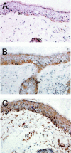
Transmembrane fractalkine is predominantly expressed in the epidermis of noninflamed human skin. Noninvolved skin samples were taken after resection of human facial skin tumors. Frozen sections (8 to 10 μm) were cut for analysis by immunohistochemistry. Immunohistochemical labeling was performed using anti-fractalkine and anti-peptide polyclonal reagents. A: The staining using the goat α-Fkn revealed positive cells restricted to the basal keratinocytes of the human epidermis, with no obvious positive cells within the dermis. B: Staining using rabbit α-C pep, which reacts to an intracellular epitope of fractalkine, revealed a similarly restricted staining pattern, again with no obvious positive cells within the dermis. C: Staining using the rabbit α-N-pep reagent showed discrete positive cell staining within the epidermis with morphology characteristic of Langerhans cells (a single cell is emphasized against the background), along the basement membrane of the epithelium with characteristics of melanocytes (M), there was also discrete staining of cells within the dermis, including structures with the appearance of blood vessels (EC) and dermal dendrocytes (DD). Negative control reagents showed no background staining (data not shown). Original magnifications, ×400.
Figure 5.
Figure 5.
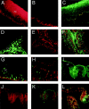
Transmembrane fractalkine is predominantly expressed in the epithelium of human large intestine and tonsil. Colon samples were taken after resection from Crohn’s or ulcerative colitis patients, whereas human tonsils were collected after routine tonsillectomy. Frozen sections (8 to 10 μm) were cut and examined for analysis using indirect immunofluorescence. A: Staining of the pharyngeal epithelium with mouse α-Fkn mAb (clone 51636.11; red) revealed a strong labeling of the epithelial cells but not underlying connective or lymphoid tissue. The staining was not uniform, with the basal layers more strongly detected than the next higher cell layers and then the strongest expression on the outer, more squamous layers. Negative control antibodies showed no background fluorescence within the pharyngeal epithelium (data not shown). No obvious DC labeling was detected. B: Staining with rabbit α-C-pep (red) that detects an intracellular epitope of fractalkine, was restricted to the basal layer of the epithelium. C: Double labeling of the pharyngeal epithelium for the expression of the chemokine domain (green) and intracellular epitope (red) revealed that transmembrane expression of fractalkine was restricted to the basal layer of the epithelium. D: Staining for fractalkine expression within the cortex of the tonsil looking for the chemokine domain of the molecule using mouse α-Fkn mAb (clone 51636.11; green) revealed that labeling was restricted to the crypt epithelium. This staining pattern was repeated using goat α-Fkn polyclonal reagent (data not shown). There was no obvious staining of cells within T or B cell areas and no obvious staining of endothelium. E: The expression of the intracellular epitope of fractalkine (α-C-pep; red) also localized to cells with a similar morphology within the crypt epithelia. Such staining was competed completely by inclusion of 10× molar excess of immunizing peptide (data not shown). F: Double labeling for the expression of the chemokine domain (mouse α-Fkn, clone 51636.11; green) and intracellular epitope (α-C-pep; red) showed a major subset of cells were strongly double-positive (orange). There were, however, other cells that were single-labeled for the chemokine domain. Strong labeling at the external surface of the crypt represents artifactual staining present in the isotype control (data not shown). G: Double-immunofluorescent labeling for the expression of CD1a-positive immature DCs (green) and the intracellular epitope of fractalkine (α-C-pep; red) showedthat although DCs are intimately associated with transmembrane expressed fractalkine, they do not express it. H: Double-immunofluorescent labeling for the expression of CD1a-positive immature DCs and the intracellular epitope of fractalkine within the cortex of the tonsil. Similar to the pharyngeal epithelium, there is a close association of single CD1a-positive immature DCs (green) with crypt epithelial cells positive for the transmembrane form of the fractalkine (red). I: Staining of human colon taken from an ulcerative colitis patient using an anti-fractalkine chemokine domain reagent (mouse α-Fkn, clone 51636.11, green) revealed strong staining within the epithelial cells of the lamina propria (LP). No obvious endothelial staining was detectable. J: Similarly, staining for the intracellular epitope (rabbit α-C-pep, red) was also restricted to the epithelial cells within the lamina propria. K: Double labeling for the chemokine domain (green) and the intracellular epitope (red) confirmed that the transmembrane fractalkine expression on epithelial cells within the lamina propria was the predominant form. L: An identical double-labeling staining pattern was seen within noninflamed large bowel sections. Human colon samples stained with the rabbit α-Fkn-pep reagent showed extensive staining of leukocytes and endothelium within the sections (data not shown). Original magnifications, ×125 (A–I), ×225 (J–L).
Figure 6.
Figure 6.
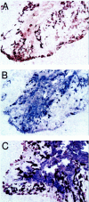
Transmembrane fractalkine is expressed by cytokeratin-positive cells with the human tonsil. Human tonsils were collected after routine tonsillectomy. Frozen sections (8 to 10 μm) were cut for immunohistochemical analysis. Adjacent serial sections were stained using the rabbit α-C-pep (A, brown) or mouse α-cytokeratin AE1/AE3 (B, blue). Another section ∼30 μmol/L further into the sample was double-labeled using both reagents (C). The rabbit α-C-pep reagent strongly detects a subpopulation of cells within the epithelial crypts. Staining for the cytokeratins AE1 and AE3 revealed staining restricted to the epithelial crypts with strong staining on cells with a similar distribution and morphology to A, with widespread but more diffuse staining throughout the crypts. Double labeling with rabbit α-C-pep (brown) and α-cytokeratin (blue) show clear double labeling (purple) of discrete cells within the crypts and especially along the internal border of the crypts. Original magnifications, ×400.

Similar articles

Cited by

References

    1. Bazan JF, Bacon KB, Hardiman G, Wang W, Soo K, Rossi D, Greaves DR, Zlotnik A, Schall TJ: A new class of membrane-bound chemokine with a CX3C motif. Nature 1997, 385:640-644 - PubMed
    1. Pan Y, Lloyd C, Zhou H, Dolich S, Deeds J, Gonzalo JA, Vath J, Gosselin M, Ma J, Dussault B, Woolf E, Alperin G, Culpepper J, Gutierrez-Ramos JC, Gearing D: Neurotactin, a membrane-anchored chemokine upregulated in brain inflammation. Nature 1997, 387:611-617 - PubMed
    1. Combadiere C, Salzwedel K, Smith ED, Tiffany HL, Berger EA, Murphy PM: Identification of CX3CR1. A chemotactic receptor for the human CX3C chemokine fractalkine and a fusion coreceptor for HIV-1. J Biol Chem 1998, 273:23799-23804 - PubMed
    1. Imai T, Hieshima K, Haskell C, Baba M, Nagira M, Nishimura M, Kakizaki M, Takagi S, Nomiyama H, Schall TJ, Yoshie O: Identification and molecular characterization of fractalkine receptor CX3CR1, which mediates both leukocyte migration and adhesion. Cell 1997, 91:521-530 - PubMed
    1. Harrison JK, Jiang Y, Chen S, Xia Y, Maciejewski D, McNamara RK, Streit WJ, Salafranca MN, Adhikari S, Thompson DA, Botti P, Bacon KB, Feng L: Role for neuronally derived fractalkine in mediating interactions between neurons and CX3CR1-expressing microglia. Proc Natl Acad Sci USA 1998, 95:10896-10901 - PMC - PubMed

Publication types

MeSH terms