. 2001 Mar;158(3):867-77.
doi: 10.1016/s0002-9440(10)64035-7.

The beta-chemokine receptor D6 is expressed by lymphatic endothelium and a subset of vascular tumors

Affiliations

The beta-chemokine receptor D6 is expressed by lymphatic endothelium and a subset of vascular tumors

R J Nibbs et al. Am J Pathol. 2001 Mar.

Abstract

The lymphatic vessels (lymphatics) play an important role in channeling fluid and leukocytes from the tissues to the secondary lymphoid organs. In addition to driving leukocyte egress from blood, chemokines have been suggested to contribute to leukocyte recirculation via the lymphatics. Previously, we have demonstrated that binding sites for several pro-inflammatory beta-chemokines are found on the endothelial cells (ECs) of lymphatics in human dermis. Here, using the MIP-1alpha isoform MIP-1alphaP, we have extended these studies to further support the contention that the in situ chemokine binding to afferent lymphatics exhibits specificity akin to that observed in vitro with the promiscuous beta-chemokine receptor D6. We have generated monoclonal antibodies to human D6 and showed D6 immunoreactivity on the ECs lining afferent lymphatics, confirmed as such by staining serial skin sections with antibodies against podoplanin, a known lymphatic EC marker. In parallel, in situ hybridization on skin with antisense D6 probes demonstrated the expression of D6 mRNA by lymphatic ECs. D6-immunoreactive lymphatics were also abundant in mucosa and submucosa of small and large intestine and appendix, but not observed in several other organs tested. In lymph nodes, D6 immunoreactivity was present on the afferent lymphatics and also in subcapsular and medullary sinuses. Tonsilar lymphatic sinuses were also D6-positive. Peripheral blood cells and the ECs of blood vessels and high endothelial venules were consistently nonreactive with anti-D6 antibodies. Additionally, we have demonstrated that D6 immunoreactivity is detectable in some malignant vascular tumors suggesting they may be derived from, or phenotypically similar to, lymphatic ECs. This is the first demonstration of chemokine receptor expression by lymphatic ECs, and suggests that D6 may influence the chemokine-driven recirculation of leukocytes through the lymphatics and modify the putative chemokine effects on the development and growth of vascular tumors.


Figures

Figure 1.
Figure 1.
In situ binding of radioiodinated MIP-1αP to lymphatic vessels in normal human skin. Cubes of viable human skin were incubated with 125I-MIP-1αP, sectioned, and the bound protein visualized as outlined in Materials and Methods. Tissue-bound 125I-MIP-1αP appears as black autoradiographic grains. Sections were counterstained with hemalaun. Thin arrows mark vessels with clear morphological features of lymphatics. Thick arrow indicates a resident dermal cell also labeled with 125I-MIP-1αP. The asterisk marks an unlabeled blood vessel. Original magnification, ×1,400.
Figure 2.
Figure 2.
FACS analysis of chemokine receptor transfectants with anti-D6 antibodies. Approximately 10 L1.2 cells stably expressing human chemokine receptors CCR1–5 or D6 were incubated with anti-D6 antibodies (50 μl of hybridoma supernatant from clone 4A5), and then antibody-containing complexes detected using a fluorescein isothiocyanate-coupled goat anti-mouse IgG and FACS analysis.
Figure 3.
Figure 3.
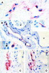
D6 immunoreactivity on lymphatic ECs in skin. Frozen sections of human skin immunostained with anti-D6 antibodies (A and D) or anti-podoplanin antibodies (C). In B, D6 immunoreactivity was detected on paraffin sections. Antibodies are visualized using an alkaline phosphatase-anti-alkaline phosphatase kit producing a red stain; all of the sections are counterstained with hemalaun. A: ECs lining two lymphatic capillaries in upper dermis display D6 immunoreactivity. Original magnification, ×2,000. B: Large collective lymphatic is stained by anti-D6 antibody, “a” marks an artery, “v” a vein, both immunonegative. Original magnification, ×900. Two insets contain resident non-ECs that show D6 immunoreactivity (both original magnifications, ×1,400). The asterisk indicates the epidermis. C and D are taken from adjacent serial sections and show a lymphatic vessel with podoplanin and D6 immunoreactivity, respectively. Original magnifications, ×1,200.
Figure 4.
Figure 4.
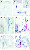
D6 mRNA expression by lymphatic ECs in the skin. Images taken from sections of human skin hybridized to DIG-labeled sense (A), or antisense (B, C, and E), D6 RNA and visualized with alkaline phosphatase-conjugated anti-DIG antibodies and BCIP/NBT solution. The presence of hybridized probe is indicated by a blue stain. D and F are sections adjacent to those used in C and E, respectively, and are immunostained with anti-podoplanin antibodies, visualized with an alkaline phosphatase-anti-alkaline phosphatase kit producing a red stain, and counterstained with hemalaun. A–D: Images taken from the upper dermis, with epidermis visible. B (inset), E and F: Images of deep dermal lymphatic vessels. Original magnifications: ×240 (A–D); ×100) (E and F).
Figure 5.
Figure 5.
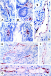
D6-immunoreactive lymphatic vessels in the gut. Paraffin-embedded sections of the gut immunostained with anti-D6 antibodies visualized using an avidin-biotin-peroxidase detection system and a peroxidase substrate producing a red stain. Representative D6 immunoreactivity associated with lymphatics in a longitudinal section of a small intestinal villus (A), lamina propria mucosae of the colon (B), a cross-section of two villi in the large intestine (C), muscular layer of the colon (D), lymphoid follicle in the lamina propria (E), lamina muscularis in normal appendix (F), and a patient with acute appendicitis (G). Original magnifications: ×400 (A), ×280 (B), ×530 (C), ×600 (D), ×440 (E, F, and G). The asterisk in D marks an immunonegative blood vessel. All sections counterstained with hematoxylin.
Figure 6.
Figure 6.
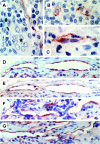
D6 immunoreactivity in secondary lymphoid organs. Representative images of paraffin-embedded sections from secondary lymphoid organs, immunostained with either anti-D6 (A–D and F), or anti-podoplanin (E and G), antibodies. Immunoreactivity is visualized using an avidin-biotin-peroxidase detection system and a peroxidase substrate producing a red stain; all sections counterstained with hematoxylin. A and B: D6 immunostaining in tonsils. Asterisk marks an HEV (both original magnifications, ×1,000). C: D6-immunoreactive vessel in red pulp of a spleen (original magnification, ×1,250). D–G: Lymph node. Adjacent sections showing subcapsular (D and E) and medullary (F and G) sinuses of the lymph node stained with anti-D6 (D and F) or anti-podoplanin (E and G) antibodies. Original magnifications: ×720 (D–G).
Figure 7.
Figure 7.
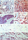
D6 immunoreactivity in vascular tumors. Paraffin-embedded sections of vascular tumors stained with anti-D6 antibodies (A–E), or anti-podoplanin antibody (F) with immunoreactivity visualized using an avidin-biotin-peroxidase detection system and a peroxidase substrate producing a red stain. All sections counterstained with hematoxylin. A, B, C, D, and E show tumors listed in Table 1 ▶ as numbers 1, 2, 3, 6, and 11, respectively. F shows a section adjacent to E stained with anti-podoplanin antibody. Only a small proportion of podoplanin-positive cells is D6-positive. Original magnifications: ×140 (A and C); ×300 (B); ×400 (D, E, and F).

References

    1. Jussila L, Valtola R, Partanen TA, Salven P, Heikkila P, Matikainen M-T, Renkonen R, Kaipainen A, Detmar M, Tschachler E, Alitalo R, Alitalo K: Lymphatic endothelium and Kaposi’s sarcoma spindle cells detected by antibodies against the vascular endothelial growth factor receptor-3. Cancer Res 1998, 58:1599-1604 - PubMed
    1. Lymboussaki A, Partanen TA, Olofsson B, Thomas-Crusells J, Fletcher CDM, de Waal RMW, Kaipainen A, Alitalo K: Expression of the vascular endothelial growth factor receptor VEGFR-3 in lymphatic endothelium of the skin and in vascular tumors. Am J Pathol 1998, 153:395-403 - PMC - PubMed
    1. Banerji S, Ni J, Wang S-X, Clasper S, Su J, Tammi R, Jones M, Jackson DG: LYVE-1, a new homologue of the CD44 glycoprotein, is a lymph-specific receptor for hyaluronan. J Cell Biol 1999, 144:789-801 - PMC - PubMed
    1. Breiteneder-Geleff S, Soleiman A, Kowalski H, Horvat R, Amann G, Kriehuber E, Diem K, Weninger W, Tschachler E, Alitalo K, Kerjaschki D: Angiosarcomas express mixed endothelial phenotypes of blood and lymphatic capillaries. Podoplanin as a specific marker for lymphatic endothelium. Am J Pathol 1999, 154:385-394 - PMC - PubMed
    1. Ebata N, Sawa Y, Ashikaga Y, Yamaoka Y, Suzuki M, Totsuka Y, Yoshida S: Lymphatic endothelium of the human tongue expresses multiple leukocyte adhesion molecules. Tissue Cell 1999, 31:34-38 - PubMed

Publication types

MeSH terms

LinkOut - more resources