. 2001 Mar;158(3):879-91.
doi: 10.1016/S0002-9440(10)64036-9.

Macrophage myeloperoxidase regulation by granulocyte macrophage colony-stimulating factor in human atherosclerosis and implications in acute coronary syndromes

Affiliations

Macrophage myeloperoxidase regulation by granulocyte macrophage colony-stimulating factor in human atherosclerosis and implications in acute coronary syndromes

S Sugiyama et al. Am J Pathol. 2001 Mar.

Abstract

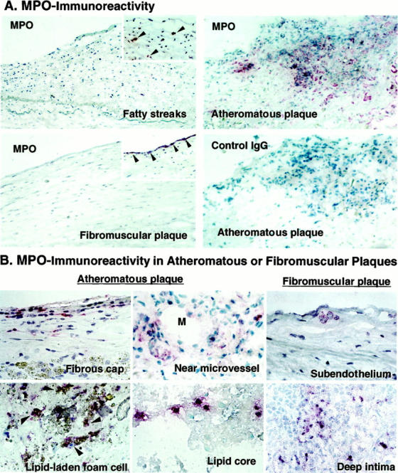
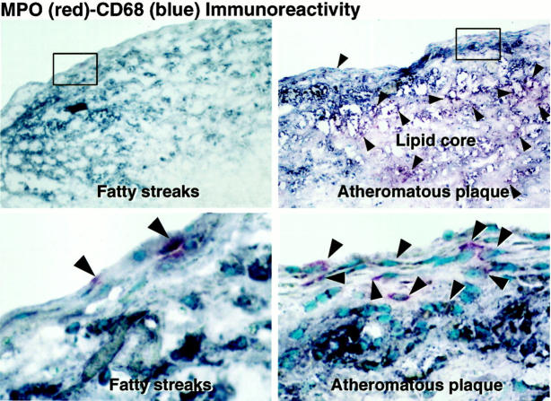
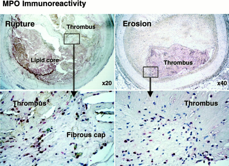
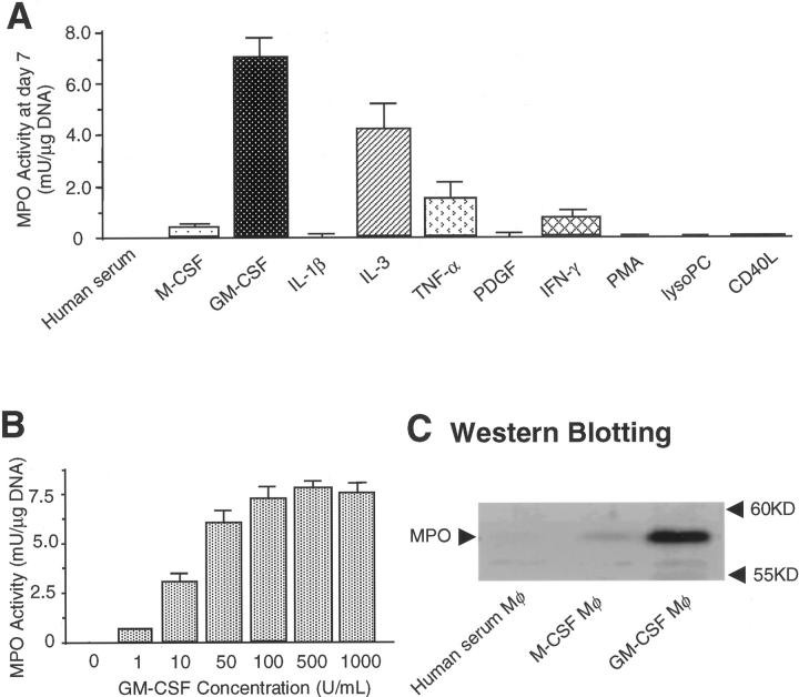
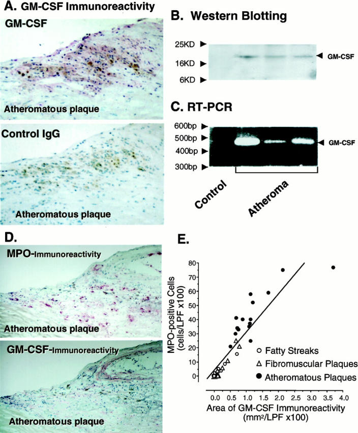
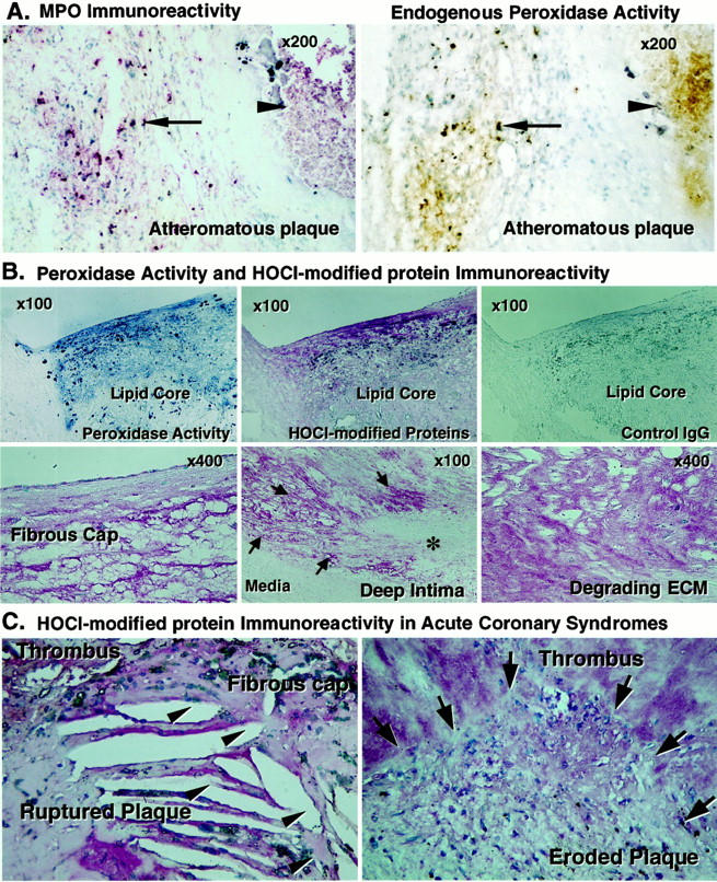
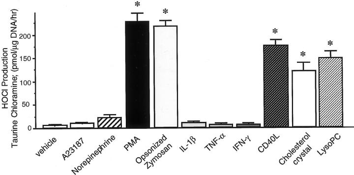
Inflammation and oxidative stress contribute to the pathogenesis of many human diseases including atherosclerosis. Advanced human atheroma contains high levels of the enzyme myeloperoxidase that produces the pro-oxidant species, hypochlorous acid (HOCl). This study documents increased numbers of myeloperoxidase-expressing macrophages in eroded or ruptured plaques causing acute coronary syndromes. In contrast, macrophages in human fatty streaks contain little or no myeloperoxidase. Granulocyte macrophage colony-stimulating factor, but not macrophage colony-stimulating factor, selectively regulates the ability of macrophages to express myeloperoxidase and produce HOCl in vitro. Moreover, myeloperoxidase-positive macrophages in plaques co-localized with granulocyte macrophage colony-stimulating factor. Pro-inflammatory stimuli known to be present in human atherosclerotic plaque, including CD40 ligand, lysophosphatidylcholine, or cholesterol crystals, could induce release of myeloperoxidase from HOCl production by macrophages in vitro. HOCl-modified proteins accumulated at ruptured or eroded sites of human coronary atheroma. These results identify granulocyte macrophage colony-stimulating factor as an endogenous regulator of macrophage myeloperoxidase expression in human atherosclerosis and support a particular role for the myeloperoxidase-expressing macrophages in atheroma complication and the acute coronary syndromes.


Figures

Figure 1.
Figure 1.
Expression of MPO immunoreactivity in various stages of human atherosclerotic arteries and localization in advanced plaques. Frozen sections were incubated with pAb-MPO and MPO immunoreactivity was visualized with the alkaline-phosphatase ABC method (red). A: MPO immunoreactivity localized in early to advanced human atherosclerosis (arrowheads). Atheromatous plaques exhibited abundant MPO, but lacked staining by nonimmune rabbit IgG used as a negative control. Arteries with fatty streaks, and fibromuscular plaques usually exhibited little MPO (large panels), but some samples contained MPO in the intima (insets). Original magnification, ×200. These results are representative of 14 fatty streaks, 17 fibromuscular plaques, and 25 atheromatous plaques. B: MPO-containing cells were localized in all regions of advanced plaques, especially in fibrous cap, near microvessel (M, indicates lumen of microvessel), and lipid core of atheromatous plaques. Some of fibromuscular plaques contained MPO-containing cells in the subendothelial space and deep intima. Some of the lipid-laden foam cells were also MPO-positive in atheroma (arrowheads). Original magnification, ×400.
Figure 2.
Figure 2.
MPO-positive macrophages and MPO-negative macrophages in human atherosclerosis. Fatty streaks and atheromatous plaques were examined by double immunostaining with pAb-MPO (red) and mAb-CD68 (blue). Bottom: A high magnification of each of the top panels. MPO-positive cells were CD68-positive mononuclear cells in both fatty streaks and atheromatous plaques (purple, arrowheads). Fatty streaks contained many intimal CD68-positive mononuclear cells but few MPO-positive cells. In atheromatous plaques, many MPO-positive cells were CD68-positive macrophages (purple, arrowheads), although some CD68-positive macrophages lacked MPO (blue). Original magnification, ×100 (top), ×400 (bottom). These results are representative of six fatty streaks and 10 atheromatous plaques.
Figure 3.
Figure 3.
MPO immunoreactivity in ruptured and eroded coronary plaques in patients with acute coronary syndromes. Paraffin sections of ruptured or eroded plaques were incubated with pAb-MPO and MPO immunoreactivity was visualized with the alkaline-phosphatase ABC method (red), MPO-positive macrophages (red) were prominent at rupture sites in atheroma and at sites where fibromuscular plaques had eroded in victims of sudden cardiac death. Bottom: High-power views (original magnifications, ×400) of each upper rectangle area. These results are representative of six eroded and eight ruptured plaques.
Figure 4.
Figure 4.
GM-CSF, but not M-CSF, modulates macrophage phenotype to express MPO in vitro. MPO activity (guaiacol peroxidation method) and MPO protein were examined in cellular lysate. A: GM-CSF (100 U/ml) and IL-3 (10 ng/ml), but not M-CSF (100 U/ml), preserved MPO activity in monocyte-derived macrophages during 7 days in culture (n = 3). IL-1β (10 ng/ml), tumor necrosis factor-α (10 ng/ml), PDGF (10 ng/ml), interferon-γ (1,000 U/ml), PMA (100 nmol/L), lysoPC (15 μmol/L), and CD40L (2 μg/ml). Each factor was added to the medium on the 0, second, fourth, and sixth day in culture. B: The effect of GM-CSF on MPO activity depended on its concentration (10 to 500 U/ml) (n = 3). C: Extracts of cultured macrophages containing 2 μg of DNA were separated by sodium dodecyl sulfate-polyacrylamide gel electrophoresis. GM-CSF-treated macrophages, but not human serum-treated macrophages and M-CSF-treated macrophages expressed MPO protein. Each result represents three independent experiments.
Figure 5.
Figure 5.
Human atherosclerotic arteries express GM-CSF and MPO-positive macrophages localize in the GM-CSF-expressing atherosclerotic intima. Frozen sections were incubated with mAb-GM-CSF and GM-CSF immunoreactivity was visualized with the alkaline-phosphatase ABC method (red). A: Advanced atheromatous plaques expressed substantial GM-CSF immunoreactivity in the intima, but lacked staining by nonimmune mouse IgG1 used as a negative control. These results are representative of those obtained by study of 15 atheromatous plaques. Original magnification, ×200. B: Each total tissue homogenate of nondiseased aorta and advanced atheromatous carotid arteries (100 μg of protein/lane) was separated by sodium dodecyl sulfate-polyacrylamide gel electrophoresis, and GM-CSF protein was determined by Western blotting. C: GM-CSF mRNA expression was determined by RT-PCR analysis as described in Materials and Methods. The advanced atheromatous carotid arteries, but not nondiseased aorta, expressed GM-CSF protein and mRNA. D: MPO and GM-CSF immunoreactivity in the adjacent sections of atheromatous plaques (red). The MPO-positive macrophages localized in the GM-CSF-expressing intima of atheroma. These results are representative of those obtained by study of 15 atheromatous plaques. Original magnification, ×200. E: MPO-positive cells were counted in the low-power field (×100), and GM-CSF-immunoreactive areas were selected and quantitated by computer-assisted image analysis. (open circle, fatty streaks; open triangle, fibromuscular plaques, filled circle, atheromatous plaques). The areas of immunoreactive GM-CSF and the number of MPO-positive macrophages correlated significantly in human atherosclerotic lesions (R2=0.84, P < 0.001).
Figure 6.
Figure 6.
MPO, endogenous peroxidase activity, and HOCl-modified proteins co-localized in advanced atheroma and HOCl-modified proteins abound in culprit lesions of acute coronary syndromes. A: Frozen sections of advanced atheromatous plaques were incubated with pAb-MPO and MPO immunoreactivity was visualized with the alkaline-phosphatase ABC method (red), and endogenous peroxidase activity was examined using the diaminobenzidine peroxidation method (brown). The adjacent sections of atheromatous plaques show that MPO immunoreactivity (left) and endogenous peroxidase activity (right) co-localized intracellularly in the intima and extracellularly in the lipid core (arrow, cellular MPO; arrowhead, lipid core MPO). These results are representative of 12 atheromatous plaques. B: Frozen (top, adjacent sections) and paraffin sections of advanced atheromatous plaques (bottom) were incubated with mAb-HOCl-modified proteins and the immunoreactivity was visualized with the alkaline-phosphatase ABC method (red). Endogenous peroxidase activity was examined using the tetramethylbenzidine peroxidation method (blue). The endogenous peroxidase activity and immunoreactive HOCl-modified proteins co-localized in the intima of advanced atheroma, but the lesions lacked staining by nonimmune mouse IgG2b used as a negative control (top, n = 8). The thin fibrous cap (left),areas of matrix fragmentation near the lipid core (middle, arrows; degrading area), and fragmented ECM in atheroma (right), but not the area of intact ECM (asterisk) and media (middle), contain HOCl-modified protein epitopes. These results are representative of 12 atheromatous plaques. C: Paraffin sections of ruptured or eroded coronary plaques were incubated with mAb-HOCl-modified proteins and HOCl-modified protein immunoreactivity was visualized with the alkaline-phosphatase ABC method (red). Shown is substantial staining of HOCl-modified protein immunoreactivity at the site of ruptured fibrous cap and erosion. (arrowhead, ruptured site; arrow, eroded site). These results are representative of six eroded and eight ruptured plaques.
Figure 7.
Figure 7.
MPO-positive macrophages in vitro produce HOCl in response to atherogenic stimuli. Bar graph indicates HOCl production from GM-CSF (100 U/ml)-treated macrophages on day 7 measured by taurine chloramine generation. PMA (300 nmol/L), opsonized zymosan (OZ) (0.2 mg/ml), lysoPC (15 μmol/L), CD40L (5 μg/ml), or cholesterol crystals (1 mg/ml) induced HOCl production in macrophages, but A23187 (1 μmol/L), norepinephrine (10 μmol/L), IL-1β (10 ng/ml), tumor necrosis factor-α (50 ng/ml), and interferon-γ (1,000 U/ml) had no effect (*, P < 0.01 versus vehicle, n = 3).

References

    1. Ross R: Atherosclerosis: an inflammatory disease. N Engl J Med 1999, 340:115-126 - PubMed
    1. Daugherty A, Dunn JL, Rateri DL, Heinecke JW: Myeloperoxidase, a catalyst for lipoprotein oxidation, is expressed in human atherosclerotic lesions. J Clin Invest 1994, 94:437-444 - PMC - PubMed
    1. Hazell LJ, Arnold L, Flowers D, Waeg G, Malle E, Stocker R: Presence of hypochlorite-modified proteins in human atherosclerotic lesions. J Clin Invest 1996, 97:1535-1544 - PMC - PubMed
    1. Hazen SL, Heinecke JW: 3-Chlorotyrosine, a specific marker of myeloperoxidase-catalyzed oxidation, is markedly elevated in low density lipoprotein isolated from human atherosclerotic intima. J Clin Invest 1997, 99:2075-2081 - PMC - PubMed
    1. Leeuwenburgh C, Rasmussen JE, Hsu FF, Mueller DM, Pennathur S, Heinecke JW: Mass spectrometric quantification of markers for protein oxidation by tyrosyl radical, copper, and hydroxyl radical in low density lipoprotein isolated from human atherosclerotic plaques. J Biol Chem 1997, 272:3520-3526 - PubMed

Publication types

MeSH terms