. 2001 Mar;158(3):905-19.
doi: 10.1016/S0002-9440(10)64038-2.

Expression of hypoxia-inducible cell-surface transmembrane carbonic anhydrases in human cancer

Affiliations

Expression of hypoxia-inducible cell-surface transmembrane carbonic anhydrases in human cancer

S Ivanov et al. Am J Pathol. 2001 Mar.

Abstract

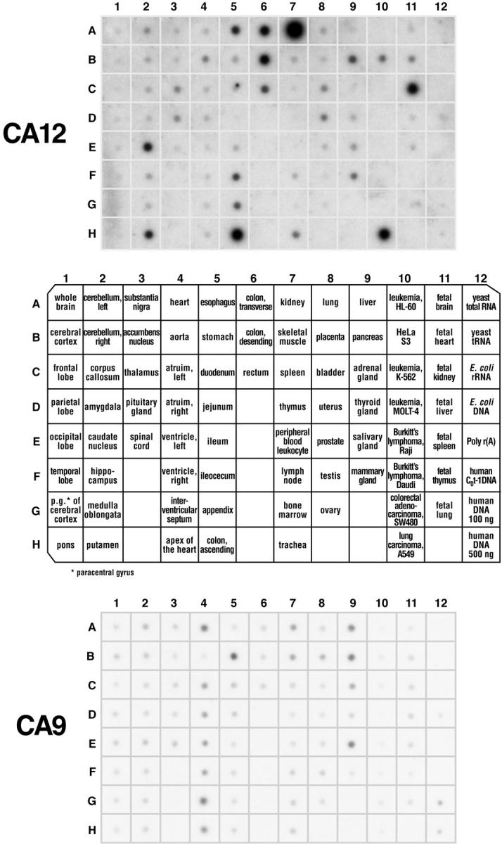
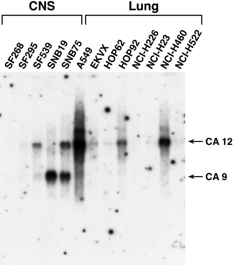
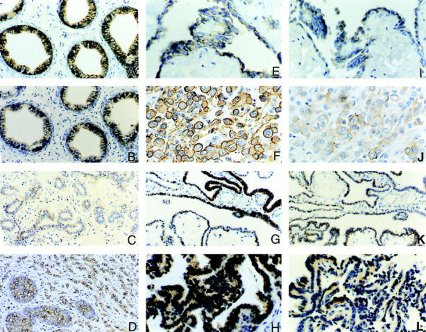
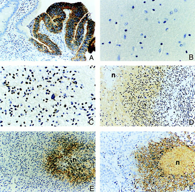
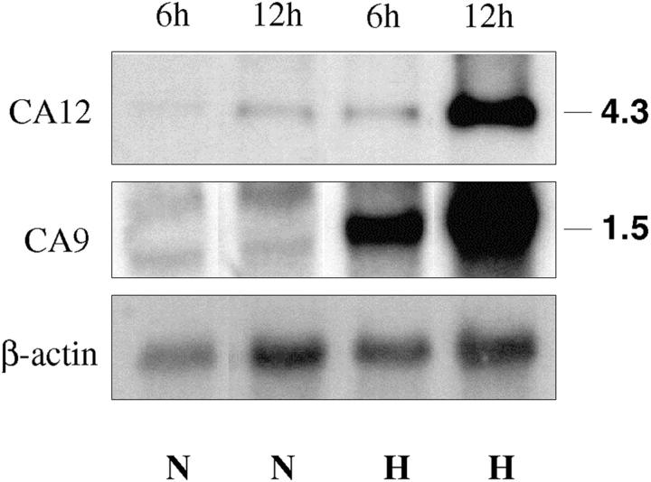
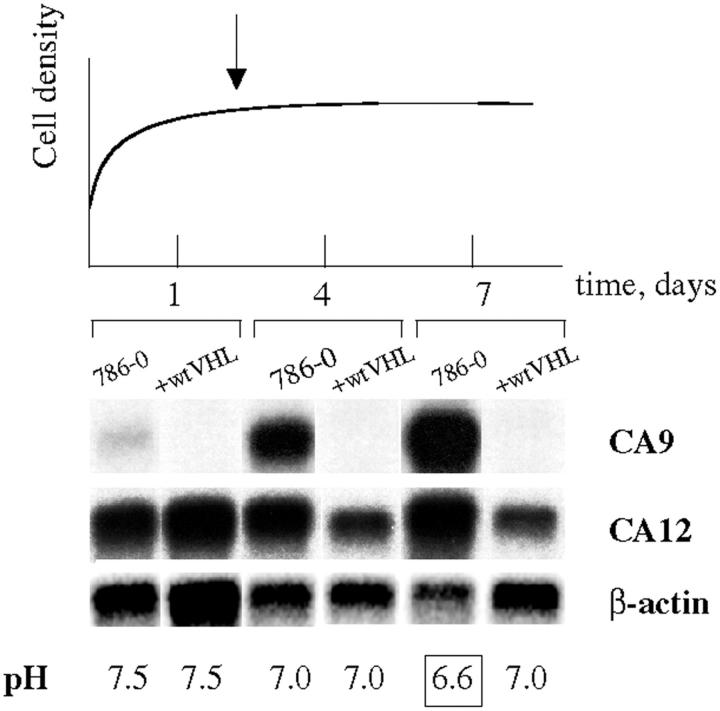
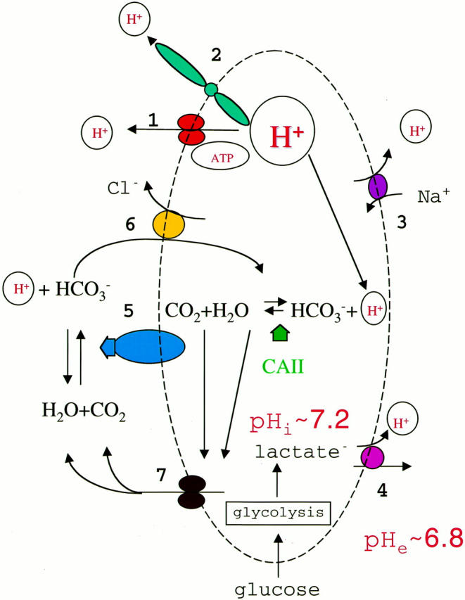
An acidic extracellular pH is a fundamental property of the malignant phenotype. In von Hippel-Lindau (VHL)-defective tumors the cell surface transmembrane carbonic anhydrase (CA) CA9 and CA12 genes are overexpressed because of the absence of pVHL. We hypothesized that these enzymes might be involved in maintaining the extracellular acidic pH in tumors, thereby providing a conducive environment for tumor growth and spread. Using Northern blot analysis and immunostaining with specific antibodies we analyzed the expression of CA9 and CA12 genes and their products in a large sample of cancer cell lines, fresh and archival tumor specimens, and normal human tissues. Expression was also analyzed in cultured cells under hypoxic conditions. Expression of CA IX and CA XII in normal adult tissues was detected only in highly specialized cells and for most tissues their expression did not overlap. Analysis of RNA samples isolated from 87 cancer cell lines and 18 tumors revealed high-to-moderate levels of expression of CA9 and CA12 in multiple cancers. Immunohistochemistry revealed high-to-moderate expression of these enzymes in various normal tissues and multiple common epithelial tumor types. The immunostaining was seen predominantly on the cell surface membrane. The expression of both genes was markedly induced under hypoxic conditions in tumors and cultured tumor cells. We conclude that the cell surface trans-membrane carbonic anhydrases CA IX and CA XII are overexpressed in many tumors suggesting that this is a common feature of cancer cells that may be required for tumor progression. These enzymes may contribute to the tumor microenvironment by maintaining extracellular acidic pH and helping cancer cells grow and metastasize. Our studies show an important causal link between hypoxia, extracellular acidification, and induction or enhanced expression of these enzymes in human tumors.


Figures

Figure 1.
Figure 1.
Transcription analysis of CA9/CA12 genes in normal tissues. Two identical membranes (No. 7775-1, Multiple tissue expression array; Clontech) contained human poly-A+ RNA samples extracted from 68 different normal tissues, eight cancer cell lines, and control samples of DNA and RNA. CA12 mRNA is expressed predominantly in the kidney (A7) and colon (H5), whereas CA9 is expressed mainly in the stomach (B5), liver (A9), pancreas (B9), salivary gland (E9), and heart (A4).
Figure 2.
Figure 2.
Activation of CA9 and CA12 transcription in cell lines derived from CNS and lung tumors. A typical Northern blot analysis of mRNA samples isolated from cultured cells is shown. Both probes were used together in the same hybridization experiment. Loading was monitored by staining the membranes with methylene blue and hybridization with an actin probe. The data extracted from different membranes are summarized in Table 1 ▶ .
Figure 3.
Figure 3.
Examples of diffuse immunostaining of CA IX and CA XII proteins in normal and neoplastic tissues. Immunostaining for CA IX is illustrated in A, E, F, G, and H. Immunostaining for CA XII is illustrated in B, C, D, I, J, K, and L. In normal adult tissues, diffuse plasma membrane immunostaining for both CA IX and CA XII is seen in the ductular efferens (A and B), mesothelial cells of the body cavity (E and I), and the choroid plexus of brain ventricles (G and K). Variable degrees of expression of CA XII but not CA IX are also observed in the epithelium of the breast (C). Persistent expression of CA IX and CA XII proteins is also seen in tumors derived from the normal tissues that express these proteins. For example, a lobular-type breast carcinoma expressing CA XII is shown in D, mesotheliomas of the pleura in F and J, and choroid plexus papillomas in H and L. Original magnifications, ×200 (A, B, G, and K) and ×400 (D–F, H–J, and L).
Figure 4.
Figure 4.
Examples of ectopic expression of CA IX and CA XII proteins in various tumors, including regions of necrosis/hypoxia. The glandular cells of the colon are normally CA IX-negative but diffuse immunoreactivity is seen in colon carcinoma (A). The normal glial tissues of the brain show no expression of either CA IX or CA XII protein (B). However, variable degrees of positive staining for CA XII are seen in low-grade glioma (C). In contrast, high levels of CA IX protein expression are observed in glioblastoma multiforme (D) and anaplastic ependymoma (E). Expression of the CA IX protein is seen in necrotic/hypoxic regions of the tumors. This induced expression associated with necrotic/hypoxic regions is also seen in high-grade ductal breast carcinoma (F). The symbol n = area of necrosis. Original magnifications, ×200 (A, D–F) and ×400 (B and C).
Figure 5.
Figure 5.
Immunostaining of normal kidney and renal cell tumors for CA IX/CA XII expression. The normal kidney is negative for CA IX (A) whereas CA XII is expressed in the distal convoluted tubules (arrow) and intercalated cells of the collecting ducts (arrowhead) (B). Co-expression of CA IX and CA XII is seen in renal cell carcinomas of the clear cell type (CA IX = C; CA XII = D). However, chromophobe cell carcinomas (E) and oncocytomas (G) do not express CA IX whereas they exhibit diffuse expression of CA XII (F and H, respectively). Original magnifications, ×200 (A and B) and ×400 (C–H).
Figure 6.
Figure 6.
Up-regulation of CA IX/CA XII expression by hypoxia in the glioblastoma cell line, U87. Levels of transcripts under normoxic (N) and hypoxic (H) conditions at different time points were assessed by Northern blot analysis with total RNA samples. After 6 hours of hypoxia an ∼4- and 1.6-fold increase in CA IX and CA XII mRNA expression, respectively, was observed. Twelve hours of hypoxia caused ∼13- and 11-fold up-regulation of CA IX and CA XII, respectively.
Figure 7.
Figure 7.
Up-regulation of CA9 and CA12 mRNA expression correlates with media acidification. 786-0 cells were harvested at different stages of the growth curve and levels of CA9 and CA12 transcripts were assessed by Northern hybridization. In parallel, glucose consumption (not shown) and changes in media pH were monitored. The onset of confluent stage (day 2) is indicated by an arrow. Enhanced acidification of the media by the VHL-deficient cell line 786-0 is pronounced at the late confluent stage (day 7, boxed) and is paralleled by up-regulation in CA9 and CA12 mRNA expression.
Figure 8.
Figure 8.
Molecular mechanisms of proton extrusion/secretion from cancer cells. To maintain intracellular neutrality excessive protons are extruded from the cancer cell by several protein machines, including plasma membrane proton pump (H+-ATPase, no. 1), proton channels (no. 2), sodium/proton exchangers (no. 3), and monocarboxylate carriers (no. 4). CA IV, CA IX, or CA XII (no. 5) may produce bicarbonate ions outside the cell that are transported inside by bicarbonate/chloride exchangers (no. 6) and used by the cytosolic CA II to titrate protons. Buffering protons facilitates proton secretion and protects the cell from intracellular acidification. Water and carbon dioxide then are transported out through aquaporins (no. 7) and used by cell-surface CAs to produce bicarbonate.

References

    1. Semenza GL: Hypoxia, clonal selection, and the role of HIF-1 in tumor progression. Crit Rev Biochem Mol Biol 2000, 35:71-103 - PubMed
    1. Hanahan D, Folkman J: Patterns and emerging mechanisms of the angiogenic switch during tumorigenesis. Cell 1996, 86:353-364 - PubMed
    1. Warburg O: The Metabolism of Tumours. 1930. Constable, London
    1. Dang CV, Semenza GL: Oncogenic alterations of metabolism. Trends Biochem Sci 1999, 24:68-72 - PubMed
    1. Stubbs M, McSheehy PM, Griffiths JR, Bashford CL: Causes and consequences of tumour acidity and implications for treatment. Mol Med Today 2000, 6:15-19 - PubMed

Publication types

MeSH terms

LinkOut - more resources