. 2001 Mar;158(3):955-66.
doi: 10.1016/S0002-9440(10)64042-4.

HOX genes in human lung: altered expression in primary pulmonary hypertension and emphysema

Affiliations

HOX genes in human lung: altered expression in primary pulmonary hypertension and emphysema

H A Golpon et al. Am J Pathol. 2001 Mar.

Abstract

HOX genes belong to the large family of homeodomain genes that function as transcription factors. Animal studies indicate that they play an essential role in lung development. We investigated the expression pattern of HOX genes in human lung tissue by using microarray and degenerate reverse transcriptase-polymerase chain reaction survey techniques. HOX genes predominantly from the 3' end of clusters A and B were expressed in normal human adult lung and among them HOXA5 was the most abundant, followed by HOXB2 and HOXB6. In fetal (12 weeks old) and diseased lung specimens (emphysema, primary pulmonary hypertension) additional HOX genes from clusters C and D were expressed. Using in situ hybridization, transcripts for HOXA5 were predominantly found in alveolar septal and epithelial cells, both in normal and diseased lungs. A 2.5-fold increase in HOXA5 mRNA expression was demonstrated by quantitative reverse transcriptase-polymerase chain reaction in primary pulmonary hypertension lung specimens when compared to normal lung tissue. In conclusion, we demonstrate that HOX genes are selectively expressed in the human lung. Differences in the pattern of HOX gene expression exist among fetal, adult, and diseased lung specimens. The altered pattern of HOX gene expression may contribute to the development of pulmonary diseases.


Figures

Figure 1.
Figure 1.
HOX gene expression pattern in normal mouse lung (top) and human adult lung tissue (bottom) assessed by the microarray technique (Affymetrix gene chip). Each solid box marks a HOX gene represented on the array, whereas the dotted line boxes represent the genes that are not included on the array. The order of genes within a cluster is depicted in the 5′ to 3′ orientation and paralogous genes are aligned vertically. The shaded boxes represent the genes that were expressed.
Figure 2.
Figure 2.
HOX gene expression pattern in the fetal lung (12 weeks of gestation age, pooled sample), normal adult (n = 2), and diseased human lungs (emphysema, n = 3; PPH, n = 3) assessed by RT-PCR using degenerate primers for the HOX domain. The products were cloned into bacteria and sequenced. Each box represents a known HOX gene. The order of genes within a cluster is depicted in the 5′ to 3′ orientation and paralogous genes are aligned vertically. The open boxes represent HOX genes that were not identified by RT-PCR, whereas the shaded boxes represent genes that were detected. The frequency at which positive clones were identified is expressed by the number in the shaded box. Twenty-seven clones originating from the fetal lung were sequenced (normal adult lung, 47 clones; emphysema, 51 clones; PPH, 47 clones).
Figure 3.
Figure 3.
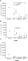
Expression of HOXA5 (A), HOXB2 (B), and HOXB6 (C) in human lung tissues assessed by quantitative RT-PCR. Each data point represents the average result for duplicate samples from the same tissue. The initial amount of RNA was assessed using TaqMan ribosomal RNA control reagents. Samples were quantified by comparison against standard curves with known copy numbers (plasmids containing cloned HOXA5, HOXB2, and HOXB6 sequences). Fetal lung (pool of lungs with gestation age of 12 weeks), PPH (n = 6), emphysema (n = 6), normal (histologically normal human adult lung tissue, n = 6). The mean ± SD of expression for each group is also shown.
Figure 4.
Figure 4.
A: HOXA5 expression in pseudoglandular stage fetal lung, showing a strong signal in septal epithelial cells (arrows), and a somewhat weaker expression in mesenchymal cells (asterisks). B: Abundant HOXA5 expression in bronchial epithelial cells in a canalicular-stage fetal lung (arrows). Note a weaker hybridization signal in mesenchymal cells (asterisks) and bronchial cartilage (arrowheads, right upper corner). C: Abundant HOXA5 expression in alveolar duct epithelial cells, with a signal similar to that expressed by epithelial cells shown in A in a canalicular-stage fetal lung (arrows). Note a weaker hybridization signal in several of the epithelial and septal mesenchymal cells (asterisks). D: Hybridization control with the sense probe for HOXA5 mRNA in the same lung field shown in B. E: Negative control with RNase digestion before hybridization with the antisense probe for HOXA5 mRNA in a similar lung field as shown in C. Original magnifications: ×400 (A, C, and E), ×300 (B and D).
Figure 5.
Figure 5.
A: Normal lung shows HOXA5 mRNA expression in septal epithelial cells (arrows), including type II pneumocytes (arrowhead). The hybridization signal appears as a dark-blue reaction in both the cytoplasm and overlying the nuclei. Inset a shows a low-power view, which highlights the overall HOXA5 expression both in alveolar septa and pulmonary vascular wall (V). B: Hybridization control with the sense probe for HOXA5 mRNA in the same lung field shown in A. C: Emphysema lung shows overall decreased HOXA5 mRNA expression, both in alveolar septal cells (arrows), and in vascular wall (inset c). D: PPH lung demonstrates intense hybridization signal for HOXA5 along alveolar septa (arrows), including type II pneumocytes (arrowhead), and vascular structures (inset d). Original magnification: ×400; insets: ×100 (A, C, D: antisense, digoxigenin-labeled probe; B: sense). E: HOXB2 mRNA expression in alveolar septal cells in normal adult lung tissue. Inset e shows the overall expression pattern of HOXB2 mRNA. The expression pattern is similar to that seen with HOXA5. F: HOXB6 mRNA expression in alveolar septal cells in normal adult lung tissue. Inset f shows the overall expression pattern of HOXB6 mRNA, which is markedly reduced when compared with that shown for HOXA5 and HOXB2 mRNA. Original magnifications: ×400; insets, ×100 (A, C, D, E, F: antisense, digoxigenin-labeled probe; B: sense).
Figure 6.
Figure 6.
A: Bronchiolar lining cells in a terminal bronchiole (b) of a normal lung express abundant HOXA5 mRNA (arrows). B: A strong hybridization signal for HOXA5 mRNA was noted in endothelial cells (arrows) of normal lung tissue. The smooth muscle cells exhibit a weaker, but clear signal (arrowheads). An overview of the vessel wall is shown in the insert. C: Localization of HOXA5 mRNA in a concentric lesion from a patient with PPH. Note the intense hybridization signal for cells filling the lumen of the vessel (arrows, probably endothelial cells) and the less intense hybridization signal in myofibroblasts (arrowheads). Original magnifications, ×100 (inset) and ×400 (A, B, and C). Antisense, digoxigenin-labeled probe. V = vessel.

References

    1. Mark M, Rijli FM, Chambon P: Homeobox genes in embryogenesis and pathogenesis. Pediatr Res 1997, 42:421-429 - PubMed
    1. Bogue CW, Lou LJ, Vasavada H, Wilson CM, Jacobs HC: Expression of Hoxb genes in the developing mouse foregut and lung. Am J Respir Cell Mol Biol 1996, 15:163-171 - PubMed
    1. Krumlauf R: Hox genes in vertebrate development. Cell 1994, 78:191-201 - PubMed
    1. Boncinelli E: Homeobox genes and disease. Curr Opin Genet Dev 1997, 7:331-337 - PubMed
    1. Stein S, Fritsch R, Lemaire L, Kessel M: Checklist: vertebrate homeobox genes. Mech Dev 1996, 55:91-108 - PubMed

Publication types

MeSH terms