. 2001 Mar;158(3):1091-100.
doi: 10.1016/S0002-9440(10)64056-4.

High glucose-induced hypertrophy of mesangial cells requires p27(Kip1), an inhibitor of cyclin-dependent kinases

Affiliations

High glucose-induced hypertrophy of mesangial cells requires p27(Kip1), an inhibitor of cyclin-dependent kinases

G Wolf et al. Am J Pathol. 2001 Mar.

Abstract

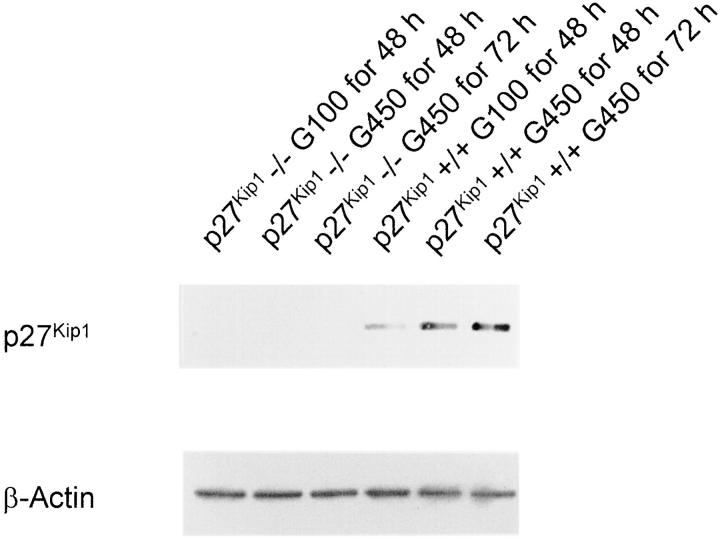
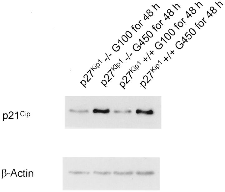
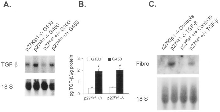
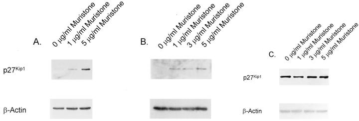
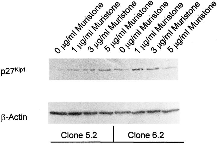
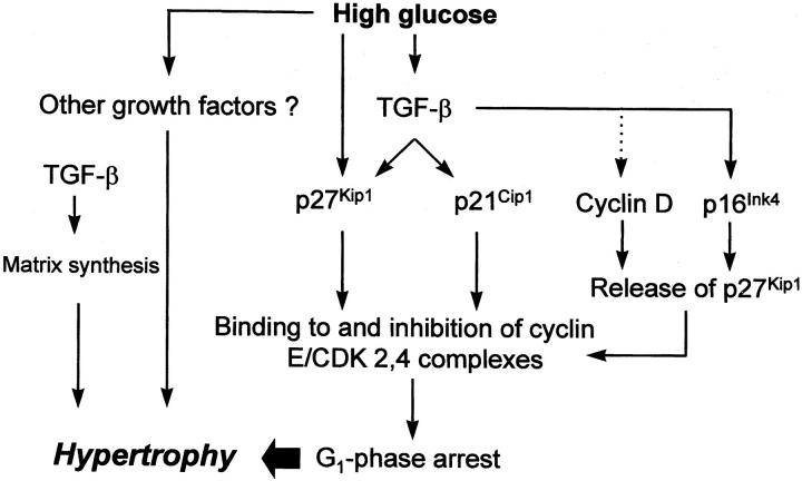
Hypertrophy of mesangial cells is one of the earliest morphological alterations in the kidney after the onset of diabetes mellitus. We have previously shown that cultured mesangial cells exposed to high ambient glucose arrest in the G1 phase of the cell cycle and that this is associated with an increased expression of inhibitors of the cyclin-dependent kinase (CDK)-inhibitors p21(Cip) and p27(Kip1). To further investigate a potential role of p27Kip1 in the development of glucose-induced hypertrophy, mesangial cells from p27Kip1 wild-type (+/+) and knockout (-/-) mice were established. High glucose medium (450 mg/dl) increased p21(Cip1) protein in p27Kip1+/+ and -/- mesangial cells, and increased p27Kip1 protein levels in p27Kip1+/+ cells. In contrast to high glucose increasing de novo protein synthesis in p27Kip1+/+ cells, high glucose did not increase protein synthesis in p27Kip1-/- cells. High glucose also reduced DNA synthesis and caused cell cycle arrest in p27Kip1+/+ cells. In contrast, despite an increase in transforming growth factor (TGF)-beta mRNA and protein expression, DNA synthesis and cell cycle progression were increased by high glucose in p27Kip1-/- cells. Exogenous TGF-beta comparably induced fibronectin mRNA in p27Kip1+/+ and -/- cells suggesting intact TGF-beta receptor transduction. In addition, high glucose failed to increase the total protein/cell number ratio in p27Kip1-/- cells. However, in the presence of high glucose, reconstituting p27Kip1 expression by transient or stable transfection in p27Kip1-/- cells, using an inducible expression system, increased the de novo protein synthesis and restored G1-phase arrest. These results show that p27Kip1 is required for glucose-induced mesangial cell hypertrophy and cell cycle arrest.


Figures

Figure 1.
Figure 1.
Western blot of total cell lysates incubated with an antibody against p27Kip1. p27Kip1 expression was, as predicted, not induced in p27Kip1−/− mesangial cells incubated in serum-free medium with high glucose (G450; 450 mg/dl) for up to 72 hours. However, a strong high glucose-mediated induction of p27Kip1 was detectable in wild-type (p27Kip1+/+) cells. The membrane was re-incubated with an antibody against β-actin to demonstrate that these changes were not because of unequal loading or transfer of proteins. This blot is representative of three independent experiments with qualitatively similar results.
Figure 2.
Figure 2.
Western blot for p21Cip1 expression. In contrast to p27Kip1, high glucose for 48 hours increased p21Cip1 protein abundance in p27Kip1−/− and +/+ mesangial cells. This blot is representative of three independent experiments with qualitatively similar results.
Figure 3.
Figure 3.
A: mRNA expression of TGF-β1. High glucose medium induces TGF-β transcripts independent of whether cells express p27Kip1 or not. This blot is representative of three independent experiments with qualitatively similar results. B: High glucose also leads to an equal amount of TGF-β1 protein synthesis in p27Kip1+/+ and −/− mesangial cells. *, P < 0.01, n = 5. C: Exogenous TGF-β1 induced fibronectin mRNA expression in p27Kip1+/+ and −/− cells indicating that both cell lines express TGF-β receptors and exhibit appropriate signal transduction systems downstream of the receptors. This blot is representative of three independent experiments with qualitatively similar results
Figure 4.
Figure 4.
A-C: Inducible p27Kip1 expression in transient transfected mesangial cells. A: p27Kip1−/− cells were transiently transfected with pINDp27Kip1/pVgRXR and grown in normal glucose medium without serum. One and 5 μg/ml of the synthetic ecdysone analog muristone for 24 hours strongly induced p27Kip1 expression as detected in this Western blot. B: However, no further induction of p27Kip1 was obtained when grown in high glucose indicating that the expression of the transgene is not under control of glucose. C: No additional induction of p27Kip1 was obtained with muristone in p27Kip1+/+ mesangial cells grown in high glucose medium that were transiently transfected with pINDp27Kip1/pVgRXR. These blots are representative of four independent experiments with qualitatively similar results.
Figure 5.
Figure 5.
Western blot for p27Kip1 in clones 5.2 and 6.2, two stable transfected clones. Muristone induced p27Kip1 expression in both cell lines. However, in contrast to transient-transfected cells, both clones demonstrated a minimal basal expression in the absence of the inducer indicating that genomic integration had occurred distal to a minimal promoter. Cells were incubated in normal glucose medium. These blots are representative of four independent experiments with qualitatively similar results.
Figure 6.
Figure 6.
Proposed molecular events leading to high glucose-induced hypertrophy of mesangial cells. High glucose induces TGF-β. TGF-β in turn stimulates the expression of the CDK inhibitors p27Kip1 and p21Cip1. We have previously shown that the high glucose-mediated induction of p27Kip1 is to some extent independent of TGF-β and is mediated by protein kinase C. Both CDK inhibitors, likely in concert, mediate G1-phase arrest by binding to and inhibiting G1-phase CDK 2, 4-cyclin E complexes. However, for the full development of cellular hypertrophy of G1-phase-arrested cells other high glucose-induced factors are necessary such as other hypertrophic growth factors and/or cell-cycle-independent effects of TGF-β including stimulated matrix synthesis. Moreover, it has been shown that TGF-β leads to a down-regulation of cyclin D and induces p16Ink4 with a potential release of p27Kip1 from cyclin D-containing complexes that could now bind to cyclin E and further reinforce the G1-phase arrest. —➤ = induction; · · · ·➤ = inhibition.

References

    1. Wolf G, Ziyadeh FN: Molecular mechanisms of diabetic renal hypertrophy. Kidney Int 1999, 56:393-405 - PubMed
    1. Mogensen CE, Andersen MJ: Increased kidney size and glomerular filtration rate in early juvenile diabetes. Diabetes 1973, 22:706-712 - PubMed
    1. Seyer-Hansen K: Renal hypertrophy in streptozotocin-diabetic rats. Clin Sci Mol 1976, 51:551-555 - PubMed
    1. Osterby R, Gundersen HJG: Glomerular size and structure in diabetes mellitus. I. Early abnormalities. Diabetologica 1975, 11:225-229 - PubMed
    1. Wolf G, Sharma K, Chen Y, Ericksen M, Ziyadeh FN: High glucose-induced proliferation in mesangial cells is reversed by autocrine TGF-β. Kidney Int 1992, 42:647-656 - PubMed

Publication types

MeSH terms