Investigation into the mechanism of the loss of laminin 5 (alpha3beta3gamma2) expression in prostate cancer
- PMID: 11238061
- PMCID: PMC1850351
- DOI: 10.1016/s0002-9440(10)64060-6
Investigation into the mechanism of the loss of laminin 5 (alpha3beta3gamma2) expression in prostate cancer
Abstract
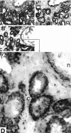
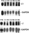
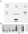
Laminin 5 is a pivotal hemidesmosomal protein involved in cell stability, migration, and anchoring filament formation. Protein and gene expression of the alpha3, beta3, and gamma2 chains of laminin 5 were investigated in normal and invasive prostate carcinoma using immunohistochemistry, Northern analysis, and in situ hybridization. Laser capture microdissection of normal and carcinomatous glands, in conjunction with RNA amplification and reverse Northern analysis, were used to confirm the gene expression data. Protein and mRNA expression of all three laminin 5 chains were detected in the basal cells of normal glands. In contrast, invasive prostate carcinoma showed a loss of beta3 and gamma2 protein expression with variable expression of alpha3 chains. Despite the loss of protein expression, there was retention of beta3 and gamma2 mRNA expression as detected by in situ hybridization, Northern and reverse Northern analysis. Our findings imply that an altered mechanism of translation of beta3 or gamma2 mRNAs into functional proteins contributes to failure of anchoring filaments and hemidesmosomal formation. The resultant hemidesmosome instability or loss would suggest a less stable epithelial-stromal junction, increased invasion and migration of malignant cells, and disruption of normal integrin signaling pathways.
Figures


Similar articles
-
Differential expression of laminin 5 (alpha 3 beta 3 gamma 2) by human malignant and normal prostate.Am J Pathol. 1996 Oct;149(4):1341-9. Am J Pathol. 1996. PMID: 8863681 Free PMC article.
-
Co-expression of laminin β3 and γ2 chains and epigenetic inactivation of laminin α3 chain in gastric cancer.Int J Oncol. 2011 Sep;39(3):593-9. doi: 10.3892/ijo.2011.1048. Epub 2011 May 20. Int J Oncol. 2011. PMID: 21617852
-
Laminin 5 beta3 and gamma2 chains are frequently coexpressed in cancer cells.Pathol Int. 2004 Sep;54(9):688-92. doi: 10.1111/j.1440-1827.2004.01681.x. Pathol Int. 2004. PMID: 15363037
-
Laminin-5 in epithelial tumour invasion.J Mol Histol. 2004 Mar;35(3):277-86. doi: 10.1023/b:hijo.0000032359.35698.fe. J Mol Histol. 2004. PMID: 15339047 Review.
-
Defining the role of laminin-332 in carcinoma.Matrix Biol. 2009 Oct;28(8):445-55. doi: 10.1016/j.matbio.2009.07.008. Epub 2009 Aug 15. Matrix Biol. 2009. PMID: 19686849 Free PMC article. Review.
Cited by
-
Regulation of Kinase Signaling Pathways by α6β4-Integrins and Plectin in Prostate Cancer.Cancers (Basel). 2022 Dec 27;15(1):149. doi: 10.3390/cancers15010149. Cancers (Basel). 2022. PMID: 36612146 Free PMC article. Review.
-
Unique Biological Activity and Potential Role of Monomeric Laminin-γ2 as a Novel Biomarker for Hepatocellular Carcinoma: A Review.Int J Mol Sci. 2019 Jan 8;20(1):226. doi: 10.3390/ijms20010226. Int J Mol Sci. 2019. PMID: 30626121 Free PMC article. Review.
-
Targeted approaches for the management of metastatic prostate cancer.Curr Oncol Rep. 2006 May;8(3):206-12. doi: 10.1007/s11912-006-0021-9. Curr Oncol Rep. 2006. PMID: 16618385 Review.
-
Laminin-332-rich tumor microenvironment for tumor invasion in the interface zone of breast cancer.Am J Pathol. 2011 Jan;178(1):373-81. doi: 10.1016/j.ajpath.2010.11.028. Epub 2010 Dec 23. Am J Pathol. 2011. PMID: 21224074 Free PMC article.
-
The role of laminins in cancer pathobiology: a comprehensive review.J Transl Med. 2025 Jan 17;23(1):83. doi: 10.1186/s12967-025-06079-0. J Transl Med. 2025. PMID: 39825429 Free PMC article. Review.
References
-
- Colognato F, Yurchenco PD: Form and function: the laminin family of heterotrimers. Dev Dyn 2000, 218:213-234 - PubMed
-
- Burgeson RE, Chiquet M, Deutzmann, Ekblom P, Engel J, Kleinman H, Martin GR, Meneguzzi G, Paulsson M, Sanes J, Timple R, Tryggvason K, Yamada Y, Yurchenco PD: A new nomenclature for the laminins. Matrix Biol 1994, 14:209-211 - PubMed
-
- Ferletta M, Ekblom P: Identification of laminin-10/11 as a strong cell adhesive complex for a normal and malignant human epithelial cell line. J Cell Science 1999, 112:1-10 - PubMed
Publication types
MeSH terms
Substances
Grants and funding
LinkOut - more resources
Full Text Sources
Other Literature Sources
Medical

