. 2001 Mar;158(3):1161-72.
doi: 10.1016/S0002-9440(10)64063-1.

Intrachoroidal neovascularization in transgenic mice overexpressing vascular endothelial growth factor in the retinal pigment epithelium

Affiliations

Intrachoroidal neovascularization in transgenic mice overexpressing vascular endothelial growth factor in the retinal pigment epithelium

C Schwesinger et al. Am J Pathol. 2001 Mar.

Abstract

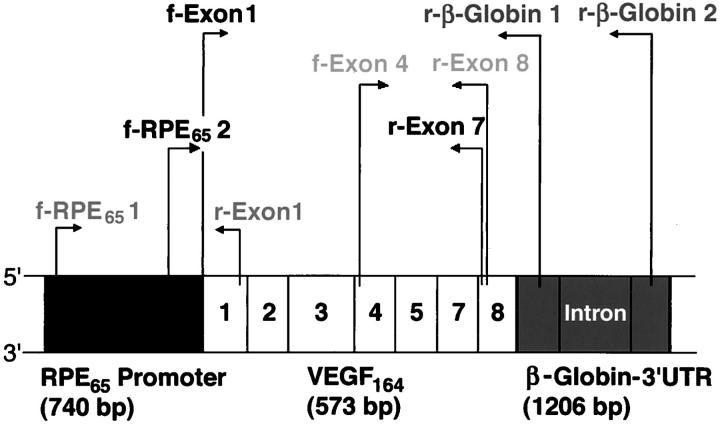
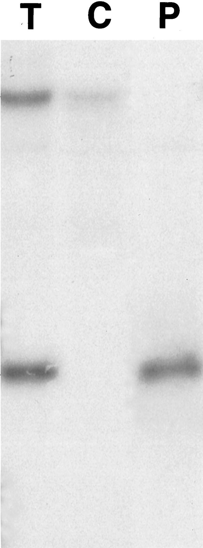
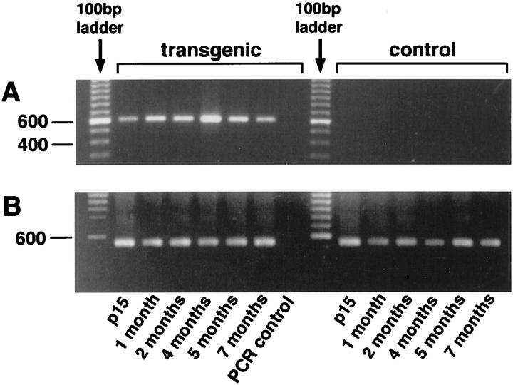
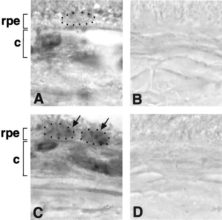
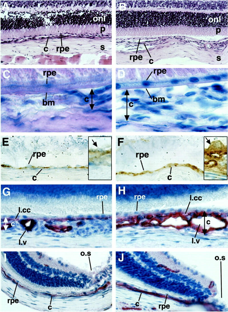
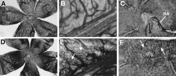
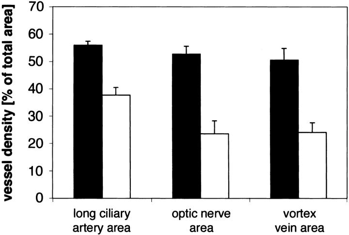
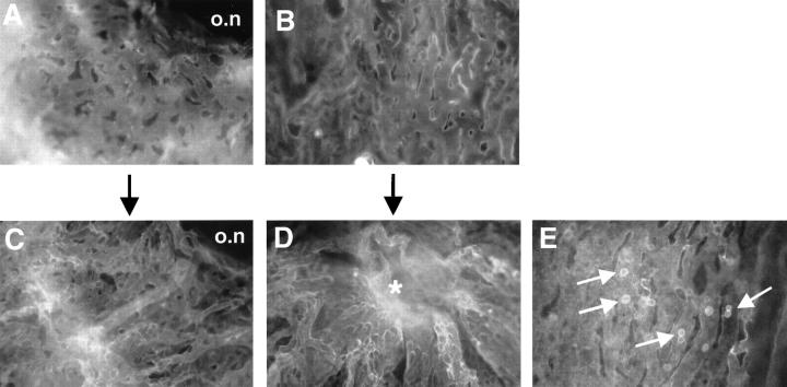
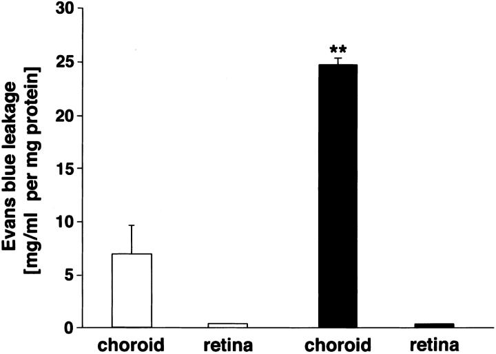
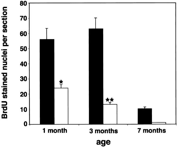
Choroidal neovascularization in age-related macular degeneration is a frequent and poorly treatable cause of vision loss in elderly Caucasians. This choroidal neovascularization has been associated with the expression of vascular endothelial growth factor (VEGF). In current animal models choroidal neovascularization is induced by subretinal injection of growth factors or vectors encoding growth factors such as VEGF, or by disruption of the Bruch's membrane/retinal pigment epithelium complex with laser treatment. We wished to establish a transgenic murine model of age-related macular degeneration, in which the overexpression of VEGF by the retinal pigment epithelium induces choroidal neovascularization. A construct consisting of a tissue-specific murine retinal pigment epithelium promoter (RPE(65) promoter) coupled to murine VEGF(164) cDNA with a rabbit beta-globin-3' UTR was introduced into the genome of albino mice. Transgene mRNA was expressed in the retinal pigment epithelium at all ages peaking at 4 months. The expression of VEGF protein was increased in both the retinal pigment epithelium and choroid. An increase of intravascular adherent leukocytes and vessel leakage was observed. Histopathology revealed intrachoroidal neovascularization that did not penetrate through an intact Bruch's membrane. These results support the hypothesis that additional insults to the integrity of Bruch's membrane are required to induce growth of choroidal vessels into the subretinal space as seen in age-related macular degeneration. This model may be useful to screen for inhibitors of choroidal vessel growth.


Figures

Figure 1.
Figure 1.
Scheme of the transgenic construct RPE65/VEGF164/β-globin-3′ UTR (2,517 bp) and location of the primer pairs. The three primer pairs f-RPE65-2 with r-exon-7; and f-exon-4 with r-β-globin-1 or r-β-globin-2 were used for sequencing of the transgenic construct. The primer pair f-RPE65-1 and r-exon-1 was used for PCR amplification of a transgene-specific sequence (800 bp). The primer pair f-exon-4 with r-exon-8 was used in RT-PCR amplification of total VEGF cDNA isoforms, whereas f-RPE6- 2 with r-exon-7 amplified a transgene-specific VEGF164 cDNA sequence (600 bp).
Figure 2.
Figure 2.
Southern blot analysis of transgenic (T) and control (C) mice tail DNA that was digested with the restriction enzyme EcoRI. A α-32P dCTP-labeled probe to exon 3 of VEGF164 hybridized to the exon 3 sequence of the endogenous VEGF gene (≈9-kb upper band) in all samples. The probe also hybridized to the exon 3 sequence of transgenic VEGF164 in transgenic mice (T) (1,259-bp band). EcoRI digestion of the plasmid (P) that contained the transgenic construct also generated a 1,259 bp band.
Figure 3.
Figure 3.
RT-PCR of transgenic and control whole eyes aged 15 days to 7 months. A: The primer pair f-RPE65-2 with r-exon-7 amplifies transgenic VEGF164 cDNA (≈600 bp) in transgenic eyes at all ages. Control eyes show no amplification product. B: Amplification of the housekeeping gene glyceraldehyde-3-phosphate-dehydrogenase (GAPDH). All PCR control reactions are negative.
Figure 4.
Figure 4.
In situ hybridization. Original magnification, ×1,600. A: In control eyes, hybridization with an antisense probe demonstrates basal VEGF mRNA expression in the RPE cell nucleus (RPE cell layer, rpe; small dots surround a RPE cell nucleus) and in choroidal cells (choroid, c). B: A sense probe shows no hybridization product. C: In transgenic eyes, hybridization with an antisense probe demonstrates increased hybridization product in RPE cell nuclei and basal VEGF mRNA expression in choroidal cells. D: A sense probe shows no hybridization product.
Figure 5.
Figure 5.
A and B: Harris hematoxylin (blue) and eosin (pink) staining. Original magnification, ×400. Control (A) and transgenic eye (B) sections demonstrate differences in choroidal (c) architecture underneath the RPE (rpe). No morphological differences are seen in the outer nuclear layer (onl) and the photoreceptor endpieces (p) of the retina. Sclera (s). C and D: PAS (pink) and Gill’s hematoxylin (blue). Original magnification, ×1,000. Control (C) and transgenic (D) eyes show an intact Bruch’s membrane (bm) and no sub-RPE vessels. Limitation of the choroid that was measured is demonstrated by the double-headed arrow. E and F: Immunohistochemistry against VEGF (brown, diaminobenzidine precipitate) in 3-month-old mice counterstained with Gill’s hematoxylin. Original magnifications: ×630, insets ×1,600. E: The control eye shows light VEGF staining in the RPE cell layer and no staining in the choroid. The inset shows a high-power view of VEGF in the RPE cells (arrow). F: Strong VEGF staining is demonstrated in RPE cell layer and the choroid of the transgenic eye. The inset shows a high-power view of an RPE cell with basal secretion of VEGF toward the choroid. G–J: CD31 immunohistochemistry (red, fast red precipitate) in 3-month-old mice counterstained with Gill’s hematoxylin. Original magnification, ×630. Control choroid (G) and transgenic choroid (H) at the optic nerve entry. In the transgenic choroid the lamina choriocapillaris (l.ml) is pronounced and the lamina vasculosa (l.v) thickened because of an increased density of vessels with pronounced lumina because of dilatation. The RPE cell layer is not disrupted. Control choroid (I) and transgenic choroid (J) in proximity of the ora serrata (o.s). The transgenic choroid is thickened.
Figure 6.
Figure 6.
ADPase staining of control and transgenic choroidal flatmounts. A: The complete view (original magnification, ×25) of the control choroid shows light vessel staining and normal choroidal architecture. Stem of the long ciliary artery of the control choroid (B) (original magnification, ×400) and optic nerve entry of the control choroid (C). Retinal artery (RA) remnants after dissection. Original magnification, ×250. D: Complete view of the transgenic choroid with strong vessel staining and perturbed vessel architecture. E: Stem of the long ciliary artery of the transgenic choroid with tortuous vessel sprouts (arrows). F: Optic nerve entry of the transgenic choroid. A dense choriocapillary net with looping vessels (arrow) is shown.
Figure 7.
Figure 7.
Histogram of the effect of VEGF overexpression from the RPE on choroidal vessel density per total area. Transgenic choroidal vessel density (black bar) relative to control choroidal vessel density (white bar) as computed with NIH image software of ADPase stainings at corresponding regions and equal magnification. Values are calculated from different flatmounts (n ≥ 4) for each location and are expressed as means ± SEM. The values for transgenic choroidal vessel density are significantly different (P < 0.05) from control values at each location.
Figure 8.
Figure 8.
Fluorescein isothiocyanate-labeled Lycopersicon esculentum lectin staining of control and transgenic choroidal flatmounts. Original magnification, ×400. A: Choriocapillary network at the optic nerve entry (o.n) of the control choroid. B: Control choriocapillaris at 1 mm from the optic nerve entry shows a two-dimensional structure with few adherent cells (not present here). C: Choriocapillary network at the optic nerve entry of the transgenic choroid. The intercapillary distance is reduced and the vessel architecture perturbed. D: Transgenic choriocapillaris at 1 mm from the optic nerve entry shows nests of dilated and irregularly stained vessels that rise out of the two dimensional focal plane (asterisk). E: Transgenic choriocapillaris with abundant adherent intravascular cells (arrows).
Figure 9.
Figure 9.
Histogram of the effect of VEGF overexpression from the RPE on choroidal and retinal vessel leakage in 3-month-old mice. Evans blue leakage in transgenic choroids and retinae (black bars) relative to control choroids and retinae (white bars) normalized for the dry weight of the tissue and expressed as Evans blue concentration in mg/ml per mg protein. Values are calculated as means ± SEM from n = 6. Choroidal vessel Evans blue leakage is significantly different (P < 0.001).
Figure 10.
Figure 10.
Histogram of the effect of VEGF overexpression from the RPE on choroidal cell proliferation. Transgenic choroidal cell number (black bar) relative to control choroidal cell number (white bar) as observed by BrdU staining at specific ages. Values are calculated as means ± SEM from eight choroid eye sections at the optic nerve entry at 1 and 3 months (n ≥ 3) and 7 months (n = 2) of age. Choroidal cell proliferation is significantly different at 1 month (P < 0.05) and 3 months (P < 0.0001).

Similar articles

Cited by

References

    1. Adamis AP, Shima DT, Yeo KT, Yeo TK, Brown LF, Berse B, D’Amore PA, Folkman J: Synthesis and secretion of vascular permeability factor/vascular endothelial growth factor by human retinal pigment epithelial cells. Biochem Biophys Res Commun 1993, 193:631-638 - PubMed
    1. Lutty G, Grunwald J, Majji AB, Uyama M, Yoneya S: Changes in choriocapillaris and retinal pigment epithelium in age-related macular degeneration. Mol Vis 1999, 5:35. - PubMed
    1. Amin R, Puklin JE, Frank RN: Growth factor localization in choroidal neovascular membranes of age-related macular degeneration. Invest Ophthalmol Vis Sci 1994, 35:3178-3188 - PubMed
    1. Frank RN, Amin RH, Eliott D, Puklin JE, Abrams GW: Basic fibroblast growth factor and vascular endothelial growth factor are present in epiretinal and choroidal neovascular membranes. Am J Ophthalmol 1996, 122:393-403 - PubMed
    1. Kvanta A, Algvere PV, Berglin L, Seregard S: Subfoveal fibrovascular membranes in age-related macular degeneration express vascular endothelial growth factor. Invest Ophthalmol Vis Sci 1996, 37:1929-1934 - PubMed

Publication types

MeSH terms