. 2001 Mar;8(2):415-23.
doi: 10.1128/CDLI.8.2.415-423.2001.

Enzyme immunoassay detection of antigen-specific immunoglobulin g antibodies in longitudinal serum samples from patients with cryptosporidiosis

Affiliations

Enzyme immunoassay detection of antigen-specific immunoglobulin g antibodies in longitudinal serum samples from patients with cryptosporidiosis

J W Priest et al. Clin Diagn Lab Immunol. 2001 Mar.

Abstract

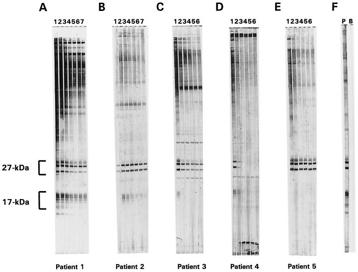
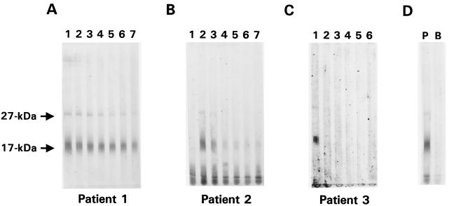
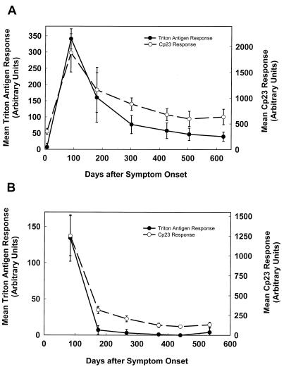
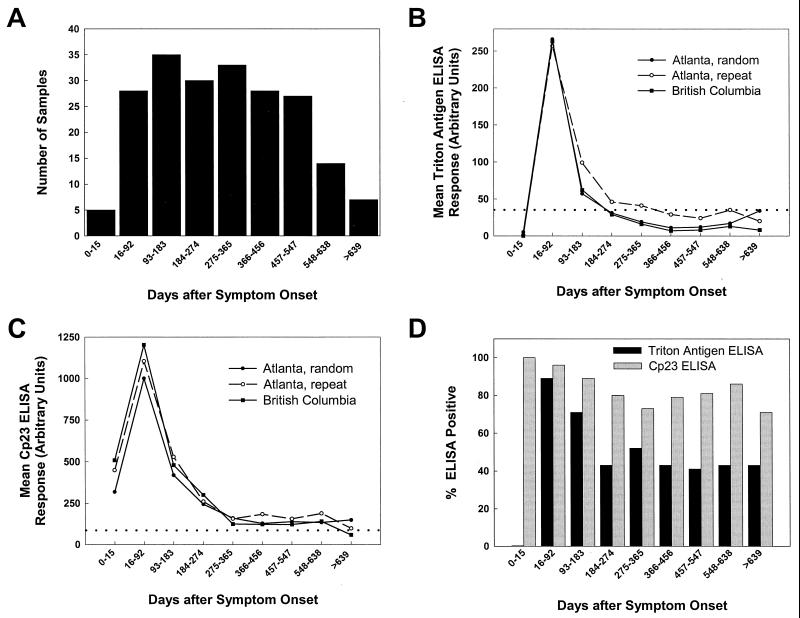
Cryptosporidium parvum is a protozoan parasite that causes diarrheal illness in a wide range of mammalian hosts, including humans. Characteristic serum immunoglobulin G (IgG) antibody responses to antigens in the 27- and 17-kDa size ranges have been shown to develop after infection, and several enzyme-linked immunosorbent assay (ELISA) and Western blot assay formats have been used to measure these IgG levels in human serum. Using a collection of serial samples from laboratory-confirmed cryptosporidiosis patients, we compared the results obtained by using two new ELISAs with those obtained with two different Western blot assays. When assayed with the large-format Western blot, 97% of the 67 patients had a demonstrable antibody response on at least one occasion. The Cp23 ELISA correctly identified 93% of the samples that had a 27-kDa response by Western blot and 100% of the negative samples. The Triton antigen ELISA detected 77% of the samples that had a 17-kDa response by Western blot and 88% of the negative samples. The sensitivity of the Triton antigen assay was higher for samples collected between 16 and 92 days after the onset of symptoms (96%). The minigel-format Western blot did not compare favorably with the large-format blot for the detection of antibodies to the 27-kDa antigen (71% sensitivity). A half-life of about 12 weeks was estimated for antibodies to both the 27- and 17-kDa antigens. We believe the Cp23 and Triton antigen ELISAs will be useful in epidemiologic studies of the prevalence of Cryptosporidium infection in the population.


Figures

FIG. 1
FIG. 1
Large-gel-format Western blots of sera collected at intervals from five cryptosporidiosis patients. Crude C. parvum antigens (600 ng of protein/mm of gel width) were resolved on an SDS-polyacrylamide (10 to 22.5%) gel under nonreducing conditions and then transferred to a PVDF membrane. Sera that had been collected from five laboratory-confirmed cryptosporidiosis patients (patients 1 to 5 represented in panels A to E, respectively) at intervals of approximately 3 months were diluted 1:100 with 0.3% Tween 20–PBS and incubated (2 ml per well) with 2-mm-wide strips of membrane overnight at 4°C. Seven serum samples were donated by patients 1 and 2 (Western blots shown in temporal order in lanes 1 to 7 of panels A and B, respectively), and six serum samples were donated by patients 3, 4, and 5 (Western blots shown in temporal order in lanes 1 to 6 of panels C, D, and E, respectively). The first sample in each panel (Western blots shown in lane 1) was collected within the first 3 months (A), 15 days (B), 3 months (C), and 1 month (D and E) after symptom onset. The blots were developed with a biotinylated mouse anti-human IgG monoclonal antibody and alkaline phosphatase-labeled streptavidin as described in Materials and Methods. The positions of the 27- and 17-kDa antigen families are indicated. Control strips that were incubated with a positive serum (P) and a buffer blank (B) are shown in panel F.
FIG. 2
FIG. 2
Minigel Western blot results for sera collected at intervals from cryptosporidiosis patients 1, 2, and 3. Crude C. parvum antigens (57 ng of protein/mm of gel width) were resolved under nonreducing conditions on an SDS-polyacrylamide (15%) minigel and transferred to a PVDF membrane. Sera from patients 1 (A), 2 (B), and 3 (C) were diluted 1:50 with 0.3% Tween 20–PBS and incubated (400 μl per channel) with separate regions of the membrane overnight at 4°C. Blots were developed as described in Materials and Methods. The positions of the 27- and 17-kDa antigens are indicated. Panel C was scanned and digitally enhanced to improve the resolution of the 27-kDa band in lane 1. Control strips that were incubated with a positive serum (P) and a buffer blank (B) are shown in panel D.
FIG. 3
FIG. 3
Mean Triton antigen and Cp23 ELISA responses for sera collected at intervals from cryptosporidiosis patients 2 and 3. Sera from cryptosporidiosis patients 2 (A) and 3 (B) were assayed independently in Atlanta (twice) and in British Columbia (once) with the Triton antigen and Cp23 ELISAs described in Materials and Methods. The optical densities were converted into an AU value based upon a 9-point standard curve that was included on each ELISA plate. The means of the ELISA values for each time point after symptom onset are plotted for both the Triton antigen ELISAs (solid line) and the Cp23 ELISAs (dashed line). Each bar indicates one standard deviation.
FIG. 4
FIG. 4
ELISA analysis of sera from cryptosporidiosis patients who donated sera on more than one occasion. Panel A shows the total number of serum samples collected during each time interval after symptom onset from 41 confirmed cryptosporidiosis patients who donated more than one specimen. Triton antigen (B) and Cp23 (C) ELISAs were performed with these serum samples on two occasions in Atlanta to assess the intralaboratory variation (Atlanta, random and Atlanta, repeat) and on one occasion in British Columbia to assess the interlaboratory variation (British Columbia). For the assays represented by the “Atlanta, random” results, the duplicate serum samples were placed in random locations on the ELISA plate to eliminate the possibility of positional bias. The geometric means (log scale) of the ELISA responses are plotted for each interval of sample collection, and the threshold for positivity is indicated in each graph by a dotted line. Panel D shows the fraction of the samples from each time interval positive for antibodies in the Triton antigen (black bars) and Cp23 (grey bars) ELISAs. A serum sample was considered ELISA positive if at least two of the responses from the three independent assays were above 35 AU for the Triton antigen assay and above 86 AU for the Cp23 assay.

Comment in

Similar articles

Cited by

References

    1. Campbell I, Tzipori A S, Hutchinson G, Angus K W. Effect of disinfectants on survival of Cryptosporidium oocysts. Vet Rec. 1982;111:414–415. - PubMed
    1. DuPont H L, Chappel C L, Sterling C R, Okhuysen P C, Rose J B, Jakubowski W. The infectivity of Cryptosporidium parvum in healthy volunteers. N Engl J Med. 1995;332:855–859. - PubMed
    1. Dworkin M S, Goldman D P, Wells T G, Kobayashi J M, Herwaldt B L. Cryptosporidiosis in Washington State: an outbreak associated with well water. J Infect Dis. 1996;174:1372–1376. - PubMed
    1. Flanigan T, Whalen C, Turneer J, Soave R, Toerner J, Havlir D, Kotler D. Cryptosporidium infection and CD4 counts. Ann Intern Med. 1992;116:840–842. - PubMed
    1. Frost F J, Calderon R L, Muller T B, Curry M, Rodman J S, Moss D M, De La Cruz A A. A two-year follow-up survey of antibody to Cryptosporidium in Jackson County, Oregon, following an outbreak of waterborne disease. Epidemiol Infect. 1998;121:213–217. - PMC - PubMed

Publication types