. 2001 Mar;8(2):460-4.
doi: 10.1128/CDLI.8.2.460-464.2001.

Use of Hoechst 33342 staining to detect apoptotic changes in bovine mononuclear phagocytes infected with Mycobacterium avium subsp. paratuberculosis

Affiliations

Use of Hoechst 33342 staining to detect apoptotic changes in bovine mononuclear phagocytes infected with Mycobacterium avium subsp. paratuberculosis

S Allen et al. Clin Diagn Lab Immunol. 2001 Mar.

Abstract

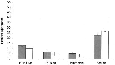
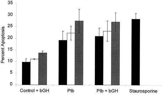
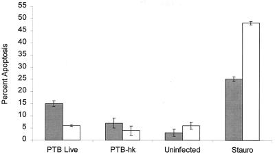
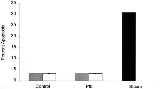
Mycobacterium avium subsp. paratuberculosis is an intracellular pathogen of macrophages that causes a chronic enteritis (Johne's disease) in ruminants. The purpose of this study was to determine whether M. avium subsp. paratuberculosis infection causes apoptosis in bovine monocytes. Using Hoechst 33342 staining, we observed increased numbers of apoptotic monocytes within 6 h of infection with M. avium subsp. paratuberculosis, and these numbers increased further at 24 and 48 h. This effect appeared to require viable bacilli, because monocytes infected with heat-killed M. avium subsp. paratuberculosis did not exhibit a significant increase in apoptosis. Preincubation of monocytes with bovine growth hormone prior to infection with M. avium subsp. paratuberculosis did not significantly alter the number of apoptotic cells.


Figures

FIG. 1
FIG. 1
Photomicrograph of apoptotic changes in control (A) or M. avium subsp. paratuberculosis-infected (B) bovine monocytes. Adherent cells were incubated in RPM 1640 medium plus 5% FBS for 6 h. The cells were then washed, fixed with 4% paraformaldehyde, and stained with Hoechst 33342 (5 μg/ml) for 20 min at 25°C. The slides were then examined by fluorescence microscopy and photographed. Cells with signs of apoptosis (fragmented nuclei) are enclosed within circles.
FIG. 2
FIG. 2
Quantitation of apoptosis in M. avium subsp. paratuberculosis-infected bovine monocytes stained with Hoechst 33342. Prior to infection, the monocytes were incubated overnight with 10 ng of BGH per ml (open bars) or with medium alone (negative control) (shaded bars). The monocytes were then infected with 106 CFU of live or heat-killed (hk) M. avium subsp. paratuberculosis (PTB) per ml. Additional monolayers of uninfected monocytes were treated with 500 nM staurosporine (Stauro) (positive control) or left untreated (negative control). The results are the mean and standard error of the mean of four experiments, using monocytes from four different donor cattle.
FIG. 3
FIG. 3
Quantitation of apoptosis in M. avium subsp. paratuberculosis-infected bovine monocytes incubated in vitro for up to 48 h. Prior to infection, the monocytes were allowed to incubate overnight with 10 ng of BGH per ml or with medium alone. Bovine monocytes were then infected with 106 CFU of live M. avium subsp. paratuberculosis (Ptb) per ml. Additional monolayers of uninfected monocytes were treated with 500 nM staurosporine (positive control) or left untreated (negative control). At 6 h (solid bars), 24 h (open bars), and 48 h (shaded bars) of incubation, triplicate monolayers were stained with Hoechst 33342 and the percentage of apoptotic cells was quantified by microscopy. The results are the mean and standard error of the mean of three experiments, using monocytes from different donor cattle.
FIG. 4
FIG. 4
Comparison of apoptosis in M. avium subsp. paratuberculosis-infected bovine monocytes and a bovine macrophage cell line stained with Hoechst 33342. Bovine monocytes (shaded bars) and a bovine macrophage cell line (open bars) were infected with 106 CFU of live or heat-killed (hk) M. avium subsp. paratuberculosis (PTB) per ml. Some uninfected wells were treated with 500 nM staurosporine (Stauro) (positive control) or left untreated (negative control) for 6 h. The results are the mean and standard error of the mean of two experiments, using monocytes obtained from separate donors.
FIG. 5
FIG. 5
A M. avium subsp. paratuberculosis-infected bovine macrophage cell line is resistant to apoptosis when incubated in vitro for 24 (shaded bars) or 48 h (open bars). Monolayers of a bovine macrophage cell line (BoMac) were infected with 106 CFU of live M. avium subsp. paratuberculosis (Ptb) per ml and incubated in vitro for up to 48 h. Some uninfected wells were treated with 500 nM staurosporine (Stauro) (positive control) or left untreated (negative control). At the indicated item points, triplicate monolayers were stained with Hoechst 33342 and apoptotic cells were quantified by microscopy. The results are the mean and standard error of the mean of two experiments.

References

    1. Chiodini R J, Van Kruiningen H J, Merkal R S. Ruminant paratuberculosis (Johne's disease): the current status and future prospects. Cornell Vet. 1984;74:218–262. - PubMed
    1. Feola R P, Collins M T, Czuprynski C J. Hormonal modulation of phagocytosis and intracellular growth of Mycobacterium avium ss. paratuberculosis in bovine peripheral blood monocytes. Microb Pathog. 1999;26:1–11. - PubMed
    1. Hayashi T, Catanzaro A, Rao S P. Apoptosis of human monocytes and macrophages by Mycobacterium avium sonicate. Infect Immun. 1997;65:5262–5271. - PMC - PubMed
    1. Keane J, Balcewicz-Sablinska M K, Remold H G, Chupp G L, Meek B B, Fenton M J, Kornfeld H. Infection by Mycobacterium tuberculosis promotes human alveolar macrophage apoptosis. Infect Immun. 1997;65:298–304. - PMC - PubMed
    1. Keane J, Romold H G, Kornfeld H. Virulent Mycobacterium tuberculosis strains evade apoptosis of infected alveolar macrophages. J Immunol. 2000;164:2016–2020. - PubMed

Publication types

Substances

LinkOut - more resources