. 2001 Mar 5;152(5):945-58.
doi: 10.1083/jcb.152.5.945.

The localization of human cyclins B1 and B2 determines CDK1 substrate specificity and neither enzyme requires MEK to disassemble the Golgi apparatus

Affiliations

The localization of human cyclins B1 and B2 determines CDK1 substrate specificity and neither enzyme requires MEK to disassemble the Golgi apparatus

V M Draviam et al. J Cell Biol. .

Abstract

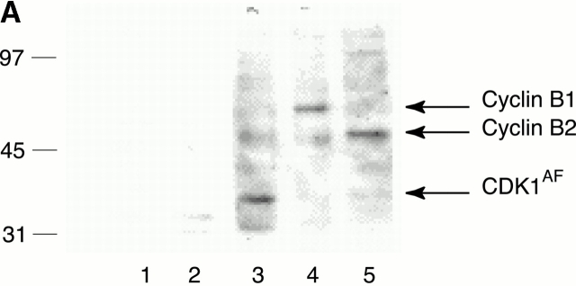
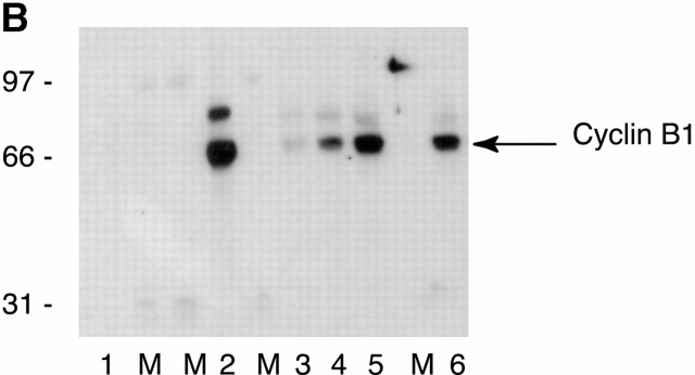
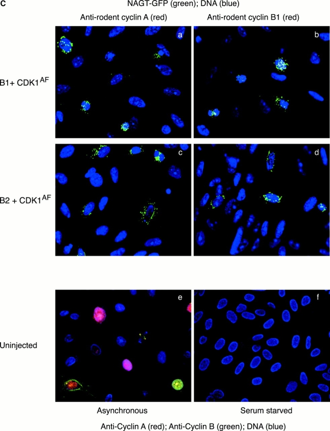
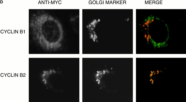
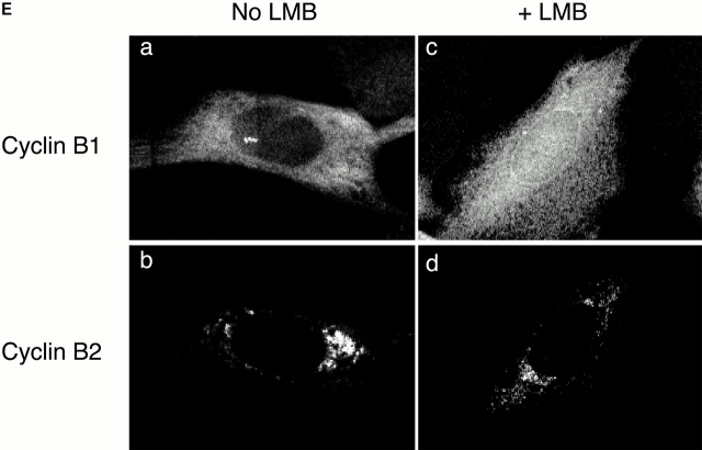
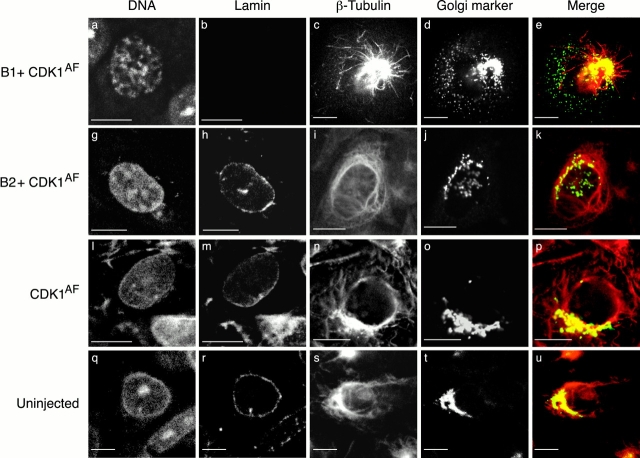
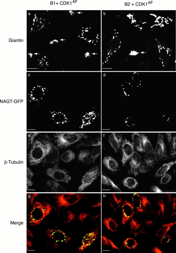
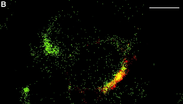
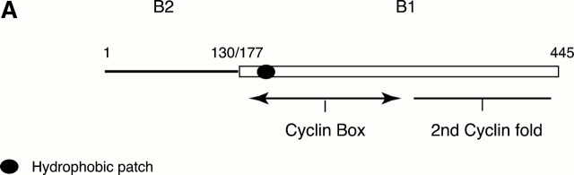
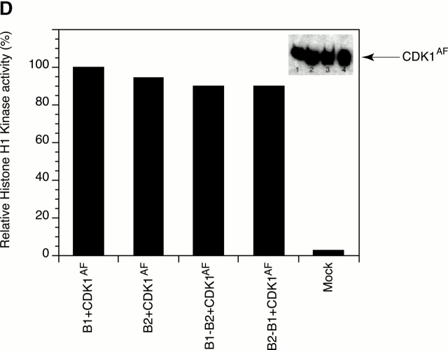
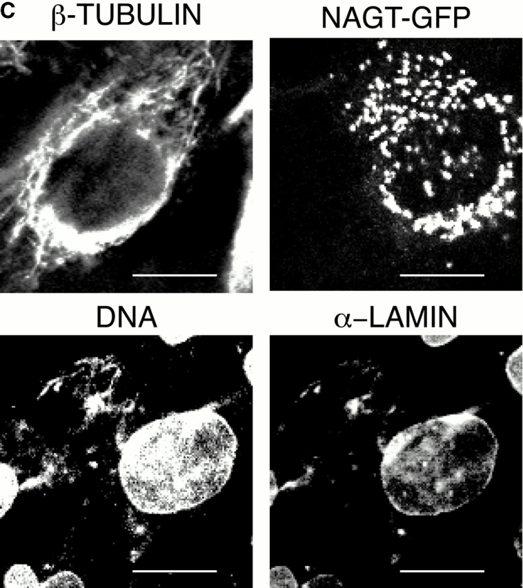
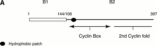
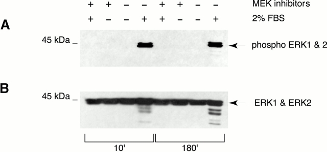
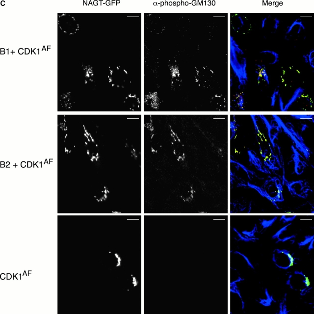
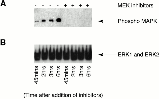
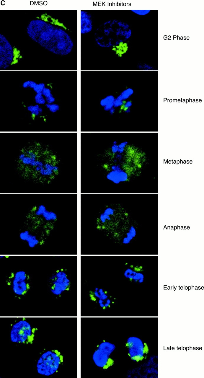
In this paper, we show that substrate specificity is primarily conferred on human mitotic cyclin-dependent kinases (CDKs) by their subcellular localization. The difference in localization of the B-type cyclin-CDKs underlies the ability of cyclin B1-CDK1 to cause chromosome condensation, reorganization of the microtubules, and disassembly of the nuclear lamina and of the Golgi apparatus, while it restricts cyclin B2-CDK1 to disassembly of the Golgi apparatus. We identify the region of cyclin B2 responsible for its localization and show that this will direct cyclin B1 to the Golgi apparatus and confer upon it the more limited properties of cyclin B2. Equally, directing cyclin B2 to the cytoplasm with the NH(2) terminus of cyclin B1 confers the broader properties of cyclin B1. Furthermore, we show that the disassembly of the Golgi apparatus initiated by either mitotic cyclin-CDK complex does not require mitogen-activated protein kinase kinase (MEK) activity.


Figures

Figure 1
Figure 1
(A) Human B-type cyclins are expressed to similar levels in CHO cells. Serum-starved CHO cells were transfected with plasmids encoding human cyclin B1 or B2 and/or CDK1AF all tagged with one copy of the myc epitope and under the “tetracyclin-OFF” promoter. Cells were incubated in the presence or absence of tetracyclin to repress or induce protein expression, respectively. 16 h after induction, cells were lysed, and the extracts were run on one-dimensional SDS-PAGE and then immunoblotted with the 9E10 mAb to detect the proteins. Lane 1, mock-transfected cells; lanes 2–5, transfected cells: (lane 2) cyclin B1 plasmid plus tetracyclin; (lane 3) Cdk1AF; (lane 4) cyclin B1 minus tetracyclin for 16 h; (lane 5) cyclin B2 minus tetracyclin for 16 h. (B) Ectopic and endogenous cyclin B1 are expressed at similar levels in CHO cells. Approximately 980 serum-starved CHO cells were microinjected with plasmids encoding myc epitope–tagged human cyclin B1 with CDK1AF. 6 h after microinjection, cells were lysed and the samples were run on one-dimensional SDS-PAGE next to lysates from 250, 500, and 1,000 mitotic cells. Proteins were immunoblotted with an anticyclin B1 monoclonal antibody V152 that recognizes both human and rodent cyclin B1 to detect the proteins (a gift from J. Gannon and T. Hunt). Lane 1, uninjected cells; lane 2, cells injected with cyclin B1–CDK1AF; lanes 3–5, 250, 500, and 1,000 mitotic HeLa cells; lane 6, 1,000 mitotic CHO cells. M, molecular mass marker lane. Results shown are representative of three independent experiments. (C) There are no detectable endogenous mitotic cyclins in serum-starved CHO cells with or without ectopic human cyclin–CDKs. (a–d) CHO cells were serum starved for 24 h and then microinjected with NAGT–GFP as a Golgi apparatus and injection marker (green) and TOTO-3 to visualize the DNA (blue) together with CDK1AF and either cyclin B1 (a and b) or cyclin B2 (c and d). 6 h after microinjection, cells were fixed and stained with an anti–mouse cyclin A antibody (red) (a gift from Dr. M. Carrington, University of Cambridge, Cambridge, UK) (a and c) or with an anti–rodent cyclin B1 monoclonal antibody V143 (red) that recognizes rodent B-type cyclins but does not cross-react with human B-type cyclins (a gift from J. Gannon and T. Hunt) (b and d). (e and f) Uninjected asynchronous (e) and serum-starved (f) CHO cells were costained with anticyclin A (red), anticyclin B1 (green), and TOTO-3 to visualize the DNA (blue). Results shown are representative of two independent experiments. (D and E) Human B-type cyclins localize correctly in CHO cells. Human cyclin B1 or B2 was tagged with one copy of the myc epitope and microinjected as cDNA under the CMV promoter into serum-starved CHO cells. 3 h after microinjection, cells were treated or not with LMB. Cells were stained with the 9E10 mAb to detect the cyclins. (D) Cells were costained with an antimannosidase II antibody to detect the Golgi apparatus (red in the merged images) and cyclin B1 (top row) or cyclin B2 (bottom row) (green in the merged images). (E) Cells expressing human cyclin B1 or B2 were fixed before (a and b) or after (c and d) treatment with 20 nM LMB for 45 min and stained for ectopically expressed cyclin B1 (a and c) or cyclin B2 (b and d).
Figure 1
Figure 1
(A) Human B-type cyclins are expressed to similar levels in CHO cells. Serum-starved CHO cells were transfected with plasmids encoding human cyclin B1 or B2 and/or CDK1AF all tagged with one copy of the myc epitope and under the “tetracyclin-OFF” promoter. Cells were incubated in the presence or absence of tetracyclin to repress or induce protein expression, respectively. 16 h after induction, cells were lysed, and the extracts were run on one-dimensional SDS-PAGE and then immunoblotted with the 9E10 mAb to detect the proteins. Lane 1, mock-transfected cells; lanes 2–5, transfected cells: (lane 2) cyclin B1 plasmid plus tetracyclin; (lane 3) Cdk1AF; (lane 4) cyclin B1 minus tetracyclin for 16 h; (lane 5) cyclin B2 minus tetracyclin for 16 h. (B) Ectopic and endogenous cyclin B1 are expressed at similar levels in CHO cells. Approximately 980 serum-starved CHO cells were microinjected with plasmids encoding myc epitope–tagged human cyclin B1 with CDK1AF. 6 h after microinjection, cells were lysed and the samples were run on one-dimensional SDS-PAGE next to lysates from 250, 500, and 1,000 mitotic cells. Proteins were immunoblotted with an anticyclin B1 monoclonal antibody V152 that recognizes both human and rodent cyclin B1 to detect the proteins (a gift from J. Gannon and T. Hunt). Lane 1, uninjected cells; lane 2, cells injected with cyclin B1–CDK1AF; lanes 3–5, 250, 500, and 1,000 mitotic HeLa cells; lane 6, 1,000 mitotic CHO cells. M, molecular mass marker lane. Results shown are representative of three independent experiments. (C) There are no detectable endogenous mitotic cyclins in serum-starved CHO cells with or without ectopic human cyclin–CDKs. (a–d) CHO cells were serum starved for 24 h and then microinjected with NAGT–GFP as a Golgi apparatus and injection marker (green) and TOTO-3 to visualize the DNA (blue) together with CDK1AF and either cyclin B1 (a and b) or cyclin B2 (c and d). 6 h after microinjection, cells were fixed and stained with an anti–mouse cyclin A antibody (red) (a gift from Dr. M. Carrington, University of Cambridge, Cambridge, UK) (a and c) or with an anti–rodent cyclin B1 monoclonal antibody V143 (red) that recognizes rodent B-type cyclins but does not cross-react with human B-type cyclins (a gift from J. Gannon and T. Hunt) (b and d). (e and f) Uninjected asynchronous (e) and serum-starved (f) CHO cells were costained with anticyclin A (red), anticyclin B1 (green), and TOTO-3 to visualize the DNA (blue). Results shown are representative of two independent experiments. (D and E) Human B-type cyclins localize correctly in CHO cells. Human cyclin B1 or B2 was tagged with one copy of the myc epitope and microinjected as cDNA under the CMV promoter into serum-starved CHO cells. 3 h after microinjection, cells were treated or not with LMB. Cells were stained with the 9E10 mAb to detect the cyclins. (D) Cells were costained with an antimannosidase II antibody to detect the Golgi apparatus (red in the merged images) and cyclin B1 (top row) or cyclin B2 (bottom row) (green in the merged images). (E) Cells expressing human cyclin B1 or B2 were fixed before (a and b) or after (c and d) treatment with 20 nM LMB for 45 min and stained for ectopically expressed cyclin B1 (a and c) or cyclin B2 (b and d).
Figure 2
Figure 2
Different effects of cyclin B1– and B2–CDKs on subcellular architecture. Serum-starved CHO cells were unperturbed (bottom row) or microinjected with expression vectors coding for a Golgi marker NAGT–GFP (d, j, and o; green in e, k, and p) and CDK1AF, alone (third row), with cyclin B1 (top row), or with cyclin B2 (second row). After 6 h, the cells were fixed and stained with TOTO-3 to visualize the DNA (a, g, l, and q) and with an antilamin antibody (b, h, m, and r) or an anti–β-tubulin antibody (c, i, n, and s; red in e, k, p, and u). Antimannosidase II was used to stain the Golgi in uninjected cells (t; green in u). Note that cyclin B1 + CDK1AF caused the nuclear lamina to disassemble, and the solubilized lamin protein was washed out of the cell during fixation. The Golgi apparatus fragmented in both cyclin B1–CDK1AF and cyclin B2–CDK1AF injected cells, but only cyclin B1 + CDK1AF caused the microtubules to become much shorter and the centrosome to nucleate more microtubules. Cells are representative of more than 120 cells analyzed in more than three separate experiments. Bars, 10 μm.
Figure 3
Figure 3
Cyclin B1–CDK1AF and cyclin B2–CDK1AF have different effects on the Golgi apparatus. Serum- starved CHO cells were microinjected with expression vectors coding for a Golgi marker NAGT–GFP (c and d) and cyclin B1 with CDK1AF (a, c, e, and g) or cyclin B2 with CDK1AF (b, d, f, and h). Cells were fixed and stained with antimannosidase II (not shown) and antigiantin (a and b, arrows denote uninjected cells) or with an anti–β-tubulin antibody (e and f). The merge shown (g and h) is between antitubulin and NAGT–GFP. Note that cyclin B1–CDK1AF causes the Golgi to break down into smaller and more numerous vesicles than does cyclin B2–CDK1AF in cells before observable changes in the cytoskeleton. Cells are representative of >250 cells analyzed in >10 separate experiments. Bars, 10 μm.
Figure 4
Figure 4
The NH2 terminus of cyclin B2 targets cyclin B1 to the Golgi and restricts its activity. (A) Schematic diagram of the chimera constructed between cyclin B2 and cyclin B1. Cyclin B2 is represented by a solid line and cyclin B1 by an open rectangle. The cyclin B2–B1 mutant exchanges at the sequence LCS (S130 in cyclin B2, and S177 in cyclin B1) in both cyclins. The hydrophobic patch is represented by the filled oval. (B) The NH2 terminus of cyclin B2 targets cyclin B1 to the Golgi apparatus. Serum-starved CHO cells were microinjected with expression vectors coding for a Golgi marker NAGT–GFP (green) and with a myc epitope–tagged cyclin B2–B1 chimera. After 6 h, the cells were fixed and stained with an anti-myc epitope antibody (red). One uninjected cell and one cell expressing the chimera are shown. (C) The NH2 terminus of cyclin B2 confers the properties of cyclin B2 on cyclin B1. Serum-starved CHO cells were microinjected with expression vectors coding for a Golgi marker NAGT–GFP and CDK1AF with a cyclin B2–B1 chimera. After 6 h, the cells were fixed and stained with an anti–β-tubulin antibody or with an antilamin antibody and TOTO-3 to visualize the DNA. Cells are representative of >100 cells analyzed in three separate experiments. (D) Cyclin B chimeras bind and activate CDK1 to a similar extent as wild-type cyclins. Human 293T cells were mock transfected with an empty vector or transfected with untagged CDK1AF and myc epitope–tagged cyclins B1, B2, or the B1–B2 or B2–B1 chimera. 12 h after transfection, cells were lysed with NP-40 lysis buffer and the transfected cyclins were immunoprecipitated with an anti-myc epitope antibody. Immunoprecipitates were processed for H1 kinase assays and the amount of phosphate incorporated was quantitated and normalized to the H1 kinase activity in the cyclin B1–CDK sample. In parallel, immunoprecipitates were immunoblotted with an anti-CDK1 monoclonal (inset) and an anti-myc epitope antibody (not shown) to demonstrate that equivalent amounts of cyclins were immunoprecipitated. Results shown are representative of two independent experiments. Bars, 10 μm.
Figure 4
Figure 4
The NH2 terminus of cyclin B2 targets cyclin B1 to the Golgi and restricts its activity. (A) Schematic diagram of the chimera constructed between cyclin B2 and cyclin B1. Cyclin B2 is represented by a solid line and cyclin B1 by an open rectangle. The cyclin B2–B1 mutant exchanges at the sequence LCS (S130 in cyclin B2, and S177 in cyclin B1) in both cyclins. The hydrophobic patch is represented by the filled oval. (B) The NH2 terminus of cyclin B2 targets cyclin B1 to the Golgi apparatus. Serum-starved CHO cells were microinjected with expression vectors coding for a Golgi marker NAGT–GFP (green) and with a myc epitope–tagged cyclin B2–B1 chimera. After 6 h, the cells were fixed and stained with an anti-myc epitope antibody (red). One uninjected cell and one cell expressing the chimera are shown. (C) The NH2 terminus of cyclin B2 confers the properties of cyclin B2 on cyclin B1. Serum-starved CHO cells were microinjected with expression vectors coding for a Golgi marker NAGT–GFP and CDK1AF with a cyclin B2–B1 chimera. After 6 h, the cells were fixed and stained with an anti–β-tubulin antibody or with an antilamin antibody and TOTO-3 to visualize the DNA. Cells are representative of >100 cells analyzed in three separate experiments. (D) Cyclin B chimeras bind and activate CDK1 to a similar extent as wild-type cyclins. Human 293T cells were mock transfected with an empty vector or transfected with untagged CDK1AF and myc epitope–tagged cyclins B1, B2, or the B1–B2 or B2–B1 chimera. 12 h after transfection, cells were lysed with NP-40 lysis buffer and the transfected cyclins were immunoprecipitated with an anti-myc epitope antibody. Immunoprecipitates were processed for H1 kinase assays and the amount of phosphate incorporated was quantitated and normalized to the H1 kinase activity in the cyclin B1–CDK sample. In parallel, immunoprecipitates were immunoblotted with an anti-CDK1 monoclonal (inset) and an anti-myc epitope antibody (not shown) to demonstrate that equivalent amounts of cyclins were immunoprecipitated. Results shown are representative of two independent experiments. Bars, 10 μm.
Figure 5
Figure 5
The NH2 terminus of cyclin B1 targets cyclin B2 to the cytoplasm and broadens its activity. (A) Schematic diagram of the chimera constructed between cyclin B1 and cyclin B2. Cyclin B1 is represented by an open rectangle and cyclin B2 by a solid line. The cyclin B1–B2 mutant exchanges at the sequence LCQ in both cyclins (Q106 in B1, and Q144 in B2). (B) The NH2 terminus of cyclin B1 targets cyclin B2 to the cytoplasm. Serum-starved CHO cells were microinjected with expression vectors coding for a Golgi marker NAGT–GFP (green) and with a myc epitope–tagged cyclin B1–B2 chimera. After 6 h, the cells were fixed and stained with an anti-myc epitope antibody (red). (C) The NH2 terminus of cyclin B1 confers some of the properties of cyclin B1 on cyclin B2. Serum-starved CHO cells were microinjected with expression vectors coding for a Golgi marker NAGT–GFP and CDK1AF with a cyclin B1–B2 chimera. After 6 h, the cells were fixed and stained with an anti–β-tubulin antibody or with an antilamin antibody and TOTO-3. Cells are representative of >150 cells analyzed in three separate experiments. Bars, 10 μm.
Figure 5
Figure 5
The NH2 terminus of cyclin B1 targets cyclin B2 to the cytoplasm and broadens its activity. (A) Schematic diagram of the chimera constructed between cyclin B1 and cyclin B2. Cyclin B1 is represented by an open rectangle and cyclin B2 by a solid line. The cyclin B1–B2 mutant exchanges at the sequence LCQ in both cyclins (Q106 in B1, and Q144 in B2). (B) The NH2 terminus of cyclin B1 targets cyclin B2 to the cytoplasm. Serum-starved CHO cells were microinjected with expression vectors coding for a Golgi marker NAGT–GFP (green) and with a myc epitope–tagged cyclin B1–B2 chimera. After 6 h, the cells were fixed and stained with an anti-myc epitope antibody (red). (C) The NH2 terminus of cyclin B1 confers some of the properties of cyclin B1 on cyclin B2. Serum-starved CHO cells were microinjected with expression vectors coding for a Golgi marker NAGT–GFP and CDK1AF with a cyclin B1–B2 chimera. After 6 h, the cells were fixed and stained with an anti–β-tubulin antibody or with an antilamin antibody and TOTO-3. Cells are representative of >150 cells analyzed in three separate experiments. Bars, 10 μm.
Figure 6
Figure 6
Cyclin B–CDK1 complexes will disassemble the Golgi apparatus in a physiologically relevant manner in the absence of MEK activity. (A and B) CHO cells were incubated in DME containing 0.1% serum for 3 d (lanes 3 and 7), after which DME with 2% FCS was added back in the presence (lanes 1 and 5) or absence (lanes 4 and 8) of the MEK inhibitors, U0126 (25 μM) and PD98059 (20 μM). MEK inhibitors were also added to cells in the absence of serum (lanes 2 and 6). Cells were lysed after 10 min (lanes 1–4) or 3 h (lanes 5–8), and whole cell extracts were probed with an anti–phospho-MAP kinase antibody ( A) and subsequently reprobed with an antibody that recognizes all forms of MAP kinase (B) as a loading control. Results shown are representative of three independent experiments. (C) CHO cells were incubated in DME containing 0.1% serum for 3 d and then microinjected with expression constructs for NAGT–GFP (left; and green, in right panels) with either cyclin B1 and CDK1AF (top panels), cyclin B2 and CDK1AF (middle panels), or CDK1AF (bottom panels) in the presence of 2% serum and MEK inhibitors as in A and B. After 3 h, cells were fixed and stained with an antibody that recognizes the phosphorylated form of GM130 (center; and red, in right panels) and an anti–β-tubulin antibody (blue in right panels). Results shown are representative of two independent experiments. Bars, 10 μm.
Figure 6
Figure 6
Cyclin B–CDK1 complexes will disassemble the Golgi apparatus in a physiologically relevant manner in the absence of MEK activity. (A and B) CHO cells were incubated in DME containing 0.1% serum for 3 d (lanes 3 and 7), after which DME with 2% FCS was added back in the presence (lanes 1 and 5) or absence (lanes 4 and 8) of the MEK inhibitors, U0126 (25 μM) and PD98059 (20 μM). MEK inhibitors were also added to cells in the absence of serum (lanes 2 and 6). Cells were lysed after 10 min (lanes 1–4) or 3 h (lanes 5–8), and whole cell extracts were probed with an anti–phospho-MAP kinase antibody ( A) and subsequently reprobed with an antibody that recognizes all forms of MAP kinase (B) as a loading control. Results shown are representative of three independent experiments. (C) CHO cells were incubated in DME containing 0.1% serum for 3 d and then microinjected with expression constructs for NAGT–GFP (left; and green, in right panels) with either cyclin B1 and CDK1AF (top panels), cyclin B2 and CDK1AF (middle panels), or CDK1AF (bottom panels) in the presence of 2% serum and MEK inhibitors as in A and B. After 3 h, cells were fixed and stained with an antibody that recognizes the phosphorylated form of GM130 (center; and red, in right panels) and an anti–β-tubulin antibody (blue in right panels). Results shown are representative of two independent experiments. Bars, 10 μm.
Figure 7
Figure 7
MEK activity is not required for Golgi disassembly in normal mitosis. (A and B) CHO cells were serum starved for 36 h and then stimulated with DME plus 10% serum. At 18 h, the MEK inhibitors U0126 (25 μM) and PD98059 (20 μM) were added to one set of cells. Samples were taken for flow cytometry analysis and for immunoblotting at 45 min and 2, 3, and 6 h after adding MEK inhibitors while cells were progressing through mitosis. Flow cytometry showed that the peak of mitosis was at 3 h after addition of inhibitors (data not shown). Samples were processed to detect phophorylated ERK1 and ERK2 (A) and total ERK1 and ERK2 (B) as in the legend to Fig. 6. Results shown are representative of three independent experiments. (C) CHO cells were serum starved and refed and the MEK inhibitors U0126 (25 μM) and PD98059 (20 μM) were added to one set of G2 phase cells as in A and B. DMSO was added to the other set. 3 h later, cells were processed for immunofluorescence with antigiantin antibodies (green) and TOTO-3 to stain the DNA (blue). Cells at various stages of mitosis are shown and are representative of >100 cells examined in three independent experiments.
Figure 7
Figure 7
MEK activity is not required for Golgi disassembly in normal mitosis. (A and B) CHO cells were serum starved for 36 h and then stimulated with DME plus 10% serum. At 18 h, the MEK inhibitors U0126 (25 μM) and PD98059 (20 μM) were added to one set of cells. Samples were taken for flow cytometry analysis and for immunoblotting at 45 min and 2, 3, and 6 h after adding MEK inhibitors while cells were progressing through mitosis. Flow cytometry showed that the peak of mitosis was at 3 h after addition of inhibitors (data not shown). Samples were processed to detect phophorylated ERK1 and ERK2 (A) and total ERK1 and ERK2 (B) as in the legend to Fig. 6. Results shown are representative of three independent experiments. (C) CHO cells were serum starved and refed and the MEK inhibitors U0126 (25 μM) and PD98059 (20 μM) were added to one set of G2 phase cells as in A and B. DMSO was added to the other set. 3 h later, cells were processed for immunofluorescence with antigiantin antibodies (green) and TOTO-3 to stain the DNA (blue). Cells at various stages of mitosis are shown and are representative of >100 cells examined in three independent experiments.

References

    1. Acharya U., Mallabiabarrena A., Acharya J.K., Malhotra V. Signaling via mitogen-activated protein kinase kinase (MEK1) is required for Golgi fragmentation during mitosis. Cell. 1998;92:183–192. - PubMed
    1. Brandeis M., Hunt T. The proteolysis of mitotic cyclins in mammalian cells persists from the end of mitosis until the onset of S phase. EMBO (Eur. Mol. Biol. Organ.) J. 1996;15:5280–5289. - PMC - PubMed
    1. Brandeis M., Rosewell I., Carrington M., Crompton T., Jacobs M.A., Kirk J., Gannon J., Hunt T. Cyclin B2-null mice develop normally and are fertile whereas cyclin B1-null mice die in utero . Proc. Natl. Acad. Sci. USA. 1998;95:4344–4349. - PMC - PubMed
    1. Chapman D.L., Wolgemuth D.J. Isolation of the murine cyclin B2 cDNA and characterization of the lineage and temporal specificity of expression of the B1 and B2 cyclins during oogenesis, spermatogenesis and early embryogenesis. Development. 1993;118:229–240. - PubMed
    1. Chen J., Jackson P.K., Kirschner M.W., Dutta A. Separate domains of p21 involved in the inhibition of cdk kinase and PCNA. Nature. 1995;374:386–388. - PubMed

Publication types

MeSH terms