. 2001 Mar 5;152(5):971-84.
doi: 10.1083/jcb.152.5.971.

Regulation of CDC42 GTPase by proline-rich tyrosine kinase 2 interacting with PSGAP, a novel pleckstrin homology and Src homology 3 domain containing rhoGAP protein

Affiliations

Regulation of CDC42 GTPase by proline-rich tyrosine kinase 2 interacting with PSGAP, a novel pleckstrin homology and Src homology 3 domain containing rhoGAP protein

X R Ren et al. J Cell Biol. .

Abstract

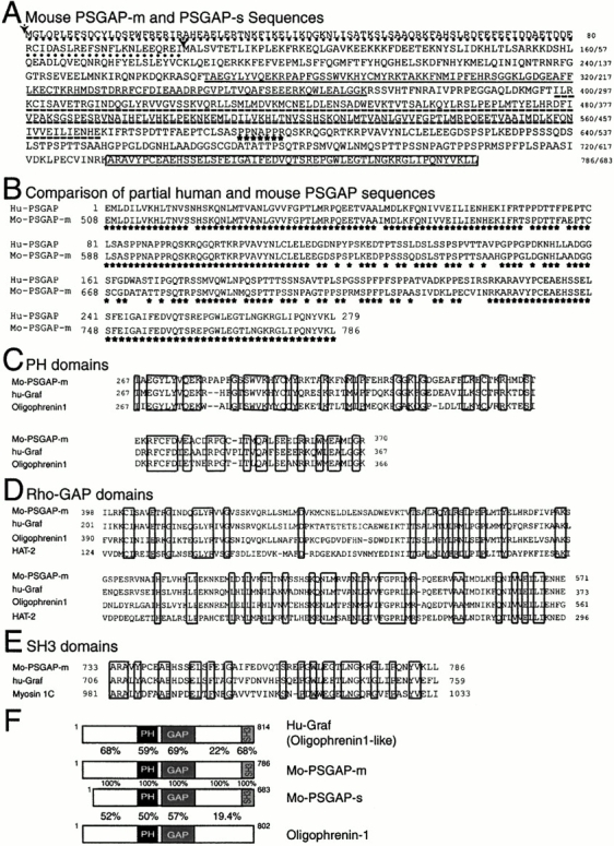
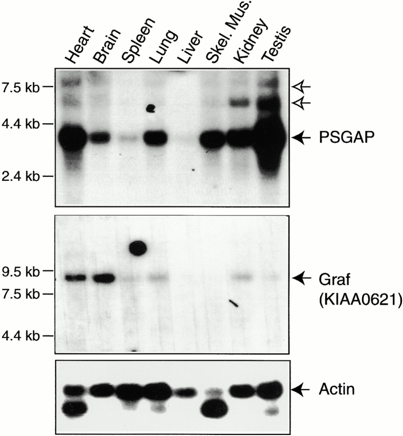
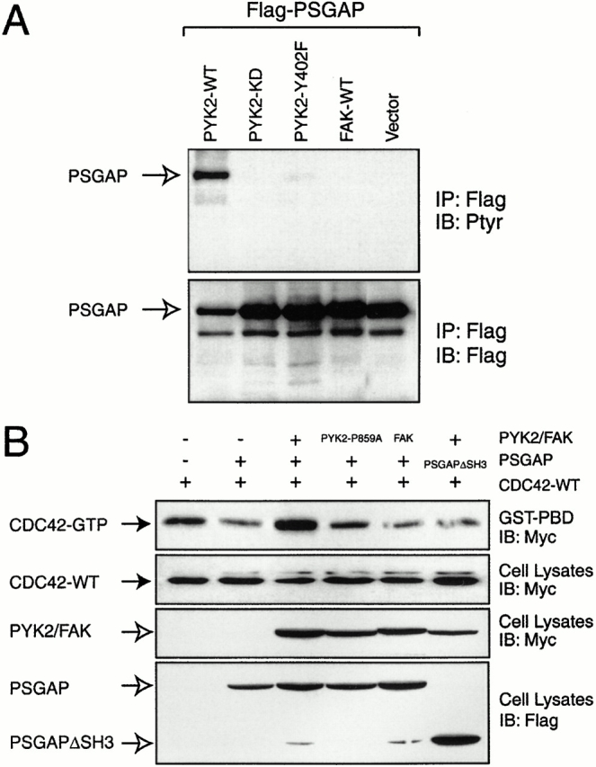
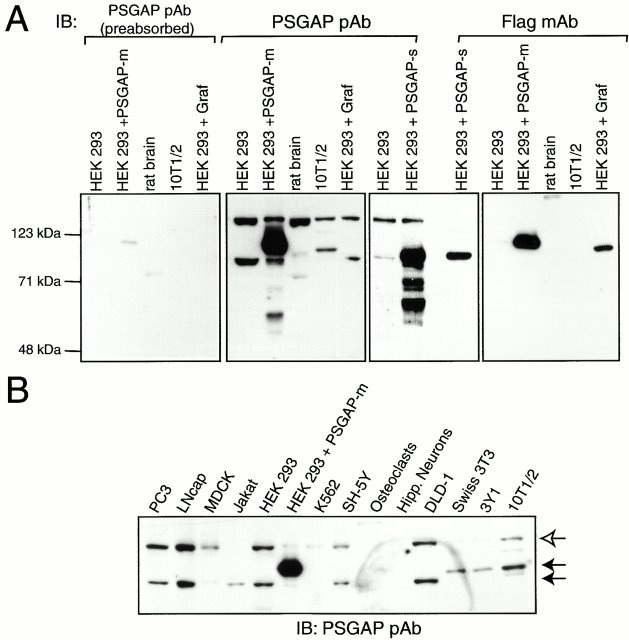
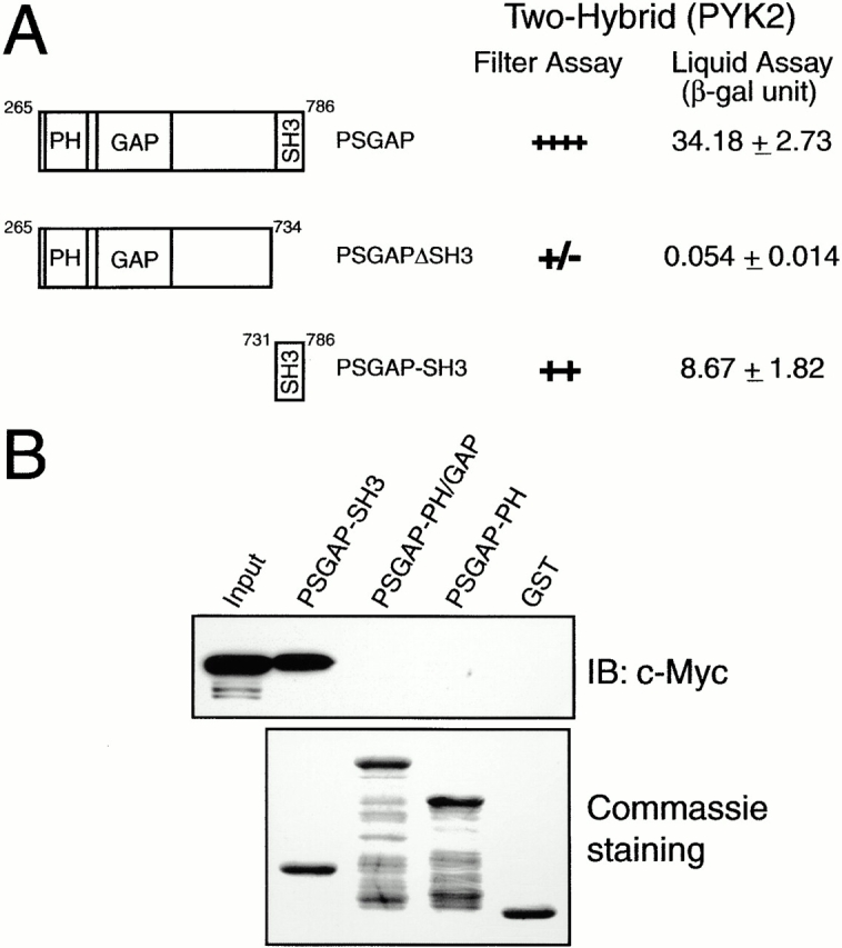
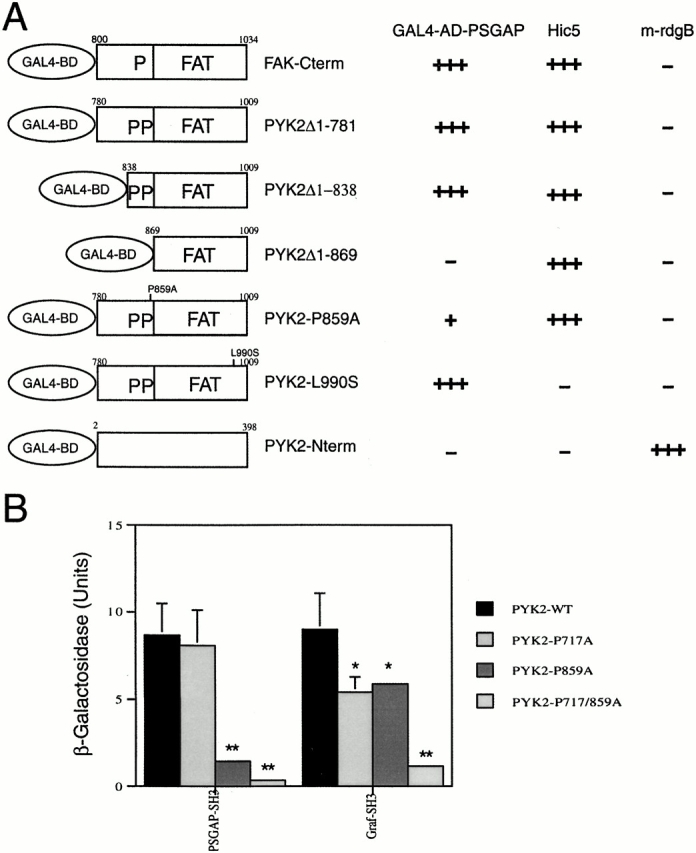
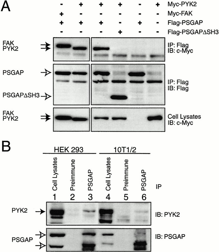
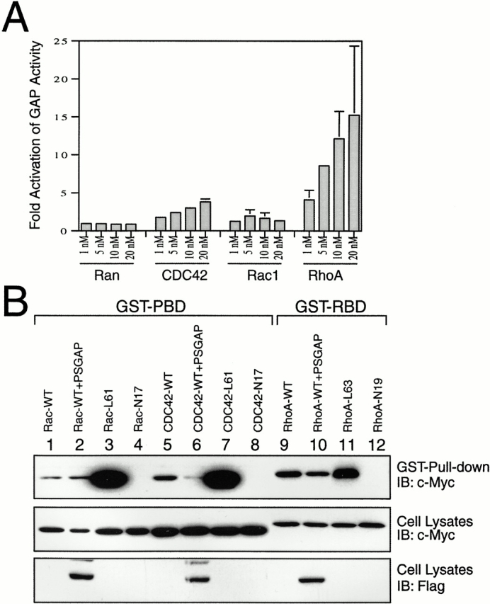
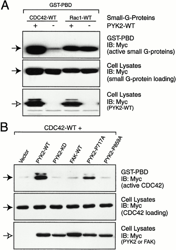
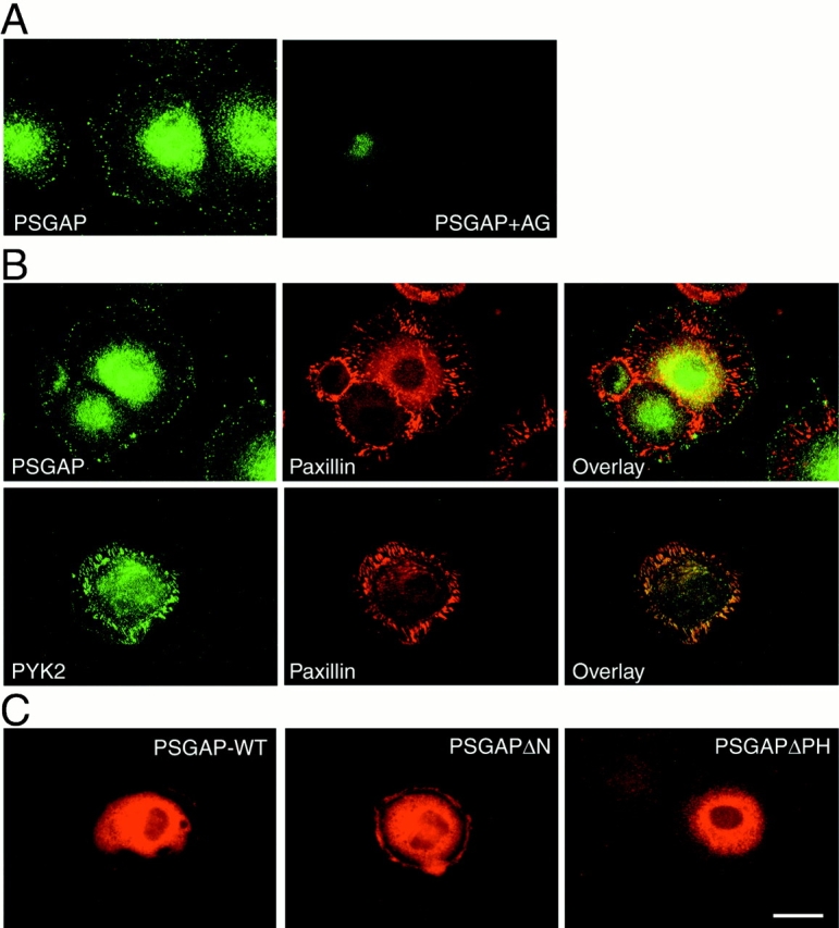
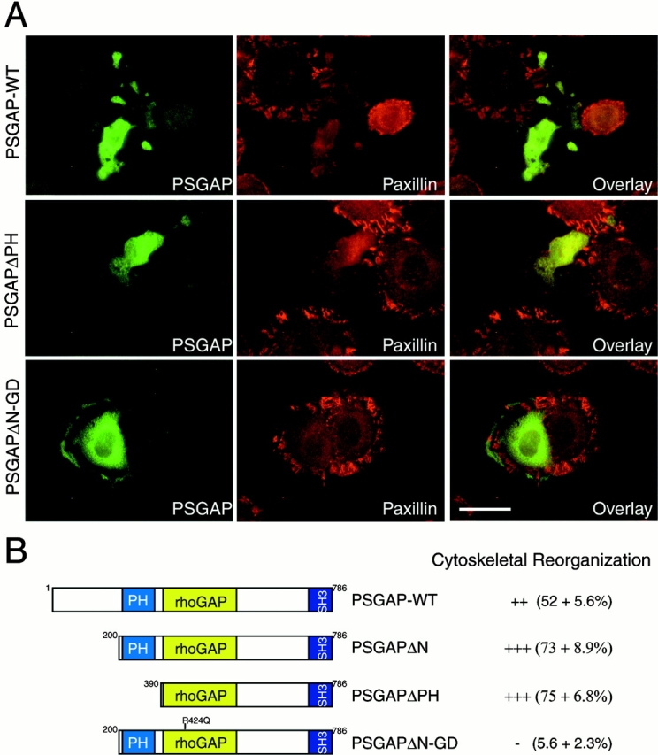
Proline-rich tyrosine kinase 2 (PYK2), a tyrosine kinase structurally related to focal adhesion kinase (FAK), is implicated in regulating cytoskeletal organization. However, mechanisms by which PYK2 participates in and regulates cytoskeletal organization remain largely unknown. Here we report identification of PSGAP, a novel protein that interacts with PYK2 and FAK and contains multiple domains including a pleckstrin homology domain, a rhoGTPase-activating protein domain, and a Src homology 3 domain. PYK2 interacts with PSGAP Src homology 3 domain via the carboxyl-terminal proline-rich sequence. PSGAP is able to increase GTPase activity of CDC42 and RhoA in vitro and in vivo. Remarkably, PYK2, but not FAK, can activate CDC42 via inhibition of PSGAP-mediated GTP hydrolysis of CDC42. Moreover, PSGAP is localized at cell periphery in fibroblasts in a pleckstrin homology domain-dependent manner. Over expression of PSGAP in fibroblasts results in reorganization of cytoskeletal structures and changes of cellular morphology, which requires rhoGTPase-activating activity. Taken together, our results suggest that PSGAP is a signaling protein essential for PYK2 regulation of cytoskeletal organization via Rho family GTPases.


Figures

Figure 1
Figure 1
Cloning of PSGAP, a novel rhoGAP-containing protein that interacts with PYK2. (A) Deduced amino acid sequences of mouse PSGAP-m and PSGAP-s. Filled arrows indicate the two methionines as the translational starting codons for the splice variants, PSGAP-m (786 amino acids) and PSGAP-s (683 amino acids), respectively. PSGAP-m contains a different amino terminus (∼103 amino acids) that is underlined with dots. The PH domain (amino acids 267–370 in PSGAP-m) is underlined, rhoGAP domain (amino acids 398–571 in PSGAP-m) is underlined with dashed lines, the proline rich motif is indicated by stars, and the SH3 domain (amino acids 733–786 in PSGAP-m) is boxed. The original clone isolated by yeast two-hybrid screen contains amino acids 265–786. (B) Comparison of the partial human and mouse PSGAP protein sequences. Partial human PSGAP (hu-PSGAP) (279 amino acids) were obtained by searching human EST databases, which corresponded to the residues of 508–786 of mouse PSGAP-m (mo-PSGAP-m) with 84% identity. Stars indicate identical residues. (C) Sequence alignment of PH domains of mouse PSGAP-m, human Graf, and oligophrenin-1. (D) Sequence alignment of the RhoGAP domains of mo-PSGAP-m, hu-Graf, Oligophrenin-1, HAT-2. (E) Sequence alignment of the SH3 domains of mo-PSGAP-m, hu-Graf, and myosin 1C. Alignments were determined using the program pileup from the Genetics Computer Group. (C, D, and E) Identical residues are boxed. (F) Comparison of domain structures of mo-PSGAP-m, mo-PSGAP-s, hu-Graf, and oligophrenin-1. The PH, rhoGAP, and SH3 domains are indicated. The amino acid identities (%) within a specific domain are shown.
Figure 2
Figure 2
Expression of PSGAP and Graf mRNAs in various tissues. Northern blot analyses were carried out using a multiple tissue blot (CLONTECH Laboratories, Inc.). Mouse cDNA fragments (796–1,358 bp for PSGAP and 1–717 bp of KIAA0621 for Graf) were labeled with [32P-α]dCTP by the random prime method and hybridized with the blot. A major transcript at 3.8 kb was detected for PSGAP (top, solid arrow). In addition, two more PSGAP transcripts, at 6 and 8 kb, respectively, were detected (top, open arrows). Graf transcript was detected at ∼9 kb (bottom, arrow). Molecular weight markers are indicated (left).
Figure 8
Figure 8
Regulation of PSGAP by PYK2, but not FAK. (A) Tyrosine phosphorylation of PSGAP by PYK2, but not FAK. HEK293 cells were transfected with indicated constructs. Cell lysates were immunoprecipitated with Flag antibodies (for PSGAP) and immunoblotted with antibodies against phosphotyrosine (Ptyr) and Flag. Open arrows indicate PSGAP. (B) Inhibition of PSGAP's effect on CDC42 by PYK2, but not FAK. HEK293 cells were transfected with indicated constructs. Cell lysates were incubated with GST-PBD immobilized on beads. Bound active CDC42 was resolved on SDS-PAGE and detected by immunoblotting. Equal amounts of CDC42 were expressed as indicated. The expression of PYK2, FAK, and PSGAP are also shown. (Solid arrows) CDC42; (open arrows) PYK2 or PSGAP.
Figure 3
Figure 3
Expression of PSGAP in various cells. (A) Characterization of anti–PSGAP antibody. A GST fusion protein containing the mouse PSGAP SH3 domain (amino acids 731–786) was used to generate rabbit polyclonal antibodies as described in Materials and Methods. Lysates (10 μg protein) of 293 cells expressing Flag-tagged PSGAP (PSGAP-m and PSGAP-s) and Graf, and other indicated cell lysates (20 μg protein) were resolved on SDS-PAGE and subjected to immunoblotting with anti–PSGAP antibodies, antibodies preabsorbed with the GST-PSGAP antigen, or an anti–Flag antibody. (B) Western blot analysis of PSGAP expression. Lysates (20 μg protein) of various cells were resolved on SDS-PAGE and subjected to immunoblotting with anti–PSGAP antibodies (6 μg protein of HEK293 cells expressing PSGAP was loaded). (Solid arrows) Transfected PSGAP and endogenously expressed PSGAP. Molecular weight markers are indicated (left).
Figure 4
Figure 4
SH3 domain of PSGAP is essential for the interaction with PYK2. (A) Mapping of the binding region in PSGAP by yeast two-hybrid assays. Yeast cells were cotransformed with a vector encoding the Gal4 binding domain fused to different PSGAP constructs and Gal4AD fused to PYK2. Transformed yeast cells were seeded in Leu, Trp, and His plates and assayed for β-Gal activity. Transformed yeast cells were also grown in Leu and Trp medium for liquid β-Gal assays. (B) Analysis of PSGAP binding to PYK2 by in vitro GST pull-down assays. HEK293 cells were transfected with Myc-tagged PYK2. Lysates of transfected cells were incubated with various GST-PSGAP fusion proteins (SH3, amino acids 731–786; PH-GAP, amino acids 265–590; and PH, amino acids 265–454) immobilized on beads. Lysate input and bound proteins were resolved on SDS-PAGE and subjected to immunoblotting with an anti–Myc antibody (top). An equal amount of GST fusion proteins was used as revealed by Coomassie staining (bottom).
Figure 5
Figure 5
Proline 859 in PYK2 is required for PSGAP binding. (A) Analyses of PYK2 domains essential for binding to PSGAP by yeast two-hybrid filter assays. Yeast cells were cotransformed with a vector encoding the Gal4 binding domain (Gal4-BD) fused to different constructs of PYK2, or FAK and Gal4AD/PSGAP. Gal4AD/Hic5 and Gal4AD/mrdgB were used as controls. Transformed yeast cells were seeded in Leu, Trp, and His plates and scored for growth and β-Gal activity (blue color). (−) No blue color in the yeast after 24 h in reaction, (+) blue reaction after 4 h, (+++) blue reaction after 30 min. (B) Reduction of the ability of P859A mutant to bind PSGAP in yeast two-hybrid liquid assays. Yeast cells were cotransformed with a vector encoding the Gal4 DB fused to SH3 domain of PSGAP or Graf and wild-type or mutant PYK2 in Gal4AD. Transformed yeast cells were grown in His, Leu, and Trp medium for liquid β-Gal assays. *P < 0.05 and **P < 0.01 in comparison with PYK2-WT, Student's t test.
Figure 6
Figure 6
Interaction of PSGAP with PYK2 and FAK in vivo. (A) Coimmunoprecipitation of PSGAP with PYK2 or FAK in mammalian-expressing cells. HEK293 cells cotransfected Myc-tagged PYK2 or FAK with Flag-tagged PSGAP or PSGAPΔSH3 were lysed. Immunoprecipitations (IP) with Flag antibody was revealed by immunoblotting (IB) with anti–Myc (top) or anti–Flag (middle) antibodies. Expression of PYK2 and FAK was demonstrated at bottom. (B) Coimmunoprecipitation of PSGAP with PYK2 in vivo. Cell lysates from HEK293 or 10T1/2 cells were immunoprecipitated with PSGAP antibodies and immunoblotted with PYK2 antibodies. (Solid arrow) PYK2; (open arrows) PSGAP.
Figure 7
Figure 7
GTPase-activating activity of PSGAP. (A) Characterization of GTPase-activating activity of PSGAP in vitro. Recombinant proteins of RhoA, Rac1, CDC42, and Ran GTPases were loaded with [γ-32P] GTP and incubated without or with increasing concentrations of PSGAP GAP domain. Hydrolyzed and GTPase-bound radioactive GTP was determined by nitrocellulose filtration assay as described in Materials and Methods. Fold activation represents the ratio of radioactivity remaining after incubation in the absence of GAP domain versus the amount of radioactivity remaining after incubation in the presence of GAP domain. (B) Analysis of the effects of PSGAP on GST-PBD or GST-RBD binding to transfected small G proteins. HEK293 cells were transfected with wild-type, active, or inactive mutants of Rac1, CDC42, or RhoA (all Myc-tagged) with or without Flag-tagged PSGAP. Cell lysates were incubated with GST-PBD or GST-RBD immobilized on beads. Bound small G proteins were resolved on SDS-PAGE and detected by immunoblotting (top). Equal amounts of small G proteins were expressed as shown in the middle. (Bottom) Expression of PSGAP.
Figure 9
Figure 9
Activation of CDC42 by PYK2, but not FAK. (A) PYK2 activation of CDC42, but not Rac. (B) PYK2, but not FAK, activation of CDC42 requires PYK2 catalytic activity and its binding region to PSGAP. (A and B) HEK293 cells were transfected with indicated constructs. Cell lysates were incubated with GST-PBD immobilized on beads. Bound active CDC42 or Rac was resolved on SDS-PAGE and detected by immunoblotting. Equal amounts of small G proteins were expressed as indicated. The expression of PYK2 and FAK are also shown. (Solid arrows) Small G proteins; (open arrows) PYK2 or FAK.
Figure 10
Figure 10
Expression of PSGAP at the cell periphery in 10T1/2 fibroblasts. 10T1/2 cells were fixed with 4% paraformaldehyde for 20 min, blocked with 10% BSA, and immunostained with indicated antibodies. (A) Immunostaining using antibodies against PSGAP or antibodies preabsorbed with the GST-PSGAP antigen. (B) Coimmunostaining with anti–PSGAP or anti–PYK2 and antipaxillin antibodies. PSGAP and PYK2 were visualized by FITC-conjugated secondary antibodies, whereas paxillin appeared in red with rhodamine-conjugated secondary antibodies. (C) Immunostaining of PSGAP wild-type (PSGAP-WT), NH2-terminal–deleted PSGAP (PSGAPΔN), and PH-domain–deleted PSGAP (PSGAPΔPH) in transfected 10T1/2 fibroblasts. Cells were transfected with the indicated constructs and stained with anti–PSGAP antibodies. Bar, 50 μm.
Figure 11
Figure 11
Cytoskeletal reorganization in fibroblasts expressing PSGAP. (A) 10T1/2 cells were transfected with the indicated constructs and stained with anti–PSGAP (green) and anti–paxillin (red) antibodies. Bar, 50 μm. (B) Schematic representation of PSGAP and PSGAP mutants. The numbers represent the amino acid residues deleted from PSGAP-m. The PH domain, rhoGAP domain, and SH3 domain are indicated. Cytoskeletal reorganization index (mean ± SEM) was determined by counting the morphologically altered PSGAP-expressing cells divided by the total number of PSGAP-expressing cells, and listed at right.

References

    1. Andreev J., Simon J.P., Sabatini D.D., Kam J., Plowman G., Randazzo P.A., Schlessinger J. Identification of a new PYK2 target protein with Arf-GAP activity. Mol. Cell. Biol. 1999;19:2338–2350. - PMC - PubMed
    1. Astier A., Avraham H., Manie S.N., Groopmen J., Canty T., Avraham S., Freedman A.S. The related adhesion focal tyrosine kinase is tyrosine-phosphorylated after β1-integrin stimulation in B cells and binds to p130Cas . J. Biol. Chem. 1997;272:228–232. - PubMed
    1. Avraham S., London R., Fu Y., Ota S., Hiregowdara D., Li J., Jiang S., Pasztor L.M., White R.A., Groopman J.E., Avraham H. Identification and characterization of a novel related adhesion focal tyrosine kinase (RAFTK) from megakaryocytes and brain. J. Biol. Chem. 1995;270:27742–27751. - PubMed
    1. Bar-Sagi D., Rotin D., Batzer A., Mandiyan V., Schlessinger J. SH3 domains direct cellular localization of signaling molecules. Cell. 1993;74:83–91. - PubMed
    1. Barrett T., Xiao B., Dodson E.J., Dodson G., Ludbrook S.B., Nurmahomed K., Gamblin S.J., Musacchio A., Smerdon S.J., Eccleston J.F. The structure of the GTPase-activating domain from p50rhoGAP. Nature. 1997;385:458–461. - PubMed

Publication types

MeSH terms

Associated data