. 2001 Mar 5;152(5):1079-85.
doi: 10.1083/jcb.152.5.1079.

Caveolin-2 is targeted to lipid droplets, a new "membrane domain" in the cell

Affiliations

Caveolin-2 is targeted to lipid droplets, a new "membrane domain" in the cell

T Fujimoto et al. J Cell Biol. .

Abstract

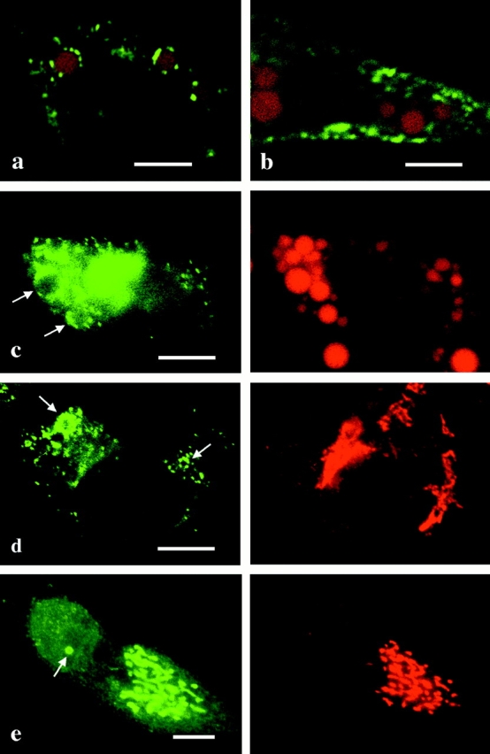
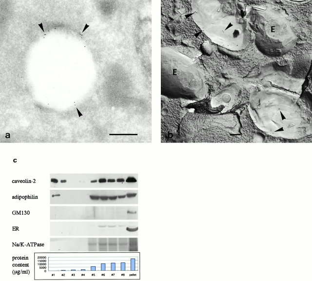
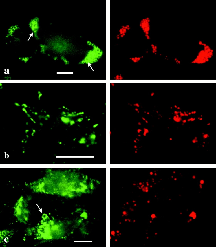
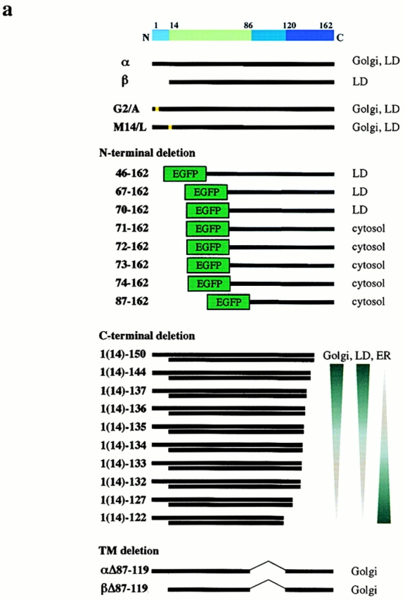
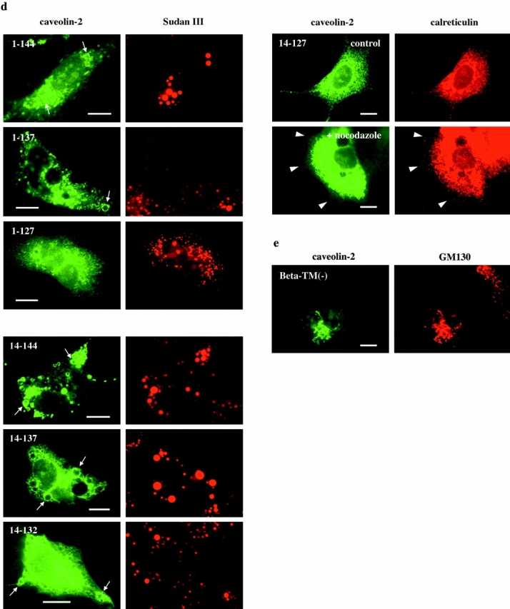
Caveolin-1 and -2 constitute a framework of caveolae in nonmuscle cells. In the present study, we showed that caveolin-2, especially its beta isoform, is targeted to the surface of lipid droplets (LD) by immunofluorescence and immunoelectron microscopy, and by subcellular fractionation. Brefeldin A treatment induced further accumulation of caveolin-2 along with caveolin-1 in LD. Analysis of mouse caveolin-2 deletion mutants revealed that the central hydrophobic domain (residues 87-119) and the NH(2)-terminal (residues 70-86) and COOH-terminal (residues 120-150) hydrophilic domains are all necessary for the localization in LD. The NH(2)- and COOH-terminal domains appeared to be related to membrane binding and exit from ER, respectively, implying that caveolin-2 is synthesized and transported to LD as a membrane protein. In conjunction with recent findings that LD contain unesterified cholesterol and raft proteins, the result implies that the LD surface may function as a membrane domain. It also suggests that LD is related to trafficking of lipid molecules mediated by caveolins.


Figures

Figure 1
Figure 1
Confocal microscopy of HepG2 stably expressing caveolin-2β (a) and caveolin-1 (b). Caveolins (green), LD stained by Sudan III (red). Caveolin-2 is seen around LD discontinuously, whereas caveolin-1 is along the cell edge, and not related to LD. (c) FRTL-5 transiently transfected with full-length caveolin-2 cDNA and cultured with OA/BSA for 2 d. Caveolin-2 (green) is observed around LD (red). HepG2 stably transfected with caveolin-2β (d) or full-length caveolin-2 (e) cDNA was labeled for caveolin-2 (green) and GM130 (red). The labeling of caveolin-2β overlapping with that of GM130 is scarce (d), whereas caveolin-2α, the predominant isoform in the cell, is mostly localized in the Golgi (e). Arrows indicate labeling around LD. Bars, 10 μm.
Figure 2
Figure 2
(a and b) Immunoelectron microscopy of HepG2 expressing caveolin-2β (clone A-8). Bar, 500 nm. (a) Ultrathin cryosection. Gold particles labeling caveolin-2 are observed in the rim of LD in small clusters (arrowheads); the content of LD appears vacant. (b) Freeze-fracture replica. Gold particles for caveolin-2 are observed in clusters (arrowheads) on the P face of LD, which show an onion-like morphology. The E face of LD (E) is devoid of labeling. (c) Western blotting of subcellular fractions obtained from HepG2 clone A-8. The graph shows the protein content of the fractions. The top two fractions (#1 and #2) contained little protein, but showed positive signals for caveolin-2 and adipophilin. GM130, 66-kD protein, and Na+/K+-ATPase (Golgi, ER, and plasma membrane markers, respectively) are found only in the bottom fractions.
Figure 3
Figure 3
(a) NRK, expressing EGFP–caveolin-2 along with endogenous caveolin-1 and -2, cultured with OA/BSA. EGFP-caveolin-2 (green) is seen around LD (red) (arrows). (b and c) HepG2 stably expressing caveolin-1 and -2 (clone 6). Caveolin-1 (red) and -2 (green) show colocalization in the plasma membrane (b), and only caveolin-2 (green) is observed around LD (red) in some cells (arrows) (c). Cells in b and c were treated with methanol and Triton X-100, respectively, to show the plasmalemmal (b) and intracellular (c) labeling. Bars, 10 μm. (d) Western blotting of subcellular fractions obtained from HepG2 clone 6. Only caveolin-2, but not caveolin-1, was detected in the LD fractions (#1 and #2).
Figure 3
Figure 3
(a) NRK, expressing EGFP–caveolin-2 along with endogenous caveolin-1 and -2, cultured with OA/BSA. EGFP-caveolin-2 (green) is seen around LD (red) (arrows). (b and c) HepG2 stably expressing caveolin-1 and -2 (clone 6). Caveolin-1 (red) and -2 (green) show colocalization in the plasma membrane (b), and only caveolin-2 (green) is observed around LD (red) in some cells (arrows) (c). Cells in b and c were treated with methanol and Triton X-100, respectively, to show the plasmalemmal (b) and intracellular (c) labeling. Bars, 10 μm. (d) Western blotting of subcellular fractions obtained from HepG2 clone 6. Only caveolin-2, but not caveolin-1, was detected in the LD fractions (#1 and #2).
Figure 4
Figure 4
(a) HepG2 stably transfected with full-length caveolin-2 cDNA was treated with BFA for 2 h. Caveolin-2 (green) became confined to LD (red) in most cells (arrows). (b and c) Human fibroblasts treated with BFA for 16 h. Not only caveolin-2 (b), but also caveolin-1 (c) (green) was observed around LD (red) (arrows). (d) HepG2 stably transfected with full-length caveolin-1 cDNA. After the BFA treatment for 7 h, caveolin-1 (green) was seen around LD (red) in occasional cells (arrows). Bars, 10 μm.
Figure 5
Figure 5
(a) A diagram of caveolin-2 mutants and their distribution on transient expression in HepG2. (b–e) Distribution of mutants was observed by anti–caveolin-2 labeling (b, d, and e) or by EGFP (c) (green), and compared with LD, Golgi, or ER markers (red). Arrows mark the labeling apparently encircling LD. Bars, 10 μm. (b) Single amino acid replacements to examine the difference between α and β isoforms. Even when methionine-14, the second translation initiation site, was replaced with leucine (M14/L), or glycine-2, a putative myristoylation site, was replaced with alanine (G2/A), distribution was not different from that of caveolin-2α. (c) NH2-terminal deletion mutants fused to the COOH terminus of EGFP. Deletion of up to 69 residues (70–162) did not affect the LD localization, but further truncation (71–162) caused cytosolic distribution. (d) COOH-terminal deletion mutants. For both α and β isoforms, the distribution was not changed when the sequence up to the 150th residue was maintained. As more amino acids were deleted, localization in LD and the Golgi became less distinct, and a network-like labeling increased. The latter labeling was similar to that of calreticulin (result of 14–127 is shown). When ER was made to retract from the cell edge (arrowheads) by nocodazole, the labeling for the mutant showed a matching redistribution. (e) Caveolin-2β lacking the central hydrophobic domain [beta-TM(−)] was localized in the Golgi, labeled by anti–GM130. Deletion of the same domain from caveolin-2α gave the same result.
Figure 5
Figure 5
(a) A diagram of caveolin-2 mutants and their distribution on transient expression in HepG2. (b–e) Distribution of mutants was observed by anti–caveolin-2 labeling (b, d, and e) or by EGFP (c) (green), and compared with LD, Golgi, or ER markers (red). Arrows mark the labeling apparently encircling LD. Bars, 10 μm. (b) Single amino acid replacements to examine the difference between α and β isoforms. Even when methionine-14, the second translation initiation site, was replaced with leucine (M14/L), or glycine-2, a putative myristoylation site, was replaced with alanine (G2/A), distribution was not different from that of caveolin-2α. (c) NH2-terminal deletion mutants fused to the COOH terminus of EGFP. Deletion of up to 69 residues (70–162) did not affect the LD localization, but further truncation (71–162) caused cytosolic distribution. (d) COOH-terminal deletion mutants. For both α and β isoforms, the distribution was not changed when the sequence up to the 150th residue was maintained. As more amino acids were deleted, localization in LD and the Golgi became less distinct, and a network-like labeling increased. The latter labeling was similar to that of calreticulin (result of 14–127 is shown). When ER was made to retract from the cell edge (arrowheads) by nocodazole, the labeling for the mutant showed a matching redistribution. (e) Caveolin-2β lacking the central hydrophobic domain [beta-TM(−)] was localized in the Golgi, labeled by anti–GM130. Deletion of the same domain from caveolin-2α gave the same result.

Comment in

Similar articles

Cited by

References

    1. Anderson R.G.W. The caveolae membrane system. Annu. Rev. Biochem. 1998;67:199–225. - PubMed
    1. Blanchette-Mackie E.J., Dwyer N.K., Barber T., Coxey R.A., Takeda T., Rondinone C.M., Theodorakis J.L., Greenberg A.S., Londos C. Perilipin is located on the surface layer of intracellular lipid droplets in adipocytes. J. Lipid Res. 1995;36:1211–1226. - PubMed
    1. Brasaemle D.L., Barber T., Kimmel A.R., Londos C. Post-translational regulation of perilipin expressionstabilization by stored intracellular neutral lipids. J. Biol. Chem. 1997;272:9378–9387. - PubMed
    1. Conrad P.A., Smart E.J., Ying Y.S., Anderson R.G.W., Bloom G.S. Caveolin cycles between plasma membrane caveolae and the Golgi complex by microtubule-dependent and microtubule-independent steps. J. Cell Biol. 1995;131:1421–1433. - PMC - PubMed
    1. Das K., Lewis R.Y., Scherer P.E., Lisanti M.P. The membrane-spanning domains of caveolins-1 and -2 mediate the formation of caveolin hetero-oligomers. Implications for the assembly of caveolae membranes in vivo. J. Biol. Chem. 1999;274:18721–18728. - PubMed

Publication types

MeSH terms