. 2001 Mar 5;152(5):1087-98.
doi: 10.1083/jcb.152.5.1087.

Mammalian sprouty-1 and -2 are membrane-anchored phosphoprotein inhibitors of growth factor signaling in endothelial cells

Affiliations

Mammalian sprouty-1 and -2 are membrane-anchored phosphoprotein inhibitors of growth factor signaling in endothelial cells

M A Impagnatiello et al. J Cell Biol. .

Abstract

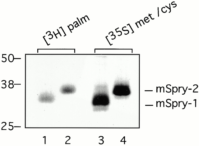
Growth factor-induced signaling by receptor tyrosine kinases (RTKs) plays a central role in embryonic development and in pathogenesis and, hence, is tightly controlled by several regulatory proteins. Recently, Sprouty, an inhibitor of Drosophila development-associated RTK signaling, has been discovered. Subsequently, four mammalian Sprouty homologues (Spry-1-4) have been identified. Here, we report the functional characterization of two of them, Spry-1 and -2, in endothelial cells. Overexpressed Spry-1 and -2 inhibit fibroblast growth factor- and vascular endothelial growth factor-induced proliferation and differentiation by repressing pathways leading to p42/44 mitogen-activating protein (MAP) kinase activation. In contrast, although epidermal growth factor-induced proliferation of endothelial cells was also inhibited by Spry-1 and -2, activation of p42/44 MAP kinase was not affected. Biochemical and immunofluorescence analysis of endogenous and overexpressed Spry-1 and -2 reveal that both Spry-1 and -2 are anchored to membranes by palmitoylation and associate with caveolin-1 in perinuclear and vesicular structures. They are phosphorylated on serine residues and, upon growth factor stimulation, a subset is recruited to the leading edge of the plasma membrane. The data indicate that mammalian Spry-1 and -2 are membrane-anchored proteins that negatively regulate angiogenesis-associated RTK signaling, possibly in a RTK-specific fashion.


Figures

Figure 1
Figure 1
Amino acid alignment of the four mSpry proteins (mSpry-1–4) with hSpry-2 and DSpry. Dashes indicate spaces introduced to maximize alignment. Shaded residues indicate residues that are identical in at least four of the aligned sequences. A putative Sprouty domain and a putative serine-rich domain are underlined by a dotted line and a gray bar, respectively.
Figure 2
Figure 2
mSpry-1 and -2 inhibit endothelial cell proliferation and differentiation. (A) Endothelial cell proliferation. HUVECs were infected with 5,000 PPC of AdmSpry-1, AdmSpry-2, or AdeGFP as indicated. 4 h after infection, the cells were starved and then stimulated with 10 ng/ml FGF2, 10 ng/ml VEGF, or 20 ng/ml EGF. The rate of DNA synthesis was determined by the incorporation of [3H]thymidine 48 h after infection. Error bars indicate the standard deviation on the average of three experiments. (B) Endothelial cell differentiation. HUVECs were infected with recombinant adenovirus as described in A and then plated on Matrigel. Representative microphotographs were taken 24 h after plating.
Figure 4
Figure 4
mSpry-1 and -2 expression is regulated by FGF2 in endothelial cells. Uninfected 1G11 cells were cultured under exponential growth conditions (exp) or serum starved for 48 h and then stimulated with 10 ng/ml FGF2 for the times indicated. Northern blot analysis was performed on total RNA isolated from each time point, using full-length cDNAs for mSpry-1 and mSpry–2 as probes. Equal loading of RNAs was confirmed by staining of the gels with ethidium bromide. The lengths of Spry mRNAs (Kb) and 18S ribosomal RNA are indicated.
Figure 3
Figure 3
Inhibition of p42/44 MAP kinase activation. HUVECs infected with AdmSpry-1, AdmSpry-2, or AdeGFP were serum starved 24 h before stimulation with 50 ng/ml VEGF for various time periods as indicated (A) or with 50 ng/ml VEGF, 50 ng/ml FGF2, or 20 ng/ml EGF for 10 or 20 min (B). Cells were lysed, and levels of activated p42/44 MAP kinase, total p42/44 MAP kinase, activated MEK, and total MEK were determined by immunoblotting with specific antibodies as indicated. Expression levels of mSpry-1 and -2 were determined by stripping and reprobing the blots with antibodies specific for mSpry-1 or -2 (A).
Figure 7
Figure 7
Colocalization with VE–cadherin. Confocal immunofluorescent doublestaining of HUVECs with antibodies specific for hSpry-1 (green) and VE–cadherin (red) as indicated. Colocalization was determined by overlaying the green and the red staining. Note the mutual exclusive localization of Spry-1 and VE–cadherin in the plasma membrane. Bars, 10 μm.
Figure 5
Figure 5
Growth factor–induced recruitment to the plasma membrane. The subcellular localization of endogenous hSpry-1 was determined in HUVECs by immunofluorescence microscopy using an affinity-purified antibody that specifically recognized mSpry-1 and hSpry-1 (green) and Hoechst staining of nuclei (blue). In actively proliferating endothelial cells (exponential growth), Spry-1 is localized to the perinuclear region, vesicular structures, and lamellipodia in the leading edge of the plasma membrane. In serum-starved cells, Spry-1 is not found in the plasma membrane (serum-starved), but a subset of it is recruited to the plasma membrane 30 min after stimulation with 10 ng/ml FGF2 (serum-starved + FGF2). Preabsorption of the affinity-purified antibodies with corresponding antigenic peptide (1 μg/ml) resulted in a complete abrogation of the immunofluorescence signal (peptide competition). Bars, 10 μm.
Figure 6
Figure 6
Colocalization with caveolin-1. The localization of endogenous hSpry-1 was determined in HUVECs by confocal immunofluorescent doublestaining with antibodies specific for hSpry-1 (green) and caveolin-1 (red) as indicated. Colocalization was determined by overlaying the green and the red staining. Note the colocalization of Spry-1 with caveolin-1 adjacent to the perinuclear region, in a subset of vesicular structures, and in the plasma membrane (visualized by yellow staining in the two bottom panels). A higher magnification of vesicular structures and plasma membrane (box, bottom left) is shown in the bottom-right panel. Bars, 10 μm.
Figure 8
Figure 8
Sprys are tightly associated with caveolin-1 and membranes. (A) mSpry-1 associates with caveolin-1. HUVECs infected with AdmSpry-1 or AdeGFP were lysed in digitonin (D) or Triton X-100 (T) buffers, as described in Materials and Methods and indicated below the lanes. Aliquots of the cell lysates were subjected to immunoprecipitations with antibodies specific for mSpry-1 (top) or unrelated rabbit immunoglobulins, as negative control (bottom), in the respective buffers. Precipitated proteins and nonprecipitated proteins (Unbound) were resolved by SDS-PAGE, and caveolin-1, mSpry-1, and annexin II were detected by immunoblotting as indicated. (B) mSpry-1 is tightly associated with membranes. HUVECs infected with AdmSpry-1 or AdeGFP were subjected to a subcellular fractionation protocol, as described in Materials and Methods. Lanes 1 and 2 (Cyto) show cytoplasmatic extract obtained by permeabilizing the cells with digitonin (0.003%). Lanes 3 and 4 (Peri) show peripheral membrane proteins washed from partially purified membranes with sodium carbonate (Na2HCO3). Lanes 5 and 6 (Int) show integral membrane proteins that were not removed from the membrane fraction by sodium carbonate treatment. Protein levels of mSpry-1, LDH, transferrin receptor (Tf-R), caveolin-1, and annexin II in the various fractions were determined by immunoblotting as indicated. (C) The majority of mSpry-1 and caveolin-1 are not in detergent-resistant lipid rafts. HUVECs infected with AdmSpry-1 or AdeGFP were lysed in a Triton X-100 buffer and sedimented on Optiprep gradients as described in Materials and Methods. mSpry-1, caveolin-1, and annexin II in the gradient fractions were visualized by immunoblotting as indicated. The Optiprep concentrations of the fractions are given below the lanes. *Indicates that half of the fractions has been loaded in these lanes.
Figure 8
Figure 8
Sprys are tightly associated with caveolin-1 and membranes. (A) mSpry-1 associates with caveolin-1. HUVECs infected with AdmSpry-1 or AdeGFP were lysed in digitonin (D) or Triton X-100 (T) buffers, as described in Materials and Methods and indicated below the lanes. Aliquots of the cell lysates were subjected to immunoprecipitations with antibodies specific for mSpry-1 (top) or unrelated rabbit immunoglobulins, as negative control (bottom), in the respective buffers. Precipitated proteins and nonprecipitated proteins (Unbound) were resolved by SDS-PAGE, and caveolin-1, mSpry-1, and annexin II were detected by immunoblotting as indicated. (B) mSpry-1 is tightly associated with membranes. HUVECs infected with AdmSpry-1 or AdeGFP were subjected to a subcellular fractionation protocol, as described in Materials and Methods. Lanes 1 and 2 (Cyto) show cytoplasmatic extract obtained by permeabilizing the cells with digitonin (0.003%). Lanes 3 and 4 (Peri) show peripheral membrane proteins washed from partially purified membranes with sodium carbonate (Na2HCO3). Lanes 5 and 6 (Int) show integral membrane proteins that were not removed from the membrane fraction by sodium carbonate treatment. Protein levels of mSpry-1, LDH, transferrin receptor (Tf-R), caveolin-1, and annexin II in the various fractions were determined by immunoblotting as indicated. (C) The majority of mSpry-1 and caveolin-1 are not in detergent-resistant lipid rafts. HUVECs infected with AdmSpry-1 or AdeGFP were lysed in a Triton X-100 buffer and sedimented on Optiprep gradients as described in Materials and Methods. mSpry-1, caveolin-1, and annexin II in the gradient fractions were visualized by immunoblotting as indicated. The Optiprep concentrations of the fractions are given below the lanes. *Indicates that half of the fractions has been loaded in these lanes.
Figure 9
Figure 9
Spry-1 and -2 are posttranslationally modified by phosphorylation on serine residues. (A) Kinetics of posttranslational modification. HUVECs infected with AdmSpry-1 were metabolically labeled with [35S]methionine/cysteine for the times indicated in minutes (pulse). Cell lysates were either collected immediately, (lanes 1–5) or after the addition of normal medium for 2 h (chase; lane 6). mSpry-1 was immunoprecipitated, and the state of its posttranslational modification was assessed by the electrophoretic mobility of the protein upon SDS-PAGE and immunoblotting. (B) Phosphatase treatment. Total cell lysate from actively proliferating 1G11 cells was treated with calf intestine phosphatase (lane 2) or with buffer alone (lane 1), and endogenous mSpry-1 was detected by immunoblotting. (C) Phosphate labeling. HUVECs infected with AdmSpry-1 or -2 were serum starved, stimulated with 10 ng/ml FGF2, and metabolically labeled with [32P]orthophosphate for the times indicated in minutes. mSpry-1 and -2 were immunoprecipitated with affinity-purified antibodies, and the incorporation of radioactivity was analyzed by SDS-PAGE and autoradiography. Exponentially growing cells were used as control (exp). (D) mSpry-1 and -2 are phosphorylated on serine residues. Phosphoamino acid analysis was performed on the immunoprecipitated mSpry-1 and -2 proteins described in C. The positions of phosphoserine (PS), phosphothreonine (PT), and phosphotyrosine (PY) markers on the thin layer chromatogram are indicated.
Figure 10
Figure 10
mSpry-1 and -2 are palmitoylated. HUVECs infected with AdmSpry-1 (lanes 1 and 3) or -2 (lanes 2 and 4) were labeled with [3H]palmitate (lanes 1 and 2) or [35S]methionine/cysteine (lanes 3 and 4) for 2 h. mSpry-1 and -2 were immunoprecipitated with affinity-purified antibodies, and radiolabeled mSpry-1 and -2 were resolved by SDS-PAGE and visualized by fluorography.

References

    1. Anderson R.G. The caveolae membrane system. Annu. Rev. Biochem. 1998;67:199–225. - PubMed
    1. Baatout S. Endothelial differentiation using Matrigel. Anticancer Res. 1997;17:451–455. - PubMed
    1. Baker A., Saltik M., Lehrman H., Killisch I., Mautner V., Lamm G., Christofori G., Cotten M. Polyethylenimine (PEI) is a simple, inexpensive and effective reagent for condensing and linking plasmid DNA to adenovirus for gene delivery. Gene Therapy. 1997;4:773–782. - PubMed
    1. Casci T., Vinos J., Freeman M. Sprouty, an intracellular inhibitor of Ras signalling. Cell. 1999;96:655–665. - PubMed
    1. Chambers D., Mason I. Expression of sprouty2 during early development of the chick embryo is coincident with known sites of FGF signalling. Mech. Dev. 2000;91:361–364. - PubMed

Publication types

MeSH terms