. 2001 Mar 5;152(5):1099-106.
doi: 10.1083/jcb.152.5.1099.

The transcription coactivator CBP is a dynamic component of the promyelocytic leukemia nuclear body

Affiliations

The transcription coactivator CBP is a dynamic component of the promyelocytic leukemia nuclear body

F M Boisvert et al. J Cell Biol. .

Abstract

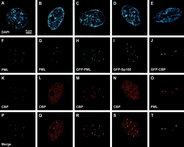
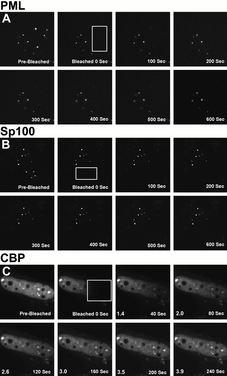
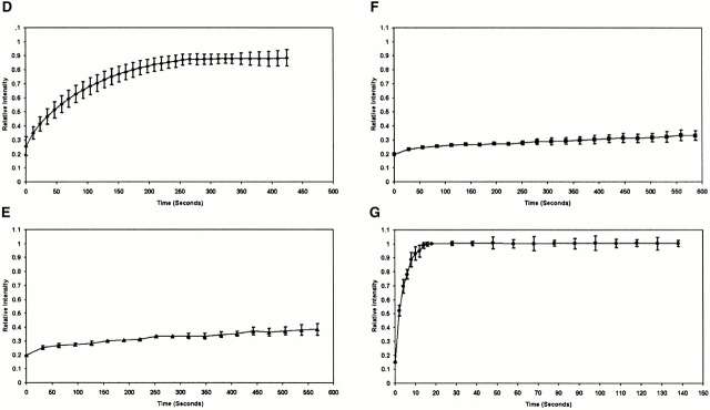
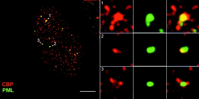
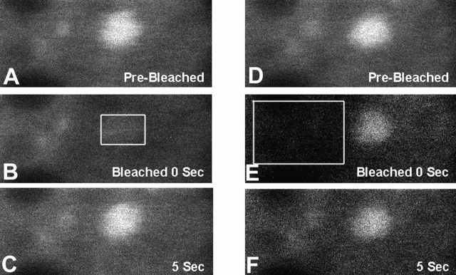
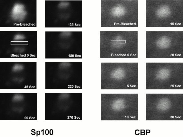
The transcription coactivator and histone acetyltransferase CAMP response element-binding protein (CBP) has been demonstrated to accumulate in promyelocytic leukemia (PML) bodies. We show that this accumulation is cell type specific. In cells where CBP does not normally accumulate in PML bodies, it can be induced to accumulate in PML bodies through overexpression of either CBP or Pml, but not Sp100. Using fluorescence recovery after photobleaching, we demonstrate that CBP moves rapidly into and out of PML bodies. In contrast, Pml and Sp100 are relatively immobile in the nucleoplasm and within PML nuclear bodies. They possess the characteristics expected of proteins that would play a structural role in the integrity of these subnuclear domains. Our results are consistent with CBP being a dynamic component of PML bodies and that the steady-state level in these structures can be modulated by Pml.


Figures

Figure 1
Figure 1
Colocalization by deconvolution immunofluorescence microscopy of CBP (NH2 terminus antibody; Santa Cruz Biotechnology, Inc.) and Pml (5E10 antibody) proteins in SK-N-SH cells (A, F, K, and P) but not in 293 cells (B, G, L, and Q). Overexpression of GFP–Pml in 293 cells (C and H) allows proper targeting into PML bodies (C) and concentrates CBP in PML bodies (M and R). Overexpression of GFP–Sp100 in 293 cells (D and I) does not concentrate CBP in PML bodies (N and S). Overexpression of GFP–CBP in 293 cells (E and J) targets this protein to PML bodies (O and T). Bar, 5 μm.
Figure 3
Figure 3
FRAP of half-nucleus of 293 cells expressing GFP–Pml (A), GFP–Sp100 (B), or GFP–CBP (C). Cells were imaged before the bleaching (Pre-Bleached), immediately after the bleaching (Bleached, time = 0 s), and during fluorescence recovery at the indicated times. A box indicates the area bleached, corresponding to half the nucleus. The fluorescence intensity in the bleached and the unbleached regions was measured and expressed as a relative intensity where a value of one is equal intensity in both halves. The relative intensity over time is shown. Fluorescence recovery curves for CBP (D), Pml (E), and Sp100 (F) show the kinetics of redistribution of the different fluorescent proteins after bleaching. As a control, recovery of GFP alone was measured (G).
Figure 3
Figure 3
FRAP of half-nucleus of 293 cells expressing GFP–Pml (A), GFP–Sp100 (B), or GFP–CBP (C). Cells were imaged before the bleaching (Pre-Bleached), immediately after the bleaching (Bleached, time = 0 s), and during fluorescence recovery at the indicated times. A box indicates the area bleached, corresponding to half the nucleus. The fluorescence intensity in the bleached and the unbleached regions was measured and expressed as a relative intensity where a value of one is equal intensity in both halves. The relative intensity over time is shown. Fluorescence recovery curves for CBP (D), Pml (E), and Sp100 (F) show the kinetics of redistribution of the different fluorescent proteins after bleaching. As a control, recovery of GFP alone was measured (G).
Figure 2
Figure 2
Localization of Pml and CBP by deconvolution immunofluorescence microscopy after exposure of these 293 cells to α-interferon (1,000 U/ml) for 72 h. Bar: (left panel) 5.5 μm; (right, panels 1–3) 1.38 μm.
Figure 4
Figure 4
Determination of the direction of movement of GFP–CBP. A whole PML nuclear body was bleached (B, box) in a 293 cell expressing GFP–CBP. 5 s after bleaching, an image showing the fluorescence recovery was recorded (5 s; C). A region just outside the PML nuclear body was then bleached (E, box), indicating that fluorescence can be drained from the PML nuclear body (E), which is followed by a rapid reequilibration (5 s; F) of the fluorescence. Bleached box in Fig. 4 B is 400 nm in length.
Figure 5
Figure 5
FRAP of subregions of PML bodies. A line of photobleaching through the middle of one PML nuclear body (boxes) was created in 293 cells expressing GFP-Sp100 (A) and GFP–CBP (B). After the bleaching, images were recorded over time. Bleached boxes are ∼300 nm in length.

References

    1. Arany Z., Newsome D., Oldread E., Livingston D.M., Eckner R. A family of transcriptional adaptor proteins targeted by the E1A oncoprotein. Nature. 1995;374:81–84. - PubMed
    1. Ascoli C.A., Maul G.G. Identification of a novel nuclear domain. J. Cell Biol. 1991;112:785–795. - PMC - PubMed
    1. Boisvert F.-M., Hendzel M.J., Bazett-Jones D.P. Promyelocytic leukemia (PML) nuclear bodies are protein structures that do not accumulate RNA. J. Cell Biol. 2000;148:282–292. - PMC - PubMed
    1. de The H., Chomienne C., Lanotte M., Degos L., Dejean A. The t(15;17) translocation of acute promyelocytic leukaemia fuses the retinoic acid receptor alpha gene to a novel transcribed locus. Nature. 1990;347:558–561. - PubMed
    1. de The H., Lavau C., Marchio A., Chomienne C., Degos L., Dejean A. The PML-RAR alpha fusion mRNA generated by the t(15;17) translocation in acute promyelocytic leukemia encodes a functionally altered RAR. Cell. 1991;66:675–684. - PubMed

Publication types

MeSH terms