

Comment
doi: 10.1083/jcb.152.5.f29.
Caveolin, cholesterol, and lipid droplets?
Affiliations
- PMID: 11238468
- PMCID: PMC2198809
- DOI: 10.1083/jcb.152.5.f29
Item in Clipboard
Comment
Caveolin, cholesterol, and lipid droplets?
J Cell Biol.
.
No abstract available
Figures
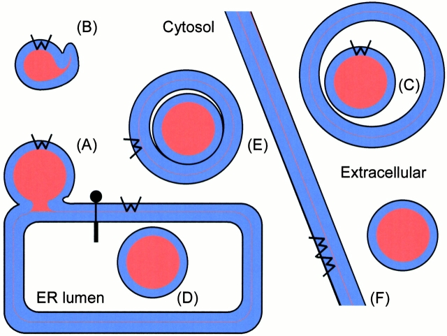
Lipid droplets in mammalian cells. Lipid droplets obtain their phospholipid monolayer by budding from the ER membrane (A). Upon lipolysis, the droplet shrinks, the surrounding monolayer folds into bilayered structures (B). The droplet can bud through the plasma membrane to yield a milk fat droplet surrounded by a lipid bilayer (C). For comparison, lipoprotein particles are assembled in the ER lumen by the microsomal triglyceride transfer protein (D), the lipoprotein is transported on the lumenal side of transport vesicles (E) to the plasma membrane, where lipoprotein is released into the extracellular medium by exocytosis (F). Caveolin is indicated by W. After cotranslational insertion into the ER membrane (Monier et al. 1995), it can diffuse onto the lipid droplet (A). Full-length caveolin leaves the ER by transport vesicles (E) to be inserted into the plasma membrane (F) by fusion of the exocytic vesicle.
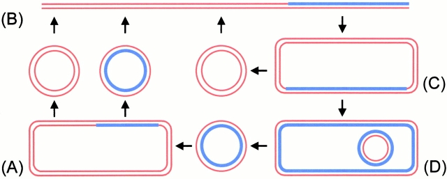
Model for cholesterol recycling through the endocytic pathway. In the lumenal leaflet of the trans-Golgi network (A), sphingolipids and cholesterol (blue) segregate from phospholipids. In epithelial cells, the sphingolipid–cholesterol rafts are transported to the apical surface, the phospholipids to the basolateral surface. In nonpolarized cells both pathways target the plasma membrane (B). From the plasma membrane the sphingolipids are endocytosed (from caveolae?) as are the phospholipids (from clathrin-coated buds?), into common early endosomes (C). There the phospholipids are sorted into a recycling pathway, whereas the sphingolipids travel to the late endosomes (D), which have obtained internal vesicles by budding from the limiting membrane. From here, the sphingolipids and cholesterol recycle to the Golgi and the plasma membrane. This budding step may be regulated by caveolin.
Comment on
-
A caveolin dominant negative mutant associates with lipid bodies and induces intracellular cholesterol imbalance.J Cell Biol. 2001 Mar 5;152(5):1057-70. doi: 10.1083/jcb.152.5.1057. J Cell Biol. 2001. PMID: 11238460 Free PMC article.
-
Accumulation of caveolin in the endoplasmic reticulum redirects the protein to lipid storage droplets.J Cell Biol. 2001 Mar 5;152(5):1071-8. doi: 10.1083/jcb.152.5.1071. J Cell Biol. 2001. PMID: 11238461 Free PMC article.
-
Caveolin-2 is targeted to lipid droplets, a new "membrane domain" in the cell.J Cell Biol. 2001 Mar 5;152(5):1079-85. doi: 10.1083/jcb.152.5.1079. J Cell Biol. 2001. PMID: 11238462 Free PMC article.
References
-
- Blanchette-Mackie E.J., Dwyer N.K., Barber T., Coxey R.A., Takeda T., Rondinone C.M., Theodorakis J.L., Greenberg A.S., Londos C. Perilipin is located on the surface layer of intracellular lipid droplets in adipocytes. J. Lipid Res. 1995;36:1211–1226. - PubMed
-
- Brasaemle D.L., Robertson A.D., Attie A.D. Transbilayer movement of cholesterol in the human erythrocyte membrane. J. Lipid Res. 1988;29:481–489. - PubMed
-
- Brasaemle D.L., Barber T., Wolins N.E., Serrero G., Blanchette-Mackie E.J., Londos C. Adipose differentiation-related protein is an ubiquitously expressed lipid storage droplet-associated protein. J. Lipid Res. 1997;38:2249–2263. - PubMed
-
- Brasaemle D.L., Levin D.M., Adler-Wailes D.C., Londos C. The lipolytic stimulation of 3T3-L1 adipocytes promotes the translocation of hormone-sensitive lipase to the surfaces of lipid storage droplets. Biochim. Biophys. Acta. 2000;1483:251–262. - PubMed
Publication types
MeSH terms
Substances
LinkOut - more resources
Full Text Sources
Medical
