. 2001 Mar;107(5):555-64.
doi: 10.1172/JCI10860.

Self-antigen-presenting cells expressing diabetes-associated autoantigens exist in both thymus and peripheral lymphoid organs

Affiliations

Self-antigen-presenting cells expressing diabetes-associated autoantigens exist in both thymus and peripheral lymphoid organs

A Pugliese et al. J Clin Invest. 2001 Mar.

Erratum in

  • J Clin Invest. 2006 Feb;116(2):548. Redondo, M [corrected to Redondo, MJ]

Abstract

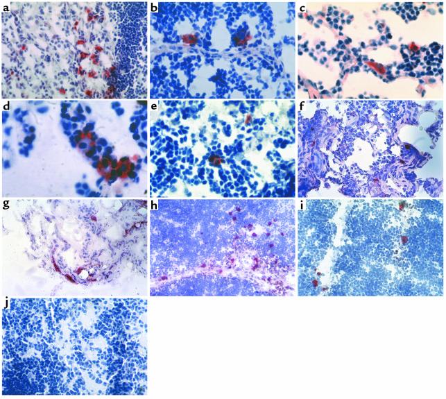
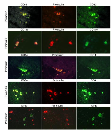
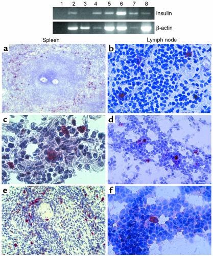
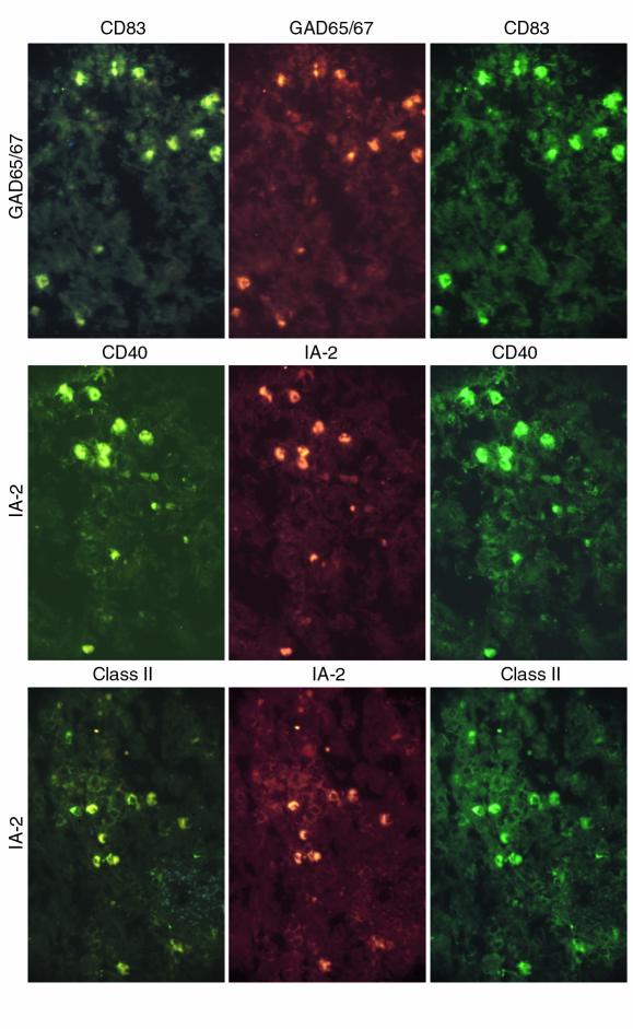
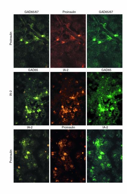
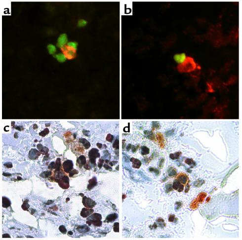
Recent reports indicate that genes with tissue-restricted expression, including those encoding the type 1 diabetes autoantigens insulin, glutamic acid decarboxylase (GAD), and the tyrosine-phosphatase-like protein IA-2 (or ICA512), are transcribed in the thymus. The reported modulation of diabetes susceptibility by genetically determined differences in thymic insulin levels and studies in transgenic mice provide correlative and functional evidence that thymic expression of peripheral proteins is crucial for immunological self-tolerance. However, there are no specific data about the existence, tissue distribution, phenotype, and function of those cells that express insulin and other self-antigens in the human thymus. We find that the human thymus harbors specialized cells synthesizing (pro)insulin, GAD, and IA-2, mainly localized in the medulla, and we demonstrate such cells also in peripheral lymphoid organs (spleen and lymph nodes). Phenotypic analysis qualifies these cells as antigen-presenting cells (APCs), including both dendritic cells and macrophages. These cells often appear surrounded by apoptotic lymphocytes, both in thymus and spleen, and may therefore be involved in the deletion of autoreactive lymphocytes. Our findings demonstrate the existence of, and define the tissue distribution and phenotype of, a novel subset of APCs expressing self-antigens in human lymphoid organs that appear to be involved in the regulation of self-tolerance throughout life.


Figures

Figure 1
Figure 1
Self-antigen–expressing cells in human thymus. Immunostaining of thymus sections using streptavidin-biotin-peroxidase and the AEC substrate (red), counterstained with hematoxylin. Sections from donors of different ages were stained for proinsulin (ag), GAD65/67 (h), IA-2 (i), and control isotype (j). (ac) Proinsulin, case 1201, 18-month-old. ∼×100, ×150, ×190. (d and e) Proinsulin, case 1303, 6-month-old. ×240 and ×190. (f) Proinsulin, case 1111, 32-year-old. ×100. (g) Proinsulin, case 1115, 56-year-old. ×50. (h) GAD65/67, case 2040, 40-week-old fetus. ×50. (i) IA-2, case 1387, 33-week-old fetus. ×100. (j) Mouse IgG1 isotype control antibody, case 1303, 6-month-old. ×75.
Figure 2
Figure 2
Self-antigen–expressing cell phenotype in thymus. Frozen thymus sections were subject to double immunofluorescence. Left column: double-stained cells exhibiting both TRITC (or Alexa-568) and FITC fluorescence often seen as variable gradations of yellow fluorescence with the tripleband filter. Center and right columns: staining for either one of the two individual markers used, as indicated above each image, using highly selective band pass filters for either TRITC (or Alexa-568) or FITC. Top to bottom: Proinsulin-CD83, case 1897. ×84. Not all cells are double stained. Proinsulin-CD11c, case 3446. ×140. Proinsulin-CD14, case 1829. ×90. Proinsulin-CD8α, case 3446. ×90. Proinsulin-AIRE, case 3446. ×90. The AIRE staining was specifically blocked with the peptide used to raise the rabbit serum. Color differences in the images showing the proinsulin staining (center panels) are due to the use of different conjugates (TRITC for the CD83 and CD14 series and Alexa-568 for the CD11c, CD8α, and AIRE series).
Figure 3
Figure 3
Self-antigen–expressing cells in peripheral lymphoid organs. Top panel: expression of insulin mRNA in peripheral lymphoid organs by RT-PCR. From left to right: no template reaction (lane 1), thymus 1278 (lane 2, positive control), spleen 1819 (lane 3), lymph node 2040 (lane 4), lymph node 1001 (lane 5), lymph node 1225 (lane 6), spleen 2073 (lane 7), and spleen 2053 (lane 8). The intensity of β-actin bands differs among samples, suggesting that significant RNA degradation had occurred for some specimens, a phenomenon that cannot always be prevented using human tissues. Bottom panel: immunostaining of spleen (left column) and lymph node (right column) sections using the streptavidin-biotin-peroxidase method and the AEC substrate (red), counterstained with hematoxylin. Sections are stained for proinsulin, GAD65/67, or IA-2, top to bottom. (a) Proinsulin staining, case 1176, 29-year-old. ×24. Note the distribution of proinsulin-positive cells around the white pulp. (b) Proinsulin staining, case 1106, 63-year-old. ×150. (c) GAD65/67 staining, case 2040, 40-week-old fetus. ×250. (d) GAD65/67 staining, case 2040, 40-week-old fetus. ×100. (e) IA-2 staining, case 2053, 39-week-old fetus. ×100. (f) IA-2 staining, case 2053, 39-week-old fetus. ×150.
Figure 4
Figure 4
Self-antigen–expressing cell phenotypes in spleen. Top to bottom: double-staining for GAD65/67-CD83, IA-2-CD40, IA-2-HLA-class II (case 1089; ∼×100; not all cells are double-stained).
Figure 5
Figure 5
Self-antigen–expressing cells express multiple self-antigens. Top to bottom: thymus sections (case 1897; ∼×100) stained for proinsulin-GAD65/67, GAD65-IA-2, and proinsulin-IA-2.
Figure 6
Figure 6
Self-antigen–expressing cells and apoptosis. Frozen sections were subject to TUNEL and proinsulin staining. (a and b) Thymus (case 1829), proinsulin (TRITC) and TUNEL (FITC) staining. ∼×320. A proinsulin-positive cell (orange fluorescence) is seen in the center of a rosette (a) surrounded by apoptotic cells (green fluorescence) or in close contact with an apoptotic cell (b). (c and d) Spleen (case 1089), proinsulin (alkaline phosphatase-AP Orange) and TUNEL staining (peroxidase-AEC). ∼×320. A large, pyramidal proinsulin-positive cell (orange) is shown in the center of a rosette surrounded by at least five cells, three of which appear TUNEL-positive (red) (c); a proinsulin-positive cell (orange) is shown in close contact with two TUNEL-positive cells (red) (d).

References

    1. Schwartz, R.H. 1993. Immunological tolerance. In Fundamental immunology. W.E. Paul, editor. Raven Press. New York, New York, USA. 677–731.
    1. Fowlkes BJ, Ramsdell F. T-cell tolerance. Curr Opin Immunol. 1993;5:873–879. - PubMed
    1. Sprent J, Webb SR. Intrathymic and extrathymic clonal deletion of T cells. Curr Opin Immunol. 1995;7:196–205. - PubMed
    1. Suss G, Shortman K. A subclass of dendritic cells kills CD4 T cells via Fas/Fas-ligand-induced apoptosis. J Exp Med. 1996;183:1789–1796. - PMC - PubMed
    1. Fraser A, Evan G. A license to kill. Cell. 1996;85:781–784. - PubMed

Publication types

MeSH terms

Substances