. 2001 Mar;107(5):575-83.
doi: 10.1172/JCI11220.

CD28-independent induction of experimental autoimmune encephalomyelitis

Affiliations

CD28-independent induction of experimental autoimmune encephalomyelitis

T Chitnis et al. J Clin Invest. 2001 Mar.

Abstract

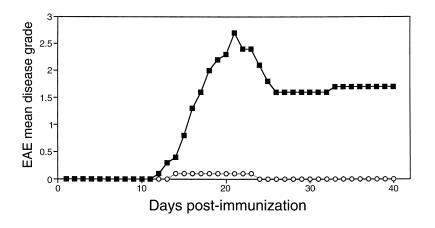
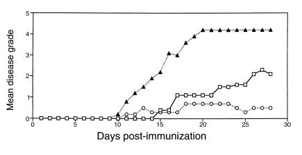
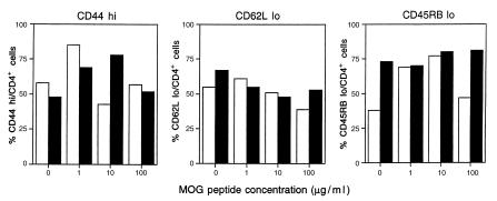
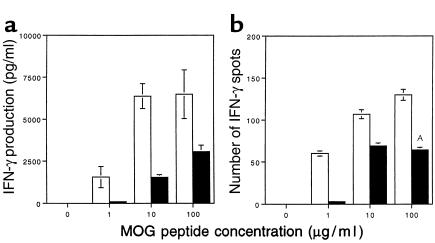
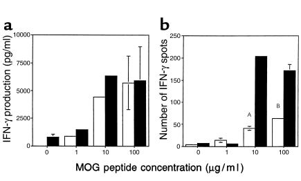
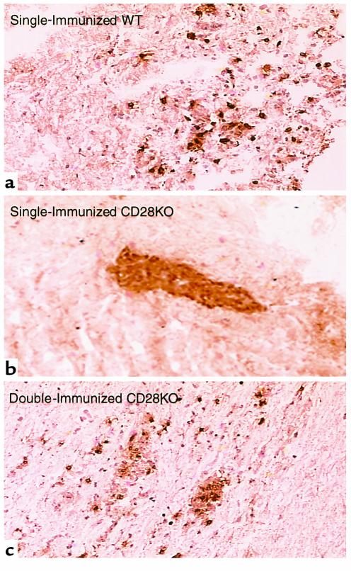
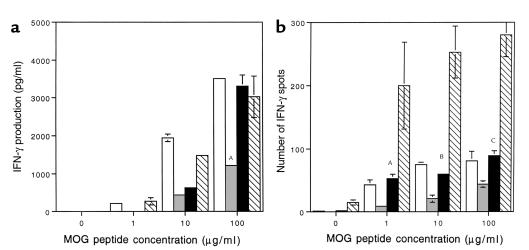
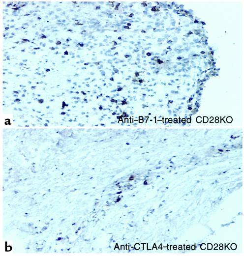
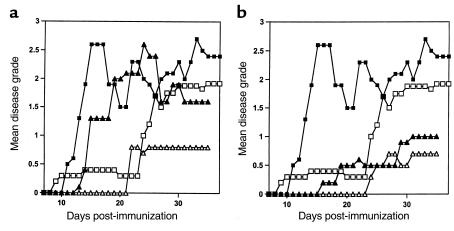
Experimental autoimmune encephalomyelitis (EAE) is a T cell-mediated disease initiated by antigen-specific CD4(+) T cells. Signaling through CD28 is a critical second signal for activation of T cells, and CD28 knockout (CD28KO) mice have been reported to be resistant to induction of EAE. We now report that CD28KO mice have no intrinsic defect in mediating disease, because they developed EAE after passive transfer of primed T cells. After immunization, peripheral T cells from CD28KO mice were primed and developed memory phenotype, but had decreased antigen-specific IFN-gamma production as compared with cells from wild-type (WT) animals. Reimmunization of CD28KO mice brought out clinical disease and increased IFN-gamma production in vitro. Pathologically, there were cellular infiltrates in the central nervous system, in contrast to single-immunized mice. We show furthermore that blocking B7-1 or CTLA4, but not B7-2, in CD28KO mice induces disease after a single immunization. Thus, EAE can be induced in animals lacking CD28-dependent costimulation, suggesting that alternative costimulatory pathways were used. Blocking the OX40-OX40L costimulatory pathway differentially affected disease induction in CD28KO mice as compared with WT controls. Our data show that EAE may develop in the absence of CD28 T-cell costimulation. These findings have implications for therapies aimed at blocking costimulatory signals in autoimmune diseases.


Figures

Figure 1
Figure 1
EAE is suppressed in CD28KO mice. A representative experiment showing disease induction in C57BL/6 WT mice (filled squares) and CD28KO mice (open circles). Mice were immunized with MOG p35-55 and graded for disease daily. The mean daily grade for each group (n = 5 mice per group) is shown. The incidence was five of five in the WT and one in five in the CD28KO mice; the mean maximal grade was 2.7 in WT and 0.1 in CD28KO (P < 0.0001).
Figure 2
Figure 2
Adoptive transfer of WT splenocytes into CD28KO mice restores EAE. Splenocytes from C57BL/6 WT mice or CD28KO mice were transferred to CD28KO mice. On day –1, 10 × 106 WT splenocytes (open squares), 100 × 106 WT splenocytes (filled triangles), or 100 × 106 CD28KO splenocytes (open circles) were transferred, and all recipients were immunized with MOG p35-55 on day 0. The mean daily score for each group (n = 5 mice per group) is shown.
Figure 3
Figure 3
Similar expression of memory cell markers on CD4+ cells from CD28KO and WT mice. Quantitation of memory cell markers on CD4+ cells from C57BL/6 WT mice (open bars) and CD28KO mice (filled bars). Splenocytes were harvested on day 14 after immunization with MOG p35-55 and cultured for 72 hours in the presence of MOG p35-55 (1, 10, or 100 μg/ml). Cells were stained and analyzed by FACS for expression of CD44, CD62 ligand, and CD45RB. All cells were counterstained for the CD4 marker, and the number of cells expressing the given memory marker per number of CD4 cells is shown as a percentage value.
Figure 4
Figure 4
Decreased IFN-γ production by MOG-reactive splenocytes in CD28KO mice. (a) IFN-γ production was measured by ELISA in the supernatants of splenocytes harvested on day 14 from C57BL/6 (open bars) or CD28KO mice (filled bars), after 48 hours of culture with MOG p35-55 at concentrations of 1, 10, or 100 μg/ml. IFN-γ production was significantly greater in the cultures from WT mice at all concentrations of MOG p35-55 (P < 0.005). (b) MOG p35-55–specific IFN-γ–producing cells were measured by ELISPOT in cultures of splenocytes from C57BL/6 WT (open bars) or CD28KO mice (filled bars). The y axis represents the number of positive cells per 2 × 10⁵ cells plated. The frequency of IFN-γ–producing cells was significantly higher in WT cultures at the 100 μg/ml antigen dose (AP = 0.006).
Figure 5
Figure 5
Increased IFN-γ production by MOG-reactive splenocytes in double-immunized CD28KO mice. (a) IFN-γ production was measured by ELISA in the supernatants of splenocytes harvested on day 14 from single-immunized C57BL/6 (open bars) or harvested on day 28 from double-immunized CD28KO mice (filled bars), after 48 hours’ culture with MOG p35-55 at concentrations of 1, 10, or 100 μg/ml. IFN-γ production was similar in both groups with no significant differences. (b) MOG p35-55–specific IFN-γ–producing cells were measured by ELISPOT in cultures of splenocytes from single-immunized C57BL/6 WT (open bars) or double-immunized CD28KO mice (filled bars). The frequency of IFN-γ–producing cells was significantly greater in cultures from double-immunized CD28KO (AP = 0.0009, BP = 0.009).
Figure 6
Figure 6
The reduction of CNS parenchymal inflammatory infiltration in single-immunized CD28KO mice is reversed in double-immunized CD28KO mice. Microphotograph (×40) of representative spinal cord section from (a) single-immunized WT mice day 17; (b) single-immunized CD28KO mice day 17; (c) double-immunized CD28KO mice day 35. Sections are immunohistochemically stained for CD4+ cells, which display a brown color. Cellular infiltrates are seen invading the brain parenchyma in WT (a) and double-immunized (c) mice, while the cells remain within blood vessels in the single-immunized CD28KO mice (b).
Figure 7
Figure 7
Increased IFN-γ production by MOG-reactive splenocytes in anti–B7-1–treated and anti-CTLA4–treated CD28KO mice. (a) IFN-γ production was measured by ELISA in the supernatants of splenocytes harvested on day 35 from C57BL/6 (open bars), CD28KO mice (gray bars), CD28KO mice treated with anti–B7-1 Ab (black bars), and CD28KO mice treated with anti-CTLA4 Ab (hatched bars). Cells were incubated with MOG p35-55 at concentrations of 1, 10, or 100 μg/ml. IFN-γ production was significantly increased in the anti–B7-1–treated CD28KO group (AP = 0.00006) and the anti-CTLA4–treated CD28KO group (P < 0.00009) compared with control Ig–treated CD28KO at the highest concentration of MOG. (b) MOG p35-55–specific IFN-γ–producing cells were measured by ELISPOT in cultures of splenocytes from C57BL/6 WT (open bars), or CD28KO mice (gray bars), or CD28KO mice treated with anti–B7-1 Ab (black bars), or CD28KO mice treated with anti-CTLA4 Ab (hatched bars), and harvested on day 35. The frequency of IFN-γ–producing cells was significantly greater in cultures from CD28KO mice treated with anti–B7-1 Ab (AP = 0.0007, BP = 0.0005, CP = 0.0011) and those treated with anti-CTLA4 than control CD28KO mice (P < 0.00001 at all concentrations of MOG peptide).
Figure 8
Figure 8
Increased inflammatory infiltration in the CNS of anti–B7-1–treated and anti–CTLA4-treated CD28KO mice. Microphotograph (×40) of representative spinal cord section from (a) CD28KO mice treated with anti–B7-1 Ab, or (b) CD28KO mice treated with anti-CTLA4, at the peak of disease day 35. The section is immunohistochemically stained for CD4+ cells, which display a brown color. Cellular infiltrates are seen invading the brain parenchyma.
Figure 9
Figure 9
Anti-OX40L reverses EAE induced in double-immunized CD28KO mice but not in WT mice. Disease course of double-immunized CD28KO mice treated with anti-OX40L (a) or anti-CD40L (b). Mice were immunized with MOG p35-55 and graded for disease daily. The mean daily grade for each group (n = 3–10 mice per group) is shown. (a) A representative experiment showing the disease course in C57BL/6 WT mice (filled squares) or in WT mice treated with anti-OX40L Ab (filled triangles) compared with double-immunized CD28KO mice (open squares) and double-immunized CD28KO mice treated with anti-OX40L Ab (open triangles). The mean maximal disease grade in WT mice was 2.7 ± 0.1; in WT treated with anti-OX40L Ab it was 2.75 ± 0.3. The mean maximal grade in double-immunized CD28KO mice was 2.35 ± 0.3, and in those treated with anti-OX40L Ab it was 0.83 ± 0.6 (P = 0.001 compared with untreated double-immunized CD28KO). (b) A representative experiment showing the disease course in C57BL/6 WT mice (filled squares) or in WT mice treated with anti-CD40L Ab (filled triangles) compared with double-immunized CD28KO mice (open squares) and double-immunized CD28KO mice treated with anti-CD40L Ab (open triangles). The mean maximal disease grade in untreated WT mice was 2.7 ± 0.1, whereas in WT treated with anti-CD40L it was 1.08 ± 1.1 (P = 0.03). The mean maximal grade in double-immunized CD28KO mice was 2.35 ± 0.3 compared with those treated with anti-CD40L (0.67 ± 1.15; P = 0.0008 compared with untreated double-immunized CD28KO mice).

Similar articles

Cited by

References

    1. Mueller DL, Jenkins MK, Schwartz RH. Clonal expansion versus functional clonal inactivation: a costimulatory signalling pathway determines the outcome of T cell antigen receptor occupancy. Annu Rev Immunol. 1989;7:445–480. - PubMed
    1. Schwartz RH. A cell culture model for T lymphocyte clonal anergy. Science. 1990;248:1349–1356. - PubMed
    1. Walunas TL, et al. CTLA-4 can function as a negative regulator of T cell activation. Immunity. 1994;1:405–413. - PubMed
    1. Krummel MF, Allison JP. CD28 and CTLA-4 have opposing effects on the response of T cells to stimulation. J Exp Med. 1995;182:459–465. - PMC - PubMed
    1. Walunas TL, Bakker CY, Bluestone JA. CTLA-4 ligation blocks CD28-dependent T cell activation. J Exp Med. 1996;183:2541–2550. - PMC - PubMed

Publication types

MeSH terms

Substances