. 2001 Mar;107(5):585-93.
doi: 10.1172/JCI11476.

Increased sensitivity to dextran sodium sulfate colitis in IRE1beta-deficient mice

Affiliations

Increased sensitivity to dextran sodium sulfate colitis in IRE1beta-deficient mice

A Bertolotti et al. J Clin Invest. 2001 Mar.

Abstract

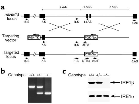
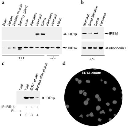
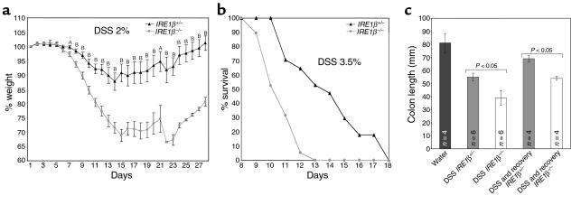
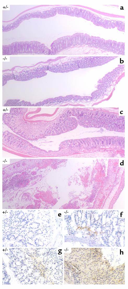
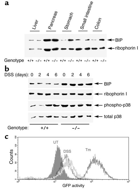
The epithelial cells of the gastrointestinal tract are exposed to toxins and infectious agents that can adversely affect protein folding in the endoplasmic reticulum (ER) and cause ER stress. The IRE1 genes are implicated in sensing and responding to ER stress signals. We found that epithelial cells of the gastrointestinal tract express IRE1beta, a specific isoform of IRE1. BiP protein, a marker of ER stress, was elevated in the colonic mucosa of IRE1beta(-/-) mice, and, when exposed to dextran sodium sulfate (DSS) to induce inflammatory bowel disease, mutant mice developed colitis 3-5 days earlier than did wild-type or IRE1beta(+/-) mice. The inflammation marker ICAM-1 was also expressed earlier in the colonic mucosa of DSS-treated IRE1beta(-/-) mice, indicating that the mutation had its impact early in the inflammatory process, before the onset of mucosal ulceration. These findings are consistent with a model whereby perturbations in ER function, which are normally mitigated by the activity of IRE1beta, participate in the development of colitis.


Figures

Figure 1
Figure 1
An induced mutation in the murine IRE1β locus. (a) Structure of the wild-type locus in the area encoding the transmembrane domain of IRE1β (top), the targeting vector (middle), and the targeted locus after homologous recombination (bottom). The deleted exons encoding the transmembrane domain are depicted as filled boxes, and intronic sequences are thin lines. The positions of the primers used to distinguish wild-type and mutant alleles are indicated, and the PCR protocols used are described in detail in Methods. (b) PCR analysis of genomic DNA from wild-type, heterozygous, and homozygous mutant mice. (c) Mutant mice do not express proteins reactive with a COOH-terminal anti-IRE1β antiserum. Lysates from colonic mucosa of mice with the indicated genotypes were immunoprecipitated with antisera to IRE1β and IRE1α and immunoblotted with the same antiserum.
Figure 2
Figure 2
Expression of IRE1β is restricted to the epithelium of the gastrointestinal tract. (a) Immunoprecipitation (IP) followed by immunoblot detection of IRE1β and IRE1α in tissue extracts from wild-type and IRE1β–/– mice. (b) Detection by immunoblotting of IRE1β in microsomes prepared from all three segments of the gastrointestinal mucosa and pancreas. The ER resident protein ribophorin I, detected by immunoblot, serves as a recovery marker. (c) Detection of IRE1β by immunoprecipitation and immunoblotting in lysates prepared from epithelial cells eluted by EDTA treatment from the colon of a wild-type mouse. Lanes 1 and 2 are from lysates prepared from the intact tissue. Lane 3 is from the cells eluted from the tissue fragment in the calcium-free EDTA-containing buffer, and lane 4 is from a lysate prepared from cells that remain associated with the tissue fragment after (partial) depletion of epithelial cells. Lane 1 was immunoprecipitated with preimmune serum (PI), whereas lanes 2–4 were immunoprecipitated with anti-IRE1β immune serum. (d) Immunostaining of cells in an aliquot of the fraction used in lane 3 of c, with mAb’s to keratins. A counterstain with the karyophilic dye H33258 reveals the nuclei of all the cells in the field. Note that almost all the cells in the field stained positive with the keratin antibodies, attesting to their epithelial identity.
Figure 3
Figure 3
Increased severity of DSS colitis in IRE1β–/– mice. (a) Body weight, expressed as percentage of weight on the day of first exposure to DSS, is plotted against time. Mice were exposed continuously to 2% DSS in the drinking water for days 1–12. Shown are the mean and SEM of a typical experiment carried out on cohort of 23 IRE1β+/– and 23 sibling IRE1β–/– 129svev mice and reproduced three times. AP < 0.05; BP < 0.0005 for the difference in mean value between the two groups (two-tailed t test). (b) Survival of a cohort of 17 age-matched IRE1β+/+ and 19 IRE1β–/– 129svev mice exposed continuously to 3.5% DSS in the drinking water for days 1–12. (c) Comparison of the colon length in untreated mice, and mice of the indicated genotypes treated with 2% DSS for 12 days (DSS) or mice treated with 2% DSS for 12 days and allowed to recover for 15 days (DSS and recovery). Shown are the means and SEM and P values calculated by two-tailed t test.
Figure 4
Figure 4
Histopathology of DSS-treated mice. Representative photomicrographs (×50) of paraffin-embedded, hematoxylin and eosin–stained longitudinal sections of the distal colon from untreated mice of the indicated genotypes (a and b) or mice that had been exposed continuously to 2% DSS for 12 days (c and d). Photomicrographs (×400) of longitudinal frozen sections of colon stained with an mAb to murine ICAM-1 revealed by diaminobenzidine (brown precipitate) and lightly counterstained with hematoxylin for orientation. Mice of the indicated genotypes were exposed to 2% DSS for 6 days (e and f) or 10 days (g and h).
Figure 5
Figure 5
Higher levels of ER stress in gastric and colonic mucosa of IRE1β–/– mice. (a) Immunoblot of BiP and ribophorin I content in tissue lysates of mice with the indicated genotypes. (b) Immunoblot of phosphorylated (active) p38 MAP kinase, total p38 MAP kinase, BiP, and ribophorin I content of colon lysates from mice of the indicated genotype exposed to 3% DSS for the indicated period. (c) Fluorescent activated cell sorting signals of CHO cells expressing an integrated transgene consisting of the CHOP promoter fused to a green fluorescent protein (GFP) reporter. Cells were left untreated (UT), exposed to 2.5 μg/mL tunicamycin for 12 hours (Tm), or cultured in the presence 3.5% DSS for 24 hours (dark gray histogram) or 4.5% DSS for 24 hours (light gray histogram).

References

    1. Sartor RB. Cytokine regulation of experimental intestinal inflammation in genetically engineered and T-lymphocyte reconstituted rodents. Aliment Pharmacol Ther. 1996;10(Suppl. 2):36–42. - PubMed
    1. Strober W, et al. Reciprocal IFN-gamma and TGF-beta responses regulate the occurrence of mucosal inflammation. Immunol Today. 1997;18:61–64. - PubMed
    1. Bhan AK, Mizoguchi E, Smith RN, Mizoguchi A. Colitis in transgenic and knockout animals as models of human inflammatory bowel disease. Immunol Rev. 1999;169:195–207. - PubMed
    1. Berg DJ, et al. Enterocolitis and colon cancer in interleukin-10-deficient mice are associated with aberrant cytokine production and CD4(+) TH1-like responses. J Clin Invest. 1996;98:1010–1020. - PMC - PubMed
    1. Sadlack B, et al. Ulcerative colitis-like disease in mice with a disrupted interleukin-2 gene. Cell. 1993;75:253–261. - PubMed

Publication types

MeSH terms