

Amelioration of collagen-induced arthritis by thrombin inhibition
- PMID: 11238564
- PMCID: PMC199423
- DOI: 10.1172/JCI11064
Amelioration of collagen-induced arthritis by thrombin inhibition
Abstract
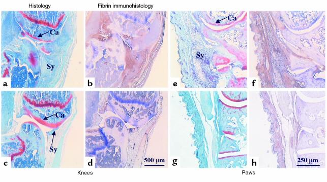
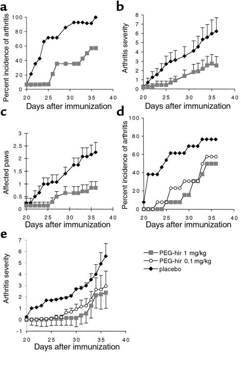
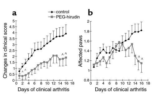
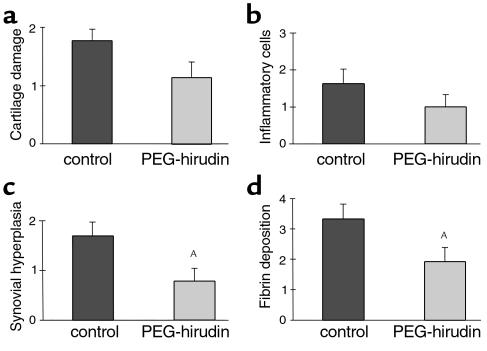
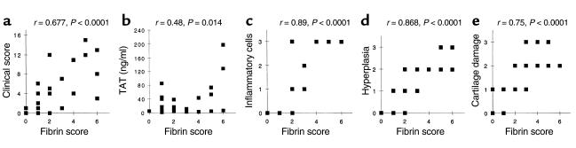
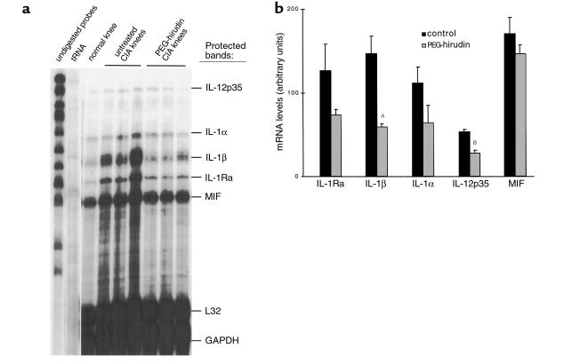
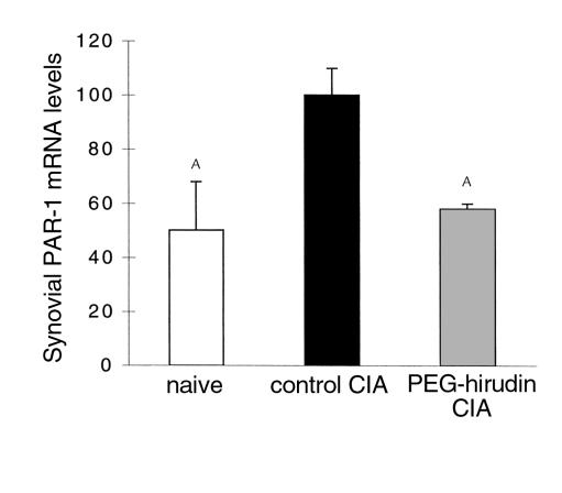
The deleterious role of fibrin deposition in arthritic joints prompted us to explore the effect of the thrombin inhibition on the course of collagen-induced arthritis (CIA) in the mouse. CIA was induced in male DBA/1J mice using native chicken type II collagen. The thrombin inhibitor polyethyleneglycol-hirudin (PEG-hirudin) was given for 16 days, starting 20 days after the first immunization (preventive treatment) or at the onset of clinical signs of arthritis (curative treatment). All the mice treated with PEG-hirudin had a significantly prolonged clotting time compared with control mice. PEG-hirudin, administered in a preventive way, led to significantly reduced incidence and severity of CIA during most of the treatment period, as assessed by clinical scoring. Accordingly, histological features showed a significant diminution of synovial hyperplasia in PEG-hirudin-treated mice compared with untreated mice. There was also a significant downmodulation of the synovial proinflammatory IL-1beta and IL-12p35 cytokine mRNAs in treated mice. Intra-articular fibrin, evaluated by immunohistochemistry, was significantly reduced in treated mice compared with control mice and correlated with both clinical and histological scorings. Most importantly, once arthritis was established, PEG-hirudin also showed a curative effect. In conclusion, PEG-hirudin can both prevent the onset of CIA in a dose-dependent manner and ameliorate established arthritis, suggesting that thrombin inhibition may offer a new therapeutic approach in arthritis.
Figures
Similar articles
-
Protection against cartilage and bone destruction by systemic interleukin-4 treatment in established murine type II collagen-induced arthritis.Arthritis Res. 1999;1(1):81-91. doi: 10.1186/ar14. Epub 1999 Oct 26. Arthritis Res. 1999. PMID: 11056663 Free PMC article.
-
Effect of thrombin inhibition on synovial inflammation in antigen induced arthritis.Ann Rheum Dis. 2000 Oct;59(10):781-7. doi: 10.1136/ard.59.10.781. Ann Rheum Dis. 2000. PMID: 11005778 Free PMC article.
-
Inhibition of collagen-induced arthritis in mice by viral IL-10 gene transfer.J Immunol. 1998 Aug 1;161(3):1516-24. J Immunol. 1998. PMID: 9686619
-
Thrombin functions and antithrombotic intervention.Thromb Haemost. 1995 Jul;74(1):493-8. Thromb Haemost. 1995. PMID: 8578512 Review. No abstract available.
-
Medicinal leeches: ancient therapy is a source of biotech drugs.J Am Pharm Assoc (Wash). 1997 May-Jun;NS37(3):285-6. doi: 10.1016/s1086-5802(16)30209-1. J Am Pharm Assoc (Wash). 1997. PMID: 9170802 Review.
Cited by
-
Essential role of platelet activation via protease activated receptor 4 in tissue factor-initiated inflammation.Arthritis Res Ther. 2008;10(2):R42. doi: 10.1186/ar2400. Epub 2008 Apr 15. Arthritis Res Ther. 2008. PMID: 18412955 Free PMC article.
-
Thrombin generation and activity in multiple sclerosis.Metab Brain Dis. 2021 Mar;36(3):407-420. doi: 10.1007/s11011-020-00652-w. Epub 2021 Jan 7. Metab Brain Dis. 2021. PMID: 33411219 Free PMC article. Review.
-
Autoantibody-mediated arthritis in the absence of C3 and activating Fcγ receptors: C5 is activated by the coagulation cascade.Arthritis Res Ther. 2012 Dec 13;14(6):R269. doi: 10.1186/ar4117. Arthritis Res Ther. 2012. PMID: 23237573 Free PMC article.
-
Analysing the effect of novel therapies on cytokine expression in experimental arthritis.Int J Exp Pathol. 2005 Oct;86(5):267-78. doi: 10.1111/j.0959-9673.2005.00443.x. Int J Exp Pathol. 2005. PMID: 16191099 Free PMC article. Review.
-
Circulating CD26 is negatively associated with inflammation in human and experimental arthritis.Am J Pathol. 2005 Feb;166(2):433-42. doi: 10.1016/S0002-9440(10)62266-3. Am J Pathol. 2005. PMID: 15681827 Free PMC article.
References
-
- Firestein, G.S. 1997. Etiology and pathogenesis of rheumatoid arthritis. In Textbook of rheumatology. W.N. Kelley, E.D. Harris, S. Ruddy, and C.B. Sledge, editors. W.B. Saunders Co. Philadelphia, Pennsylvania, USA. 851–897.
-
- Feldmann M, Brennan FM, Maini RN. Rheumatoid arthritis. Cell. 1996;85:307–310. - PubMed
-
- Camerer E, Kolsto AB, Prydz H. Cell biology of tissue factor, the principal initiator of blood coagulation. Thromb Res. 1996;81:1–4. - PubMed
-
- Weinberg JB, Pippen AM, Greenberg CS. Extravascular fibrin formation and dissolution in synovial tissue of patients with osteoarthritis and rheumatoid arthritis. Arthritis Rheum. 1991;34:996–1005. - PubMed
-
- Zacharski LR, et al. Pathways of coagulation activation in situ in rheumatoid synovial tissue. Clin Immunol Immunopathol. 1992;63:155–162. - PubMed
Publication types
MeSH terms
Substances
LinkOut - more resources
Full Text Sources
Other Literature Sources
Medical
