. 2001 Mar 5;193(5):607-20.
doi: 10.1084/jem.193.5.607.

Selective inhibition of vascular endothelial growth factor-mediated angiogenesis by cyclosporin A: roles of the nuclear factor of activated T cells and cyclooxygenase 2

Affiliations

Selective inhibition of vascular endothelial growth factor-mediated angiogenesis by cyclosporin A: roles of the nuclear factor of activated T cells and cyclooxygenase 2

G L Hernández et al. J Exp Med. .

Abstract

Cyclosporin A (CsA) is an immunosuppressive drug that inhibits the activity of transcription factors of the nuclear factor of activated T cells (NFAT) family, interfering with the induction of cytokines and other inducible genes required for the immune response. Here we show that CsA inhibits migration of primary endothelial cells and angiogenesis induced by vascular endothelial growth factor (VEGF); this effect appears to be mediated through the inhibition of cyclooxygenase (Cox)-2, the transcription of which is activated by VEGF in primary endothelial cells. Consistent with this, we show that the induction of Cox-2 gene expression by VEGF requires NFAT activation. Most important, the CsA-mediated inhibition of angiogenesis both in vitro and in vivo was comparable to the Cox-2 inhibitor NS-398, and reversed by prostaglandin E(2). Furthermore, the in vivo corneal angiogenesis induced by VEGF, but not by basic fibroblast growth factor, was selectively inhibited in mice treated with CsA systemically. These findings involve NFAT in the regulation of Cox-2 in endothelial cells, point to a role for this transcription factor in angiogenesis, and may provide a novel mechanism underlying the beneficial effects of CsA in angiogenesis-related diseases such as rheumatoid arthritis and psoriasis.


Figures

Figure 1
Figure 1
VEGF induces Cox-2 in HUVECs in a CsA-sensitive manner. HUVECs, either pretreated with or without CsA (200 ng/ml) for 2 h were left untreated (−) or were treated with 50 ng/ml VEGF (A and B), 50 ng/ml TNF-α (C), or 10 ng/ml bFGF (D), for the indicated periods of time. Cell extracts were analyzed by Western blot probed with anti–Cox-2, anti–Cox-1, or anti–β-tubulin antibodies to ensure equal loading. The data are representative of at least three independent experiments with similar results.
Figure 2
Figure 2
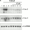
Effect of VEGF on Cox-2 transcription. HUVECs were pretreated with or without 200 ng/ml CsA for 2 h and further stimulated with 50 ng/ml VEGF for the time points indicated, and total or nuclear RNA was analyzed by Northern blot or nuclear run-on, respectively. (A) Northern blot analysis from HUVECs activated by VEGF. After isolation, 20 μg RNA was separated by agarose gel electrophoresis, blotted onto a nitrocellulose membrane, and hybridized with a human Cox-2 cDNA probe, then sequentially rehybridized with Cox-1 and GAPDH-specific cDNA probes. (B) Nuclear run-on analysis using nuclear RNA from HUVECs activated by VEGF. Nuclear RNA samples, labeled with [32P]UTP, were hybridized to nylon membrane containing 5 μg of dot-blotted linearized cDNAs for Cox-2, GAPDH, or Bluescript plasmid. The results shown in the autoradiographs (top) are represented (in arbitrary densitometric units of Cox-2/GAPDH) as fold induction over the baseline levels of unstimulated cells 1 (bottom).
Figure 2
Figure 2
Effect of VEGF on Cox-2 transcription. HUVECs were pretreated with or without 200 ng/ml CsA for 2 h and further stimulated with 50 ng/ml VEGF for the time points indicated, and total or nuclear RNA was analyzed by Northern blot or nuclear run-on, respectively. (A) Northern blot analysis from HUVECs activated by VEGF. After isolation, 20 μg RNA was separated by agarose gel electrophoresis, blotted onto a nitrocellulose membrane, and hybridized with a human Cox-2 cDNA probe, then sequentially rehybridized with Cox-1 and GAPDH-specific cDNA probes. (B) Nuclear run-on analysis using nuclear RNA from HUVECs activated by VEGF. Nuclear RNA samples, labeled with [32P]UTP, were hybridized to nylon membrane containing 5 μg of dot-blotted linearized cDNAs for Cox-2, GAPDH, or Bluescript plasmid. The results shown in the autoradiographs (top) are represented (in arbitrary densitometric units of Cox-2/GAPDH) as fold induction over the baseline levels of unstimulated cells 1 (bottom).
Figure 3
Figure 3
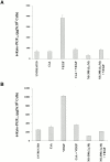
Induction of prostacyclin synthesis by VEGF. HUVECs were pretreated or not with 200 ng/ml CsA or 1 μM NS-398 for 2 h and subsequently treated with VEGF (50 ng/ml) for an additional 8-h period, and the levels of 6-keto-PGF were determined in the culture medium supernatants (A) or in the supernatants of intact cells after addition of 10 μM AA (B). Prostacyclin (determined as 6-keto-PGF1α) was quantified by using an enzyme immunoassay. Two independent experiments yielding similar results were performed in triplicate. Results are expressed as picogram per 5 × 10⁵ cells.
Figure 4
Figure 4
Activation of Cox-2 gene promoter by VEGF. HUVECs were transfected with the different Cox-2 promoter reporter plasmids for 4.5 h. 16 h later, the cells were pretreated with 200 ng/ml CsA for 2 h where indicated, and stimulated with 50 ng/ml VEGF for an additional 6 h. Experiments were performed in triplicate. Luciferase activity is expressed as fold induction over the baseline levels of transfected unstimulated cells. Results of a representative experiment out of three performed are shown. Deletions ranging from −1796 to −46 relative to the transcription start site of the Cox-2 promoter were used. The extent of the 5′ truncations is shown with numbers indicating positions relative to the transcription start site. Consensus sequences are denoted by boxes.
Figure 5
Figure 5
VEGF induces NFAT–DNA binding activity to the Cox-2 promoter. Nuclear extracts from HUVECs stimulated for 20 min with 50 ng/ml VEGF with or without 200 ng/ml CsA were analyzed by EMSA with probes including the distal NFAT site of the Cox-2 human promoter (nucleotides −117 to −91) (A) or the proximal NFAT site of the Cox-2 human promoter (nucleotides −82 to −58) (B). A 20-fold molar excess of the unlabeled NFAT consensus site oligonucleotide of the IL-2 promoter was added to the binding reaction mixtures to determine the specificity of the binding. EMSAs were performed in the presence or absence of either preimmune serum (Preimm.) or the 674 anti–all-NFAT (α-all) antiserum. The specific DNA–NFAT complexes are indicated by an arrow.
Figure 6
Figure 6
Role of NFAT for the Cox-2 promoter activity induced by VEGF. (A) Sequences of the wild-type and mutated NFAT and NF-IL6 sites of the Cox-2 gene promoter. The consensus sequences are marked in bold and the substitutions introduced are indicated by dots. (B) Reporter vectors harboring mutations in the NFAT or NF-IL6 sites were transfected into HUVECs for 4.5 h. 16 h after transfection, the cells were stimulated with 50 ng/ml VEGF for 6 h and luciferase activity was determined. The activity is expressed as fold induction over the baseline levels of transfected unstimulated cells. One out of three independent experiments performed is shown. The P2-274 (−170, +104) and P2-431 (−327, +104) constructs were used as templates for site-directed mutagenesis. Mutated sites are indicated by X.
Figure 7
Figure 7
Inhibition of in vitro endothelial cell morphogenesis by CsA. HUVECs (2 × 10⁴ cells/well) were seeded into 96-well plates precoated with 50 μl of Matrigel. HUVECs were untreated (control) or treated with CsA, a neutralizing anti-VEGF antibody, NS-398, PGE2, a neutralizing anti–TGF-β antibody alone (A), or in the presence of exogenous VEGF (B). (C) Representative photographs (original magnifications: ×100) of five different fields corresponding to the experiments showed in A and B. (D) HUVECs either pretreated with or without CsA were treated with VEGF or bFGF and tube formation was quantified. Results are expressed as the percentage of tubes formed in the presence of CsA relative to the tubes formed in the presence of VEGF (100%) or bFGF (100%). For the different treatments shown in A, B, and C, 200 ng/ml CsA, 50 ng/ml VEGF, 10 ng/ml bFGF, 1 ng/ml PGE2, 10 μg/ml of anti-VEGF antibody, 4 μg/ml of anti–TGF-β antibody, and 10 μM NS-398 were used alone or in the indicated combinations. When used, CsA was added 1 h before the cells were plated on Matrigel, and then its concentration was maintained during the treatment. Tube formation was quantified 12 h after cells were plated in Matrigel by counting the number of tubular structures in four to six fields. Results are representative of at least four independent experiments performed.
Figure 7
Figure 7
Inhibition of in vitro endothelial cell morphogenesis by CsA. HUVECs (2 × 10⁴ cells/well) were seeded into 96-well plates precoated with 50 μl of Matrigel. HUVECs were untreated (control) or treated with CsA, a neutralizing anti-VEGF antibody, NS-398, PGE2, a neutralizing anti–TGF-β antibody alone (A), or in the presence of exogenous VEGF (B). (C) Representative photographs (original magnifications: ×100) of five different fields corresponding to the experiments showed in A and B. (D) HUVECs either pretreated with or without CsA were treated with VEGF or bFGF and tube formation was quantified. Results are expressed as the percentage of tubes formed in the presence of CsA relative to the tubes formed in the presence of VEGF (100%) or bFGF (100%). For the different treatments shown in A, B, and C, 200 ng/ml CsA, 50 ng/ml VEGF, 10 ng/ml bFGF, 1 ng/ml PGE2, 10 μg/ml of anti-VEGF antibody, 4 μg/ml of anti–TGF-β antibody, and 10 μM NS-398 were used alone or in the indicated combinations. When used, CsA was added 1 h before the cells were plated on Matrigel, and then its concentration was maintained during the treatment. Tube formation was quantified 12 h after cells were plated in Matrigel by counting the number of tubular structures in four to six fields. Results are representative of at least four independent experiments performed.
Figure 8
Figure 8
Effect of CsA on endothelial cell migration. (A) Phase–contrast micrographs of migrating cells invading collagen gels and forming tubelike structures. HUVECs were seeded onto the surface of collagen gels, in the presence or absence of 50 ng/ml VEGF or 10 ng/ml bFGF, and were tested alone or in combination with 200 ng/ml CsA for 24 h (top). Migrating cells were quantified under the different conditions with respect to control cells in the presence of VEGF (100%) or bFGF (100%) (bottom). (B) Migration of dermal microvascular endothelial cells. 200 ng/ml CsA was tested for its ability to inhibit endothelial cell migration induced by 100 pg/ml VEGF or 20 ng/ml bFGF. Endothelial cells in EBM plus 0.1% BSA were plated onto the lower surface of a gelatinized 5.0-μm filter in an inverted, modified Boyden chamber. Results are expressed as the number of cells migrated by 10 high power fields. EBM supplemented with 0.1% BSA was used as a negative control (background resulting from random migration). One representative experiment out of three independent performed is shown.
Figure 8
Figure 8
Effect of CsA on endothelial cell migration. (A) Phase–contrast micrographs of migrating cells invading collagen gels and forming tubelike structures. HUVECs were seeded onto the surface of collagen gels, in the presence or absence of 50 ng/ml VEGF or 10 ng/ml bFGF, and were tested alone or in combination with 200 ng/ml CsA for 24 h (top). Migrating cells were quantified under the different conditions with respect to control cells in the presence of VEGF (100%) or bFGF (100%) (bottom). (B) Migration of dermal microvascular endothelial cells. 200 ng/ml CsA was tested for its ability to inhibit endothelial cell migration induced by 100 pg/ml VEGF or 20 ng/ml bFGF. Endothelial cells in EBM plus 0.1% BSA were plated onto the lower surface of a gelatinized 5.0-μm filter in an inverted, modified Boyden chamber. Results are expressed as the number of cells migrated by 10 high power fields. EBM supplemented with 0.1% BSA was used as a negative control (background resulting from random migration). One representative experiment out of three independent performed is shown.
Figure 9
Figure 9
Inhibition of corneal neovascularization by CsA and NS-398. VEGF or bFGF, alone or in combination with anti–TGF-β, were incorporated into pellets and implanted in corneas of mice that were treated systemically with CsA or control vehicle (A), with CsA in combination with PGE2, or with NS-398 alone (B) as indicated in Table and Table . Vigorous ingrowth of new capillaries from the limbus towards the pellets was scored as a positive response. Maximal response was observed at day 5 after implantation. (A) Photographs of representative corneas from the experiment summarized in Table . (B) Representative corneas from the experiment summarized in Table .
Figure 9
Figure 9
Inhibition of corneal neovascularization by CsA and NS-398. VEGF or bFGF, alone or in combination with anti–TGF-β, were incorporated into pellets and implanted in corneas of mice that were treated systemically with CsA or control vehicle (A), with CsA in combination with PGE2, or with NS-398 alone (B) as indicated in Table and Table . Vigorous ingrowth of new capillaries from the limbus towards the pellets was scored as a positive response. Maximal response was observed at day 5 after implantation. (A) Photographs of representative corneas from the experiment summarized in Table . (B) Representative corneas from the experiment summarized in Table .

References

    1. Ferrara N., Carver-Moore K., Chen H., Dowd M., Lu L., O'Shea K.S., Powell-Braxton L., Hillan K.J., Moore M.W. Heterozygous embryonic lethality induced by targeted inactivation of the VEGF gene. Nature. 1996;380:439–442. - PubMed
    1. Ferrara N. Molecular and biological properties of vascular endothelial growth factor. J. Mol. Med. 1999;77:527–543. - PubMed
    1. Carmeliet P., Ferreira V., Breier G., Pollefeyt S., Kieckens L., Gertsenstein M., Fahrig M., Vandenhoeck A., Harpal K., Eberhardt C. Abnormal blood vessel development and lethality in embryos lacking a single VEGF allele. Nature. 1996;380:435–439. - PubMed
    1. Gerber H.P., Hillan K.J., Ryan A.M., Kowalski J., Keller G.A., Rangell L., Wright B.D., Radtke F., Aguet M., Ferrara N. VEGF is required for growth and survival in neonatal mice. Development. 1999;126:1149–1159. - PubMed
    1. Ferrara N., Davis-Smyth T. The biology of vascular endothelial growth factor. Endocr. Rev. 1997;18:4–25. - PubMed

Publication types

MeSH terms

Substances