. 2001 Mar 5;193(5):631-6.
doi: 10.1084/jem.193.5.631.

Essential role of nuclear factor (NF)-kappaB-inducing kinase and inhibitor of kappaB (IkappaB) kinase alpha in NF-kappaB activation through lymphotoxin beta receptor, but not through tumor necrosis factor receptor I

Affiliations

Essential role of nuclear factor (NF)-kappaB-inducing kinase and inhibitor of kappaB (IkappaB) kinase alpha in NF-kappaB activation through lymphotoxin beta receptor, but not through tumor necrosis factor receptor I

A Matsushima et al. J Exp Med. .

Abstract

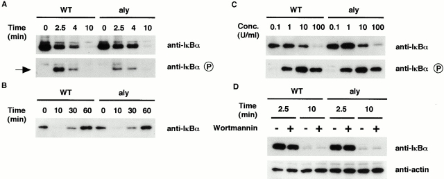
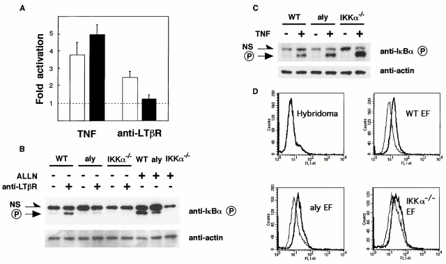
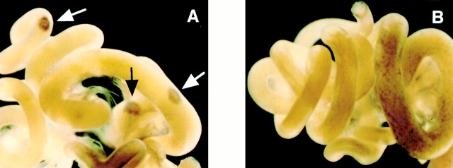
Both nuclear factor (NF)-kappaB-inducing kinase (NIK) and inhibitor of kappaB (IkappaB) kinase (IKK) have been implicated as essential components for NF-kappaB activation in response to many external stimuli. However, the exact roles of NIK and IKKalpha in cytokine signaling still remain controversial. With the use of in vivo mouse models, rather than with enforced gene-expression systems, we have investigated the role of NIK and IKKalpha in signaling through the type I tumor necrosis factor (TNF) receptor (TNFR-I) and the lymphotoxin beta receptor (LTbetaR), a receptor essential for lymphoid organogenesis. TNF stimulation induced similar levels of phosphorylation and degradation of IkappaBalpha in embryonic fibroblasts from either wild-type or NIK-mutant mice. In contrast, LTbetaR stimulation induced NF-kappaB activation in wild-type mice, but the response was impaired in embryonic fibroblasts from NIK-mutant and IKKalpha-deficient mice. Consistent with the essential role of IKKalpha in LTbetaR signaling, we found that development of Peyer's patches was defective in IKKalpha-deficient mice. These results demonstrate that both NIK and IKKalpha are essential for the induction of NF-kappaB through LTbetaR, whereas the NIK-IKKalpha pathway is dispensable in TNFR-I signaling.


Figures

Figure 1
Figure 1
NF-κB activation in response to TNF is retained in EFs from NIK-mutant mice. EFs from wild-type mice (WT) and aly mice (aly) were stimulated with TNF (100 U/ml), and cells were harvested at the indicated time points (A and B). IκBα degradation (detected by anti-IκBα Ab) and IκBα phosphorylation (detected by phospho-specific anti-IκBα Ab) was assessed by Western blot analysis. Arrow indicates phosphorylated IκBα (A). EFs were stimulated with different concentrations (Conc.) of TNF ranging from 0.1 to 100 U/ml as indicated, and cells were harvested 7 min after stimulation (C). EFs were stimulated with TNF (100 U/ml) with or without prior treatment with 1 μM wortmannin (D). The same blot was probed with anti-actin Ab (bottom).
Figure 2
Figure 2
Impaired NF-κB activation in response to LTβR stimulation in NIK-mutant and IKKα-deficient EFs. Wild-type EFs and aly mouse EFs were transfected with NF-κB reporter plasmids and stimulated with TNF or agonistic anti-LTβR mAb. 8 h later, NF-κB activation was assessed by the measurement of luciferase activities. Data were expressed as fold activation compared with stimulation by control mAb, and the results were plotted as the mean ± SEM for a total of four independent experiments. White and black bars represent wild-type EFs and aly mouse EFs, respectively (A). EFs from wild-type, aly, and IKKα-deficient mice (IKKα−/−) were stimulated with agonistic anti-LTβR mAb for 1 h, and IκBα phosphorylation was assessed by Western blot analysis (B, top). For the assessment of the basal level of NF-κB activation, EFs were treated with ALLN alone. Phosphorylated IκBα is indicated as P. NS, nonspecific bands. The same blot was probed with anti-actin Ab (B, bottom). TNF stimulation induced IκBα phosphorylation in IKKα-deficient EFs as in wild-type and aly EFs (C). LTβR expression assessed by flow-cytometric analysis with anti-LTβR mAb (D, thick line) was similar among wild-type, aly, and IKKα-deficient EFs (D). Anti-KLH mAb Ha4/8 (D, thin line) and mouse hybridoma cells (top left) were used as negative control.
Figure 3
Figure 3
Lack of PP development in IKKα-deficient mice. Embryonic intestines isolated from control littermates (A) and IKKα-deficient mice (B) were stained with anti–VCAM-1 mAb. Arrows indicate the sites of PPs. Original magnification, ×10.
Figure 4
Figure 4
Disruption of interaction with IKKα by the aly-type NIK mutation. Protein extracts from COS-7 cells transfected with the indicated cDNAs were immunoprecipitated with anti-Myc mAb and detected with either anti-Flag mAb (top) or anti-Myc mAb (center). Expression of NIK was verified by Western blot analysis of total cell lysates with anti-Flag mAb (bottom). aly-type NIK (aly) has an amino acid substitution (G860R) in the COOH-terminal region. Minus (−) indicates transfection with empty vectors.

References

    1. Ghosh S., May M.J., Kopp E.B. NF-κB and Rel proteinsevolutionarily conserved mediators of immune responses. Annu. Rev. Immunol. 1998;16:225–260. - PubMed
    1. Van Antwerp D.J., Martin S.J., Verma I.M., Green D.R. Inhibition of TNF-induced apoptosis by NF-κB. Trends Cell Biol. 1998;8:107–111. - PubMed
    1. Baldwin A.S. The NF-κB and IκB proteinsnew discoveries and insights. Annu. Rev. Immunol. 1996;14:649–681. - PubMed
    1. Maniatis T. Catalysis by a multiprotein IκB kinase complex. Science. 1997;278:818–819. - PubMed
    1. Stancovski I., Baltimore D. NF-κB activationthe IκB kinase revealed? Cell. 1997;91:299–302. - PubMed

Publication types

MeSH terms