. 2001 Apr;75(7):3077-88.
doi: 10.1128/JVI.75.7.3077-3088.2001.

Herpes simplex virus-induced keratitis: evaluation of the role of molecular mimicry in lesion pathogenesis

Affiliations

Herpes simplex virus-induced keratitis: evaluation of the role of molecular mimicry in lesion pathogenesis

S P Deshpande et al. J Virol. 2001 Apr.

Abstract

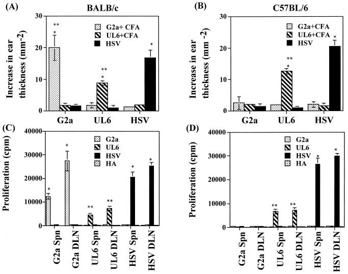
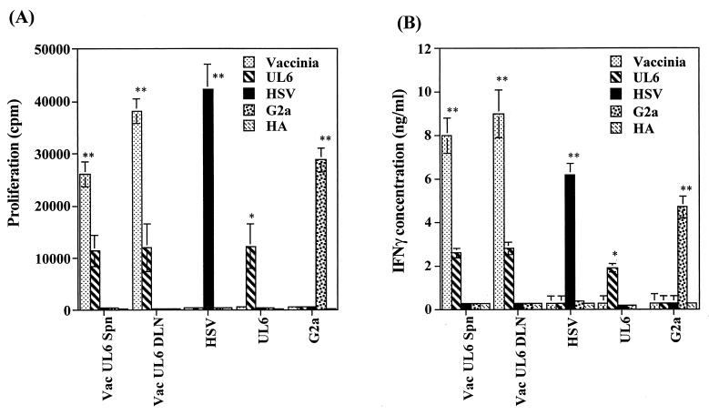
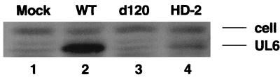
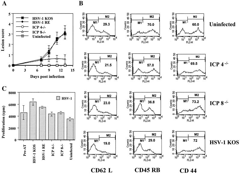
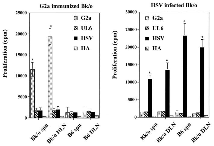
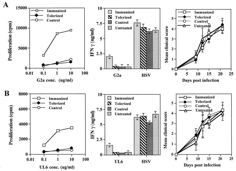
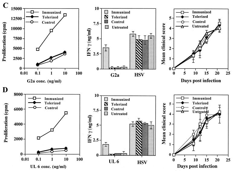
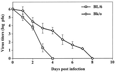
Viruses are suspected but usually unproven triggering factors in autoimmunity. One favored mechanism to explain the role of viruses in the genesis of autoimmunity is molecular mimicry. An immunoinflammatory blinding lesion called herpetic stromal keratitis (HSK) that follows ocular infection with herpes simplex virus (HSV) is suggested to result from a CD4(+) T-cell response to a UL6 peptide of HSV that cross-reacts with a corneal autopeptide shared with the immunoglobulin G2a(b) (IgG2a(b)) isotype. The present report reevaluates the molecular mimicry hypothesis to explain HSK pathogenesis. Our results failed to reveal cross-reactivity between the UL6 and IgG2a(b) peptides or between peptide reactive T cells and HSV antigens. More importantly, animals infected with HSV failed to develop responses that reacted with either peptide, and infection with a recombinant vaccinia UL6 vector failed to cause HSK, in spite of generating UL6 reactivity. Other lines of evidence also failed to support the molecular mimicry hypothesis, such as the failure to affect HSK severity upon tolerization of susceptible BALB/c and B-cell-deficient mice with IgG2a(b) or UL6 peptides. An additional study system revealed that HSK could be induced in mouse strains, such as the OT2 x RAG1(-/-) mice (T cell receptor transgenic recognizing OVA(323-339)) that were unable to produce CD4(+) T-cell responses to any detectable HSV antigens. Our results cast doubt on the molecular mimicry hypothesis as an explanation for the pathogenesis of HSK and indicate that if autoimmunity is involved its likely proceeds via a bystander activation mechanism.


Figures

FIG. 1
FIG. 1
Analyses of cross-reactivity between G2a peptide, UL6 peptide, and HSV in lymphoid cells following peptide immunization or HSV-1 RE ocular infection. Groups of B/c and B6 mice were immunized subcutaneously with 100 μg of peptide in CFA at the base of neck or were infected with 5 × 10⁵ PFU or 107 PFU of HSV-1 RE, respectively, on scarified corneas. (A and B) At day 15 postinfection, a DTH reaction was elicited in the ear pinnae of mice to 10 μg of peptides per ml or UV-inactivated HSV-1 (105 PFU) and 1× PBS or Vero extract in a 20-μl volume in the right and left ear pinnae, respectively. The increase in the ear thickness was measured after 48 h as described in Materials and Methods. The data are expressed as the difference in ear thickness ± the standard deviation (in mm−2) and represents the mean of two experiments, each including six to seven mice. Statistically significant differences between groups (∗∗, P < 0.001) and within groups (∗, P < 0.05) are indicated. (C and D) At days 9 to 15 post-peptide immunization and at days 15 to 18 post-HSV-1 ocular infection, mice (n = 4) were sacrificed, and cervical and submandibular DLN or spleen cells were used as responders in a lymphoproliferation assay as described in Materials and Methods. Responders were stimulated with irradiated syngeneic splenocytes pulsed with a range of peptide concentrations (10, 1.0, and 0.1 μg/ml), UV-irradiated virus (MOI = 5.0), or no stimulation. Data are represented only for peptide stimulations with a 10-μg/ml concentration. Polyclonal stimulator, ConA (2 μg/ml), was used as a positive control (data not shown). Results are the mean of three independent experiments. The experiment was repeated at least six times with similar results. Statistically significant differences between HSV-stimulated responders and peptide-stimulated responders (∗, P < 0.0001; ∗∗, P < 0.05) are indicated.
FIG. 2
FIG. 2
Recombinant vaccinia virus expressing UL6 protein fails to induce reactivity to HSV or G2a peptide. Groups of B/c mice (n = 6) were infected with 2 × 10⁶ PFU of vaccinia virus expressing UL6 (Vac UL6) on scarified corneas. At day 14 postinfection mice were sacrificed, and pooled popliteal lymph nodes and individual splenocytes were used as responders in a lymphoproliferation assay (A) and an IFN-γ cytokine ELISA (B) as described in Materials and Methods. Responders were stimulated with irradiated syngeneic splenocytes pulsed with UV-irradiated vaccinia virus (MOI = 5.0), UV-irradiated HSV (MOI = 5.0), and the UL6, G2a, and HA peptides (10, 1.0, and 0.1 μg/ml). The data represent peptide stimulation with a 10-μg/ml concentration. The polyclonal stimulator, ConA (2 μg/ml), was used as a positive control (data not shown). The results of two independent experiments are expressed as the means ± the standard deviations. Statistically significant differences within groups (∗∗, P < 0.001) and between groups (∗, P < 0.05) are indicated.
FIG. 3
FIG. 3
Analysis of UL6 protein expression by various HSV strains. Infected cell lysates from Hep-2 cells infected with the indicated viruses were analyzed for UL6 expression by Western blotting as described in Materials and Methods. Shown in figure is the image of Western blotting. The top band provides a loading control, and the bottom band is the UL6 protein. Lanes: 1, mock infected; 2, KOS wild-type (WT) virus; 3, KOS d120 (ICP4) virus; 4, KOS HD-2 (ICP8) virus.
FIG. 4
FIG. 4
Infection with replication-defective HSV (UL6 positive or negative) fails to induce HSK in SCID mice reconstituted with HSV immune T cells. SCID mice (n = 5/group) were infected with ICP4−/−, ICP8−/−, and HSV-1 KOS (5 × 10⁵ PFU) on scarified corneas at days 0, 2, and 4. At day 1 postinfection, SCID mice were reconstituted with 107 HSV immune splenocytes in a 400-μl volume of 1× PBS given i.v. The data represents the results from one of two independent experiments with similar results. (A) Mean clinical scores of SCID mice at days 7, 9, 11, and 12 postinfection. Mice were scored by using a slit-lamp microscope as described in Materials and Methods. (B and C) Mice were terminated at day 13 postinfection, and cervical and submandibular DLN cells were used as responders in an HSV-specific lymphoproliferation assay (C) or were stained for activation markers CD62L, CD45 RB, and CD44 by flow cytometry assay (B) as described in Materials and Methods. The percentage of cell surface expression of activation markers under marker M2 is indicated in the histograms. The data indicate the presence of activated HSV-specific CD4+ T cells in all of the wild-type and mutant virus-infected reconstituted SCID mice groups irrespective of the development of HSK lesions shown in panel A.
FIG. 5
FIG. 5
Peptide G2a immunization or HSV infection in Bk/o fails to generate cross-reactivity. Groups of Bk/o and B6 mice (n = 4) were infected with 107 PFU of HSV-1 RE on scarified corneas or were immunized with 100 μg of UL6, G2a, or HA peptides in CFA. Mice were terminated from days 9 to 15 post-peptide immunization or days 15 to 18 post-HSV infection. DLN and spleen cells were used in lymphoproliferation assays with various concentrations of G2a, UL6, and HA peptides (10, 1.0, or 0.1 μg/ml; the data shown here represent peptide stimulation with a 10-μg/ml concentration) or UV-irradiated HSV (MOI = 5.0) as described previously. The data are representative of one of three experiments with similar results. Statistically significant differences between groups are indicated (∗, P < 0.001).
FIG. 6
FIG. 6
Bk/o mice are highly susceptible to herpetic encephalitis and HSK. Bk/o and B6 mice (n = 10 to 12) were infected on scarified corneas with a range of infectious doses and then examined for survival and induction of HSK lesions. The data represent one of two similar experiments. (A) Lesions were scored using a slit-lamp microscope and were recorded as described in Materials and Methods. The data represent the percentage of mice developing lesion scores at day 15 postinfection of ≥3.0. (B) Mice were examined daily for signs of herpetic encephalitis. The results are expressed as the percent survival of mice per time point.
FIG. 7
FIG. 7
Tolerization of peptides fails to induce resistance to HSK in susceptible Bk/o or B/c mice. Groups of Bk/o mice and B/c mice (n = 12) were tolerized i.v. with 50 μg of soluble UL6 or G2a peptides at days 0 and 7. At day 15 posttolerization, groups of peptide tolerized mice (n = 6) were infected with HSV-1 RE alone (tolerized) or were infected with HSV-1 RE (107 PFU for Bk/o mice and 106 PFU for B/c mice) on scarified corneas, as well as immunized with 100 μg of the same peptide in CFA (control). Groups of age- and sex-matched mice (n = 6) were immunized with 100 μg of the same peptide in CFA and then infected with HSV-1 RE (immunized) or infected with HSV-1 RE alone (untreated). Mice were examined for lesions by using a slit-lamp microscope, and the mean clinical scores at days 9, 12, 15, and 21 are shown. Mice were terminated at day 18 postinfection, and DLN cells were used in a lymphoproliferation assay to tolerogenic peptides and in an IFN-γ cytokine assay to peptides and UV-irradiated HSV (MOI = 5.0), as described previously. The data represent one of three experiments with similar results. (A) G2a-tolerized Bk/o mice (B) UL6-tolerized Bk/o mice (C) G2a-tolerized B/c mice. (D) UL6-tolerized B/c mice.
FIG. 7
FIG. 7
Tolerization of peptides fails to induce resistance to HSK in susceptible Bk/o or B/c mice. Groups of Bk/o mice and B/c mice (n = 12) were tolerized i.v. with 50 μg of soluble UL6 or G2a peptides at days 0 and 7. At day 15 posttolerization, groups of peptide tolerized mice (n = 6) were infected with HSV-1 RE alone (tolerized) or were infected with HSV-1 RE (107 PFU for Bk/o mice and 106 PFU for B/c mice) on scarified corneas, as well as immunized with 100 μg of the same peptide in CFA (control). Groups of age- and sex-matched mice (n = 6) were immunized with 100 μg of the same peptide in CFA and then infected with HSV-1 RE (immunized) or infected with HSV-1 RE alone (untreated). Mice were examined for lesions by using a slit-lamp microscope, and the mean clinical scores at days 9, 12, 15, and 21 are shown. Mice were terminated at day 18 postinfection, and DLN cells were used in a lymphoproliferation assay to tolerogenic peptides and in an IFN-γ cytokine assay to peptides and UV-irradiated HSV (MOI = 5.0), as described previously. The data represent one of three experiments with similar results. (A) G2a-tolerized Bk/o mice (B) UL6-tolerized Bk/o mice (C) G2a-tolerized B/c mice. (D) UL6-tolerized B/c mice.
FIG. 8
FIG. 8
Persistence of virus for longer durations in the absence of Ig in Bk/o mice. Groups of Bk/o and B6 mice (n = 6) were infected with 107 PFU on scarified corneas. Ocular swabs were taken on days 0 to 10 postinfection and tested for the presence of virus by the standard plaque assay described in Materials and Methods. The data are represented as the means ± the standard deviations.
FIG. 9
FIG. 9
Viral antigen and CD4+ KJ+ T cells in the corneal stroma of OT2xRAG1−/− HSV ocularly infected mice. OT2xRAG1−/− mice were infected with 2 × 10⁶ PFU virus on scarified corneas. At days 10 to 13 postinfection, mice were sacrificed, and the eyes were snap-frozen in OCT compound. (A) Histopathology of infiltrating cells in the corneal stroma. (B) Immunohistochemistry for viral antigens in the corneal stroma (magnification, × 200). (C and D) Immunohistochemistry for CD4+ cells (magnification, × 400) (C) and for OVA323–339 TCR clonotypic antibody KJ1-26.1+ cells (magnification, ×400) (D).

References

    1. Atkinson M A, Bowman M A, Campbell L, Darrow B L, Kaufman D L, Maclaren N K. Cellular immunity to a determinant common to glutamate decarboxylase and coxsackie virus in insulin-dependent diabetes. J Clin Investig. 1994;94:2125–2129. - PMC - PubMed
    1. Avery A C, Zhao Z S, Rodriguez A, Bikoff E K, Soheilian M, Foster C S, Cantor H. Resistance to herpes stromal keratitis conferred by an IgG2a derived peptide. Nature. 1995;276:431–434. - PubMed
    1. Babu J S, Thomas J, Kanangat S, Morrison L A, Knipe D M, Rouse B T. Viral replication is required for induction of ocular immunopathology by herpes simplex virus. J Virol. 1996;70:101–107. - PMC - PubMed
    1. Buchmeier M J, Lane T E. Viral induced neurodegenerative disease. Curr Opin Microbiol. 1999;2:398–402. - PMC - PubMed
    1. Chun S, Daheshia M, Kuklin N A, Rouse B T. Modulation of viral immunoinflammatory responses with cytokine DNA administered by different routes. J Virol. 1998;72:5545–5551. - PMC - PubMed

Publication types