. 2001 Apr;75(7):3220-9.
doi: 10.1128/JVI.75.7.3220-3229.2001.

Purification and characterization of West Nile virus nucleoside triphosphatase (NTPase)/helicase: evidence for dissociation of the NTPase and helicase activities of the enzyme

Affiliations

Purification and characterization of West Nile virus nucleoside triphosphatase (NTPase)/helicase: evidence for dissociation of the NTPase and helicase activities of the enzyme

P Borowski et al. J Virol. 2001 Apr.

Abstract

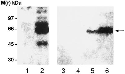
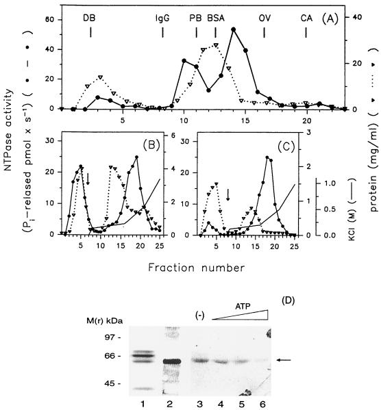
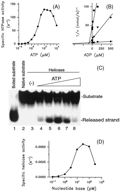
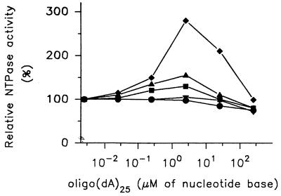
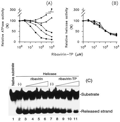
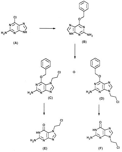
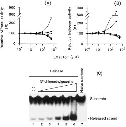
The nucleoside triphosphatase (NTPase)/helicase associated with nonstructural protein 3 of West Nile (WN) virus was purified from cell culture medium harvested from virus-infected Vero cells. The purification procedure included sequential chromatography on Superdex-200 and Reactive Red 120 columns, followed by a concentration step on an Ultrogel hydroxyapatite column. The nature of the purified protein was confirmed by immunoblot analysis using a WN virus-positive antiserum, determination of its NH(2) terminus by microsequencing, and a binding assay with 5'-[(14)C]fluorosulfonylbenzoyladenosine. Under optimized reaction conditions the enzyme catalyzed the hydrolysis of ATP and the unwinding of the DNA duplex with k(cat) values of 133 and 5.5 x 10(-3) s(-1), respectively. Characterization of the NTPase activity of the WN virus enzyme revealed that optimum conditions with respect to the Mg(2+) requirement and the monovalent salt or polynucleotide response differed from those of other flavivirus NTPases. Initial kinetic studies demonstrated that the inhibition (or activation) of ATPase activity by ribavirin-5'-triphosphate is not directly related to changes in the helicase activity of the enzyme. Further analysis using guanine and O(6)-benzoylguanine derivatives revealed that the ATPase activity of WN virus NTPase/helicase may be modulated, i.e., increased or reduced, with no effect on the helicase activity of the enzyme. On the other hand the helicase activity could be modulated without changing the ATPase activity. Our observations show that the number of ATP hydrolysis events per unwinding cycle is not a constant value.


Figures

FIG. 1
FIG. 1
Analysis of SFII and WN virus immunoreactivity of cells and cell culture medium infected with WN virus. Vero E6 cells were infected with WN virus as described in Materials and Methods. Five days after the infection the cells were precipitated with TCA and an aliquot (10 μg) of the collected proteins was subjected to SDS-PAGE. The separated proteins were transferred onto a nitrocellulose filter and reacted with NS3/HEL(1&2) antibody. Simultaneously, portions of the cell culture medium were removed immediately (lane 4), 2 days (lane 5), or 5 days (lane 6) after infection. The proteins were precipitated with TCA, and aliquots (50 μg of protein) were separated by SDS-PAGE followed by immunoblotting with NS3/HEL(1&2) antibody. Immunoblots were prepared using uninfected cells (lane 1), infected cells (lane 2), and cell culture supernatant from uninfected cells (lane 3). The nitrocellulose filters were autoradiographed for 14 h. Molecular mass markers are indicated at the left. Arrow, position of WN virus NTPase/helicase.
FIG. 2
FIG. 2
Elution profiles of ATPase activity of WN virus NTPase/helicase from consecutive chromatographic columns and analysis of the purified protein obtained from the last purification step by SDS-PAGE. Aliquots (20 μl) of the tested fractions were assayed for NTPase activity and protein content as described in Materials and Methods. (A) Superdex-200 column chromatography of the concentrated medium harvested from an infected-cell culture. The column was calibrated with dextran blue (DB; 2,000 kDa) and with the following marker proteins: immunoglobulin G (IgG; 160 kDa), phosphorylase b (PB; 97 kDa), BSA (66 kDa), ovalbumin (OV; 45 kDa), and carbonic anhydrase (CA; 30 kDa). (B) Reactive Red 120 chromatography of pooled Superdex-200 fractions 10, 11, 14, and 15. (C) Reactive Red 120 rechromatography of pooled fractions 16 to 20 obtained from panel B. Arrows (B and C), start of the salt gradients. (D) Aliquots of the final enzyme preparation (10 μg of protein) were separated by SDS-PAGE and visualized by staining with Coomassie blue (lane 1) or transferred onto nitrocellulose and immunoblotted with the NS3/HEL(1&2) antibody followed by incubation with rabbit anti-human antiserum and 125I-protein A (lane 2). The blot was exposed for 12 h. Aliquots (20 μg) of the final enzyme preparation were incubated with [14C]FSBA in the absence (lane 3) or presence of ATP added at 10 μM (lane 4), 100 μM (lane 5), or 1 mM (lane 6). The samples were subjected to SDS-PAGE, and the gel was dried and exposed for 4 days. The molecular masses of the investigated proteins were estimated by reference to protein standards (left). Arrow, position of the WN virus NTPase/helicase.
FIG. 3
FIG. 3
Substrate and product inhibition of the NTPase and helicase reactions mediated by WN virus NTPase/helicase. (A) The ATPase reaction as a function of increasing concentrations of ATP was investigated as shown. The product of the reaction (33Pi) was quantified using the charcoal adsorption method described in Materials and Methods. (B) The plots, performed as described by Dixon, demonstrate the competitive type of inhibition of the ATPase activity by ADP. The reaction was carried out in the presence of ATP adjusted to concentrations equal to 1/10 (▪), 1 (▾), or 10 (⧫) times of the Km value (9.5 μM) and the indicated ADP concentrations. (C) The strand-displacing activity of the WN virus NTPase/helicase was determined with a 4.7 pM concentration of the nucleotide bases of the DNA duplex as the substrate in the absence (lane 3) or presence of ATP adjusted to 0.95 μ (lane 4), 9.5 μM (lane 5), 95 μM (lane 6), 950 μM (lane 7), and 9.5 mM (lane 8). Lanes 1 and 2, boiled and native substrates, respectively. The samples were separated in a TBE-polyacrylamide gel, and the levels of 32P radioactivity associated with the substrate and the released strand were visualized by exposition of the dried gels for 10 h. (D) The helicase activity of the WN virus NTPase/helicase as a function of increasing concentrations of the DNA duplex as the substrate was determined at the saturating ATP concentration (90 μM). The samples were separated in a TBE-polyacrylamide gel, and the levels of 32P radioactivity associated with the substrate and the released strand were quantified as described in Materials and Methods. The data obtained were presented as the sum of the nucleotide bases of the unwound DNA duplex. The results shown are representative of three independent experiments.
FIG. 4
FIG. 4
Modulation of the ATPase activity of the WN virus NTPase/helicase by oligo(dA)25. The ATPase reaction was investigated as a function of increasing concentrations of oligo(dA)25. The reaction was performed at ATP concentrations equal to 1/100 (ℑ), 1/10 (▾), 1 (▪), 10 (∓), and 100 (⧫) times the Km value (9.5 μM). The ATPase activity measured for each ATP concentration in the absence of oligo(dA)25 was taken as 100%. The results shown are representative of three independent experiments.
FIG. 5
FIG. 5
Inhibition of the ATPase and helicase activities of WN virus NTPase/helicase by ribavirin and ribavirin-TP. The ATPase (A) and helicase (B) reactions were performed at ATP concentrations equal to 1/100 (ℑ), 1/10 (▾), 1 (▪), 10 (∓), and 100 (⧫) times of the Km value (9.5 μM) as a function of increasing amounts of ribavirin-TP as the inhibitor. The ATPase activity of the enzyme was determined by the charcoal adsorption method described in Materials and Methods. The helicase activity was assayed at a 4.7 pM concentration of nucleotide bases of the DNA duplex as the substrate. The samples were separated in a TBE-polyacrylamide gel, and the 32P radioactivity associated with the released strand was quantified as described in Materials and Methods. The activity of the enzyme measured for each ATP concentration in the absence of the ribavirin-TP was taken as 100%. (C) Comparison of the inhibitory effects of ribavirin and ribavirin-TP on the helicase activity of WN virus NTPase/helicase. The reaction took place in the absence (lanes 2 and 7) or presence of ribavirin (lanes 3 to 6) or ribavirin-TP (lanes 8 to 11). The concentrations of both compounds were adjusted to 5 μM (lanes 3 and 8), 50 μM (lanes 4 and 9), 500 μM (lanes 5 and 10), and 5 mM (lanes 6 and 11). The assay was performed at 9.5 μM ATP and a DNA duplex concentration equal to the Km value (4.7 pM concentration of nucleotide bases). The substrate and released strand were separated in a TBE-polyacrylamide gel and visualized by exposition of dried gel onto X-ray film for 14 h. The results shown are representative of three independent experiments.
FIG. 6
FIG. 6
Structures of guanine and O6-benzylguanine derivatives used in this study. The synthesis and purification of the compounds were performed according to procedures presented in Materials and Methods.
FIG. 7
FIG. 7
Comparison of the modulating effects of the guanine and O6-benzylguanine derivatives on the ATPase and helicase activities of the WN virus NTPase/helicase. The ATPase (A) and helicase (B) activities of the enzyme were investigated as a function of increasing amounts of O6-benzylguanine (ℑ), O6-benzyl-N9-chloroethylguanine (▾), O6-benzyl-N7-chloroethylguanine (▪), N9-chloroethylguanine (∓), and N7-chloroethylguanine (⧫). The ATPase reaction was performed in the presence of ATP adjusted to a concentration equal to the Km value, and the activity of the enzyme was determined by using the charcoal adsorption method described in Materials and Methods. The helicase activity of the enzyme was investigated at 9.5 μM ATP and a 4.7 pM concentration of nucleotide bases of DNA duplex as the substrate. The 32P radioactivity of the released strand was quantified as described in Materials and Methods. The ATPase and helicase activities measured in the absence of the inhibitors were taken as 100%. (C) Autoradiography of a TBE-polyacrylamide gel demonstrating the activating effect of N7-chloroethylguanine on the helicase activity of WN virus NTPase/helicase. The reaction took place in the absence (lane 1) or presence of the compound added at 2.2 (lane 2), 6.6 (lane 3), 22 (lane 4), 66 (lane 5), or 220 μM (lane 6). The native substrate is presented in lane 7. The dried gel was exposed for 14 h without an intensifying screen. The results shown are representative of three independent experiments.

References

    1. Ali J A, Lohman T M. Kinetic measurement of the step size of DNA unwinding by Escherichia coli UvrD helicase. Science. 1997;275:377–380. - PubMed
    1. Allison S L, Stadler K, Mandl C W, Kunz C, Heinz F X. Synthesis and secretion of recombinant tick-borne encephalitis virus protein E in soluble and particulate form. J Virol. 1995;69:5816–5820. - PMC - PubMed
    1. Bazan J F, Fletterick R. Detection of a trypsin-like serine protease domain in flaviviruses and pestiviruses. Virology. 1989;171:637–639. - PubMed
    1. Borowski P, Heiland M, Oehlmann K, Becker B, Kornetzky L, Feucht H-H, Laufs R. Non-structural protein 3 of hepatitis C virus inhibits phosphorylation mediated by cAMP-dependent protein kinase. Eur J Biochem. 1996;237:611–618. - PubMed
    1. Borowski P, Kühl R, Laufs R, Schulze zur Wiesch J, Heiland M. Identification and characterization of a histone binding site of nonstructural protein 3 of hepatitis C virus. J Clin Virol. 1999;13:61–69. - PubMed

MeSH terms

LinkOut - more resources