. 2001 Apr;75(7):3268-76.
doi: 10.1128/JVI.75.7.3268-3276.2001.

Evidence of two Lyssavirus phylogroups with distinct pathogenicity and immunogenicity

Affiliations

Evidence of two Lyssavirus phylogroups with distinct pathogenicity and immunogenicity

H Badrane et al. J Virol. 2001 Apr.

Abstract

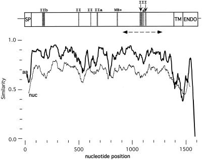
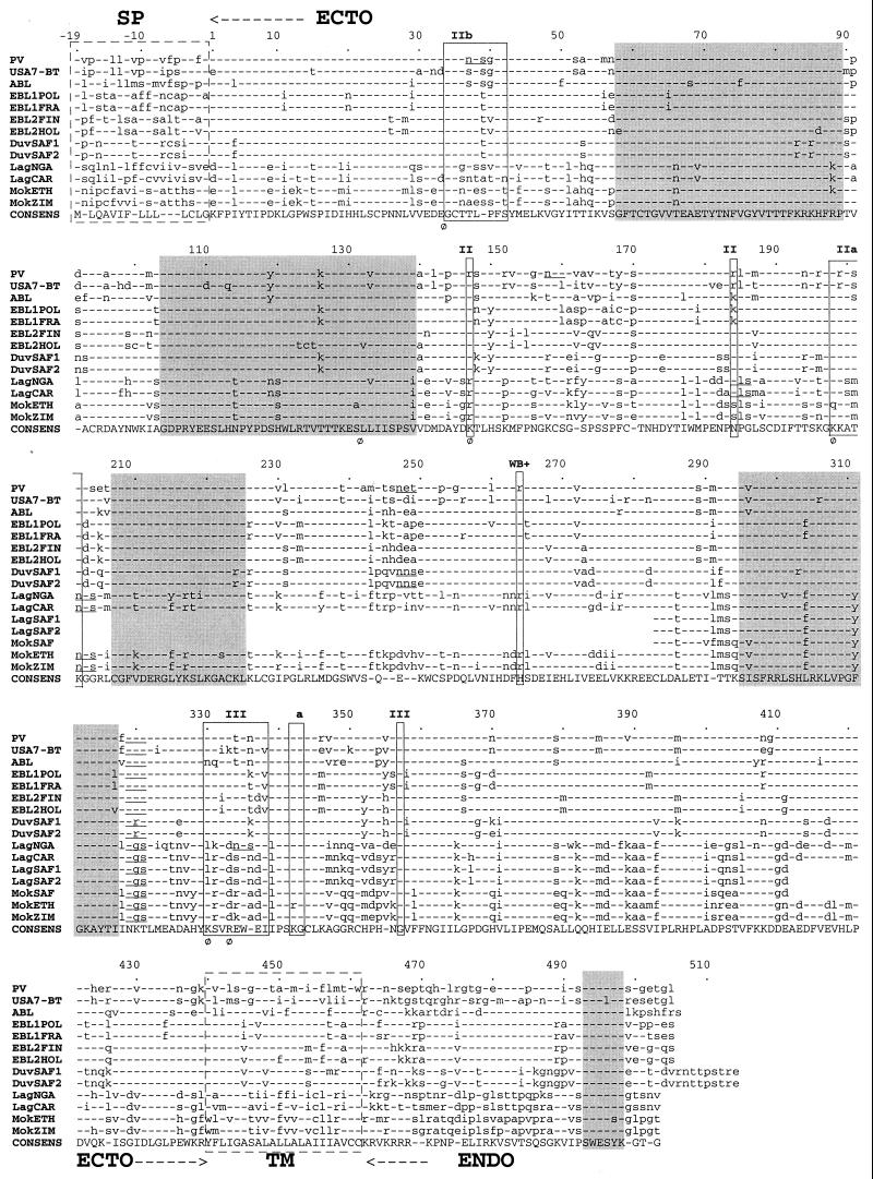
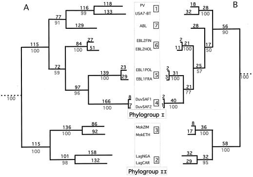
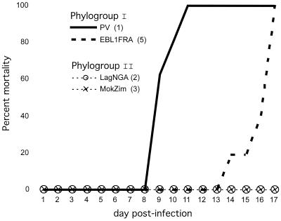
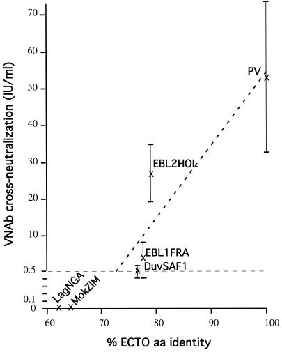
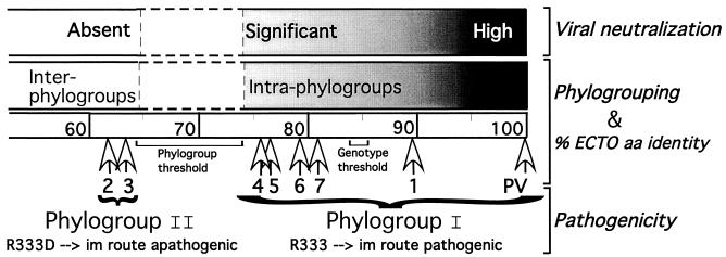
The genetic diversity of representative members of the Lyssavirus genus (rabies and rabies-related viruses) was evaluated using the gene encoding the transmembrane glycoprotein involved in the virus-host interaction, immunogenicity, and pathogenicity. Phylogenetic analysis distinguished seven genotypes, which could be divided into two major phylogroups having the highest bootstrap values. Phylogroup I comprises the worldwide genotype 1 (classic Rabies virus), the European bat lyssavirus (EBL) genotypes 5 (EBL1) and 6 (EBL2), the African genotype 4 (Duvenhage virus), and the Australian bat lyssavirus genotype 7. Phylogroup II comprises the divergent African genotypes 2 (Lagos bat virus) and 3 (Mokola virus). We studied immunogenic and pathogenic properties to investigate the biological significance of this phylogenetic grouping. Viruses from phylogroup I (Rabies virus and EBL1) were found to be pathogenic for mice when injected by the intracerebral or the intramuscular route, whereas viruses from phylogroup II (Mokola and Lagos bat viruses) were only pathogenic by the intracerebral route. We showed that the glycoprotein R333 residue essential for virulence was naturally replaced by a D333 in the phylogroup II viruses, likely resulting in their attenuated pathogenicity. Moreover, cross-neutralization distinguished the same phylogroups. Within each phylogroup, the amino acid sequence of the glycoprotein ectodomain was at least 74% identical, and antiglycoprotein virus-neutralizing antibodies displayed cross-neutralization. Between phylogroups, the identity was less than 64.5% and the cross-neutralization was absent, explaining why the classical rabies vaccines (phylogroup I) cannot protect against lyssaviruses from phylogroup II. Our tree-axial analysis divided lyssaviruses into two phylogroups that more closely reflect their biological characteristics than previous serotypes and genotypes.


Figures

FIG. 1
FIG. 1
Lyssavirus similarity profile along the G gene coding region. Nucleotide (nuc, thin line) and amino acid (aa, bold line) sequence similarity profiles among 13 complete Lyssavirus glycoproteins are shown (similarityplot program of GCG; window, 100 nucleotides or 50 aa, step 1). SP, signal peptide; TM, transmembrane domain; ENDO, endodomain. Gray boxes indicate two major antigenic sites (II and III), the minor antigenic site (a), and the neutralizing linear epitope VI (WB+, Western blot positive). The discontinuous line with arrowheads indicates the region sequenced in LagSAF1, LagSAF2, and MokSAF (Fig. 2).
FIG. 2
FIG. 2
Multiple alignment of Lyssavirus glycoproteins. Alignment of complete or partial deduced amino acid sequences of the glycoprotein from genotypes 1 (PV and USA7-BT), 2 (LagNGA, LagCAR, LagSAF1, and LagSAF2), 3 (MokSAF, MokETH, and MokZIM), 4 (DuvSAF1 and DuvSAF2), 5 (EBL1POL and EBL1FRA), 6 (EBL2FIN and EBL2HOL), and 7 (ABL). Dashes indicate amino acids agreeing with the consensus sequence (CONSENS). Boxes with discontinuous lines show the hydrophobic signal peptide (SP) and transmembrane domain (TM). ECTO, ectodomain; ENDO, endodomain. Boxes with continuous lines outline the main antigenic sites and epitopes. Underlined NX(S/T) motifs in the ectodomain are potential N-glycosylation sites. Phi (Φ) indicates residues involved in pathogenicity. Gray boxes show the five most conserved blocks.
FIG. 3
FIG. 3
Lyssavirus phylogeny. Estimated rooted phylogenetic trees using nucleotide (A) or amino acid (B) sequences of the G ectodomain. The PAUP program (parsimony) was used with the following options: branch and bound search, bootstrap with 50% majority rule consensus, and collapsed zero-length branches. Outline numbers are bootstrap values of 100 replicates, testing the robustness of their corresponding internal branches. Bold numbers are steps occurring on each branch (branch lengths). Numbers inside squares indicate the seven genotypes. Chandipura and Piry viruses from the Vesiculovirus genus were used as an outgroup.
FIG. 4
FIG. 4
Pathogenicity of lyssaviruses by the i.m. route. Eight BALB/c mice were injected by the i.m. route in the thigh with 105 to 107 LD50 i.c. of PV, EBL1FRA, LagNGA, or MokZIM. For each virus, the genotype number is indicated in parentheses. Results are expressed as the percentage of dead animals monitored up to 17 days postinfection.
FIG. 5
FIG. 5
Curve of ectodomain amino acid identity versus cross-neutralization. We correlated the cross-neutralization (mean ± standard deviation VNAb titers at day 39 post-DNA immunization) and the G ectodomain percent identity (% ECTO aa identity) between PV and four phylogroup I members (PV, EBL1FRA, EBL2HOL, and DuvSAF1) (see Table 3). No significant titers were found against two phylogroup II members (MokZIM and LagNGA). A linear correlation between VNAb cross-neutralization and % ECTO aa identity was observed (dashed oblique line) with a high coefficient (r = 0.92). The horizontal dashed line at 0.5 IU/ml corresponds to the accepted minimal titer for seroconversion. The vertical axis was enlarged between 0 and 0.5 for convenience.
FIG. 6
FIG. 6
Schematic representation of the three-axis analysis. % ECTO aa identity is a scale of distances where the seven white arrowheads show the positions of the seven genotypes (only their numbers are given) from PV. Phylogrouping is represented by a horizontal box with a directional color gradient from black to gray (intraphylogroup comparisons, ≥74% ECTO aa identity) to white (interphylogroup comparisons, ≤64% ECTO aa identity). The phylogroup and genotype thresholds are shown within ranges from 64 to 74% and 83.5 to 86% ECTO aa identity, respectively. VNAb titer is represented by a horizontal box with a directional color gradient from black (high titer) to gray (significant titer) to white (no protection, about 74% ECTO aa identity). For pathogenicity, the presence of R333 in the glycoprotein of phylogroup I members (PV and genotypes 1, 4, 5, 6, and 7) is an indication of their pathogenicity for mice by the i.m. route, whereas the R333D replacement in the glycoprotein of phylogroup II members (genotypes 2 and 3) concords with their apathogenicity for mice by the i.m. route.

References

    1. Amengual B, Whitby J E, King A, Cobo S, Bourhy H. Evolution of European bat lyssaviruses. J Gen Virol. 1997;78:2319–2328. - PubMed
    1. Atanasiu P, Tsiang H, Perrin P, Favre S. Demonstration of sialic acid in the rabies virus. Consequences of its removal on infectious and hemagglutinating properties. C R Acad Sc Hebd Seances Acad Sci D. 1976;283:111–114. - PubMed
    1. Bahloul C, Jacob Y, Tordo N, Perrin P. DNA-based immunisation for exploring the enlargement of immunological cross-reactivity against the lyssaviruses. Vaccine. 1998;16:417–425. - PubMed
    1. Benmansour A, Leblois H, Coulon P, Tuffereau C, Gaudin Y, Flamand A, Lafay F. Antigenicity of rabies virus glycoprotein. J Virol. 1991;65:4198–4203. - PMC - PubMed
    1. Bourhy H, Kissi B, Lafon M, Sacramento D, Tordo N. Antigenic and molecular characterization of bat rabies virus in Europe. J Clin Microbiol. 1992;30:2419–2426. - PMC - PubMed

Publication types

LinkOut - more resources