. 2001 Apr;75(7):3277-90.
doi: 10.1128/JVI.75.7.3277-3290.2001.

Epitope mapping porcine reproductive and respiratory syndrome virus by phage display: the nsp2 fragment of the replicase polyprotein contains a cluster of B-cell epitopes

Affiliations

Epitope mapping porcine reproductive and respiratory syndrome virus by phage display: the nsp2 fragment of the replicase polyprotein contains a cluster of B-cell epitopes

M B Oleksiewicz et al. J Virol. 2001 Apr.

Abstract

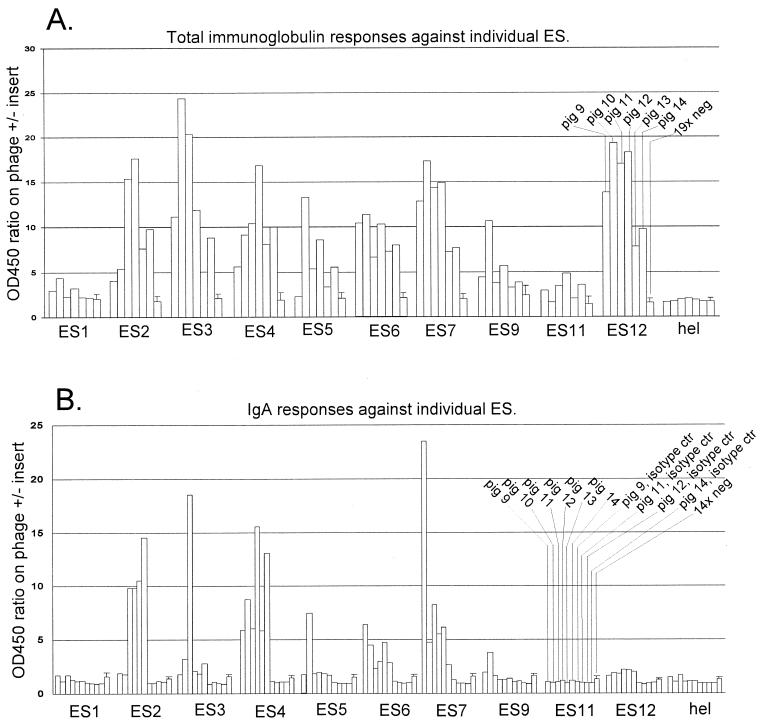
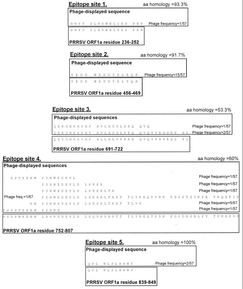
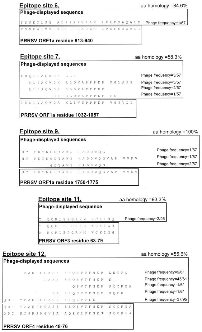
We screened phage display libraries of porcine reproductive and respiratory syndrome virus (PRRSV) protein fragments with sera from experimentally infected pigs to identify linear B-cell epitopes that are commonly recognized during infection in vivo. We identified 10 linear epitope sites (ES) 11 to 53 amino acids in length. In the replicase polyprotein, a total of eight ES were identified, six of which localized to the Nsp2 replicase polyprotein processing end product. In the structural proteins, a total of two ES were identified, in the ORF3 and ORF4 minor envelope glycoproteins. The ORF4 ES was previously identified by monoclonal antibody mapping (J. J. M. Meulenberg, A. P. van Nieuwstadt, A. van Essen-Zandenbergen, and J. P. M. Langeveld, J. Virol. 71:6061-6067, 1997), but its immunogenicity had not been examined in pigs. We found that six experimentally PRRSV-infected pigs consistently had very high antibody titers against the ORF4 ES. In some animals, sera diluted 1:62,500 still gave weak positive enzyme immunoassay reactivity against the ORF4 ES. This hitherto unrecognized immunodominance likely caused phages displaying the ORF4 ES to outcompete phages displaying other ES during library screening with porcine sera and accounted for our failure to identify more than two ES in the structural genes of PRRSV. Genetic analysis showed that variable ES were also the most immunogenic in vivo. Serological analysis indicated differences in the immunoglobulin A responses between short-term and longer-term viremic pigs towards some ES. The implications of these findings for PRRSV diagnostics and immunopathogenesis are discussed.


Figures

FIG. 1
FIG. 1
Serological confirmation of ES by ELISA. ELISA to detect total porcine Ig or porcine IgA directed against the different ES was done as described in Material and Methods. Hel is a phage displaying a 14-amino-acid fragment from the PRRSV helicase which is not an epitope, i.e., a negative-control antigen. To estimate the specific reactivity of porcine sera against the phage-displayed PRRSV peptides and to correct for any unspecific reactivity against the M13 virion, the OD450 ratio (y axis) was calculated as described in Materials and Methods. The 19× neg (A) and 14× neg (B) columns show the mean reaction of negative serum panels, with bars indicating 1 standard deviation. Experimental sera (pigs 9, 10, 11, 12, 13, and 14) were from 42-dpi PRRSV 111/92 infection. (B) Isotype control bars indicate the reactivity of the indicated pig serum using an isotype-matched MAb instead of the anti-IgA MAb. Sera from pigs 10 and 12 were used for phage library selection, as described in Table 2, footnote a.
FIG. 2
FIG. 2
Sequences of serologically confirmed ES. For each ES, phage-displayed sequences are shown in the top box. The phage-displayed sequences were derived from PRRSV isolate 111/92, which was used for library construction. The sequences shown were fused to the phage pIII protein, in the context MKKLLFAIPLVVPFYSHS∗A?AQTVQSCLA, where the pIII leader peptide is underlined, ∗ is the predicted leader peptidase cleavage site, ? is the phage-displayed PRRSV sequence shown in the figure, and naturally phage-encoded residues immediately neighboring the displayed sequence in the mature pIII protein are in italics. The published PRRSV sequence is shown in the bottom box, annotated and aligned to the phage-displayed sequences. For ES in ORF 1, only published Lelystad PRRSV sequence is shown. For ES in the structural genes (sites 11 and 12), published 111/92 and Lelystad PRRSV sequences are shown (111/92 top, Lelystad bottom). The sequences for two ES which could not be confirmed by ELISA (ES8 and ES10) are not shown. Amino acid (aa) homologies were calculated between the phage-displayed sequences and Lelystad PRRSV, in all cases omitting the first and last of the phage-displayed residues, which might have been corrupted during the cloning procedure. For ES12, the 5 C-terminal residues in the first phage sequence and the three N-terminal residues in the second sequence likely represented a ligation artifact and were omitted from the homology calculation. For each phage clone, the number of times the clone was observed (numerator) in the total number of sequenced phages from a given library (denominator) is indicated. Altogether 57, 95, and 61 phage clones were sequenced from the ORF 1, ORF 2-3 and ORF 4-7 libraries, respectively. Where more than one phage identified an ES, the phage displaying the longest PRRSV sequence was used as the ELISA antigen in all cases.
FIG. 2
FIG. 2
Sequences of serologically confirmed ES. For each ES, phage-displayed sequences are shown in the top box. The phage-displayed sequences were derived from PRRSV isolate 111/92, which was used for library construction. The sequences shown were fused to the phage pIII protein, in the context MKKLLFAIPLVVPFYSHS∗A?AQTVQSCLA, where the pIII leader peptide is underlined, ∗ is the predicted leader peptidase cleavage site, ? is the phage-displayed PRRSV sequence shown in the figure, and naturally phage-encoded residues immediately neighboring the displayed sequence in the mature pIII protein are in italics. The published PRRSV sequence is shown in the bottom box, annotated and aligned to the phage-displayed sequences. For ES in ORF 1, only published Lelystad PRRSV sequence is shown. For ES in the structural genes (sites 11 and 12), published 111/92 and Lelystad PRRSV sequences are shown (111/92 top, Lelystad bottom). The sequences for two ES which could not be confirmed by ELISA (ES8 and ES10) are not shown. Amino acid (aa) homologies were calculated between the phage-displayed sequences and Lelystad PRRSV, in all cases omitting the first and last of the phage-displayed residues, which might have been corrupted during the cloning procedure. For ES12, the 5 C-terminal residues in the first phage sequence and the three N-terminal residues in the second sequence likely represented a ligation artifact and were omitted from the homology calculation. For each phage clone, the number of times the clone was observed (numerator) in the total number of sequenced phages from a given library (denominator) is indicated. Altogether 57, 95, and 61 phage clones were sequenced from the ORF 1, ORF 2-3 and ORF 4-7 libraries, respectively. Where more than one phage identified an ES, the phage displaying the longest PRRSV sequence was used as the ELISA antigen in all cases.
FIG. 3
FIG. 3
Positions of serologically confirmed ES in the PRRSV genome. The positions of the serologically confirmed ES are shown by black boxes. Box width is proportional to the total length of the ES, as defined in Fig. 2. Only ORF 1 features of relevance to the identified ES are shown, using the protease domain and Nsp nomenclature suggested by Ziebuhr et al. (45). Arrowheads indicate predicted autoproteolytic cleavage sites in the ORF 1 replicase polyprotein. For the PCP1 α/β cleavage site, the question mark indicates that the exact position is not known. PCP1α, PCP1β, and CP2 denote accessory protease domains; the Nsp4 fragment is the main arteriviral protease. RKASLSTS is a previously identified ORF 3 epitope (30). Hopp-Woods antigenicity predicitions were made using the built-in option of the Omiga software and a 17-amino-acid sliding window.
FIG. 4
FIG. 4
Seroconversion kinetics of six experimentally infected pigs against phage-displayed PRRSV ES. Serum samples taken at 0, 14, 21, and 42 dpi from six experimentally PRRSV-infected pigs were examined in total Ig ELISA against all the ES identified in this study. Seropositive/seronegative scoring was done as described in Materials and Methods. Hel is a phage displaying a 14-amino-acid fragment from the PRRSV helicase which is not an epitope. No pigs seroconverted to this negative-control antigen. Viremia levels were examined on more closely spaced serum samples than seroconversion, as indicated.
FIG. 5
FIG. 5
Fine mapping of the immunodominant ES12. Serum samples taken at 42 dpi from six experimentally infected pigs were titrated in ELISA on each of the five phage clones defining the ORF 4 epitope site (ES12). Each data point represents the average ELISA reactivity of the six experimental sera, and bars indicate 1 standard deviation. The boxed residues in the Lelystad PRRSV ORF 4 sequence indicate a core epitope previously determined by Meulenberg et al. using murine MAbs and Pepscan (23). The boxed residues in the 111/92 ORF 4 sequence indicate a core epitope defined by the sequence overlap between the three most strongly reacting phage clones (6-1, 6-2, and 6-62). Arrows indicate two highly conserved cysteine residues, and the heavy horizontal black bar indicates PRRSV sequence which is hypervariable and deletion prone in field isolates (30). The five C-terminal residues in phage 6-2, the three N-terminal residues in phage 6-1, and the C-terminal residue in phage 8-05 do not match the 111/92 sequence and likely represent a ligation or cloning artifact during library construction.
FIG. 6
FIG. 6
For linear ES in PRRSV, antigenicity in vivo correlates with genetic variability. The amino acid homology values (x axis) are from Fig. 2, and the maximal antibody titers (y axis) are from Table 3.
FIG. 7
FIG. 7
Short-term and longer-term viremic pigs exhibit differences in humoral immune responses. Serum samples taken from six experimentally infected pigs at 0, 14, 21, 31, and 42 dpi were examined in IgA ELISA. The time of seroconversion (the earliest time when ELISA reactivity exceeded the cutoff determined by examining a negative serum panel, as described in Materials and Methods) was plotted against the duration of viremia for ES12 as well as for the ORF 1 ES (ES1 to ES9) collectively. Each data point represents a single animal. All six experimentally infected pigs, irrespective of viremia duration, were positive for PRRSV by RT-PCR on lung or tonsil material at euthanasia at 42 to 56 dpi.

References

    1. Bøtner A, Nielsen J, Bille-Hansen V. Isolation of porcine reproductive and respiratory syndrome (PRRS) virus in a Danish swine herd and experimental infection of pregnant gilts with the virus. Vet Microbiol. 1994;40:351–360. - PubMed
    1. Bøtner A, Strandbygaard B, Sørensen K J, Have P, Madsen K G, Smedegaard Madsen E, Alexandersen S. Appearance of acute PRRS-like symptoms in sow herds after vaccination with a modified live PRRS vaccine. Vet Rec. 1997;141:497–499. - PubMed
    1. Christopher-Hennings J, Nelson E A, Hines R J, Nelson J K, Swenson S L, Zimmerman J J, Chase C L, Yaeger M J, Benfield D A. Persistence of porcine reproductive and respiratory syndrome virus in serum and semen of abult boars. J Vet Diagn Investig. 1995;7:456–464. - PubMed
    1. Cleveland S M, Buratti E, Jones T D, North P, Baralle F, McLain L, McInerney T, Durrani Z, Dimmock N J. Immunogenic and antigenic dominance of a nonneutralizing epitope over a highly conserved neutralizing epitope in the gp41 envelope glycoprotein of human immunodeficiency virus type 1: its deletion leads to a strong neutralizing response. Virology. 2000;266:66–78. - PubMed
    1. Day L A. Conformations of single-stranded DNA and coat protein in fd bacteriophage as revealed by ultraviolet absorption spectroscopy. J Mol Biol. 1969;39:265–277. - PubMed

MeSH terms

Associated data

LinkOut - more resources