. 2001 Apr;75(7):3291-300.
doi: 10.1128/JVI.75.7.3291-3300.2001.

Individual contributions of mutant protease and reverse transcriptase to viral infectivity, replication, and protein maturation of antiretroviral drug-resistant human immunodeficiency virus type 1

Affiliations

Individual contributions of mutant protease and reverse transcriptase to viral infectivity, replication, and protein maturation of antiretroviral drug-resistant human immunodeficiency virus type 1

G Bleiber et al. J Virol. 2001 Apr.

Abstract

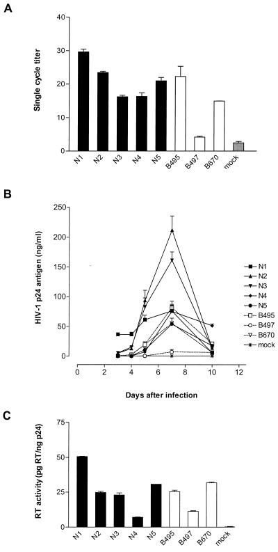
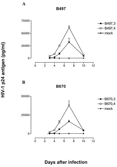
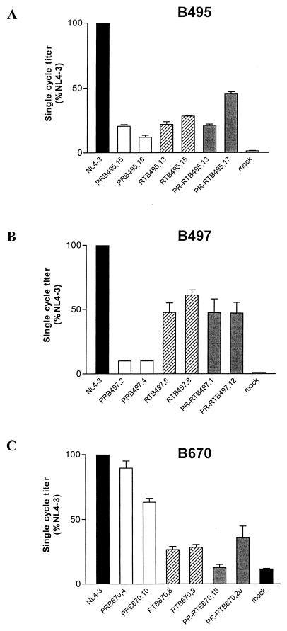
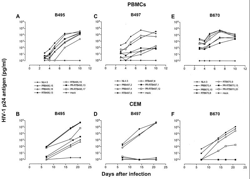
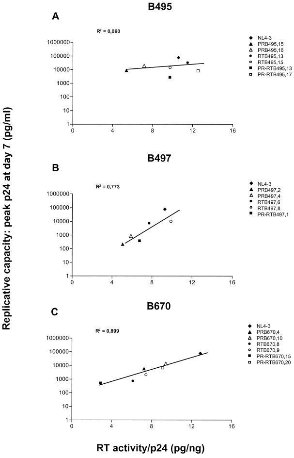
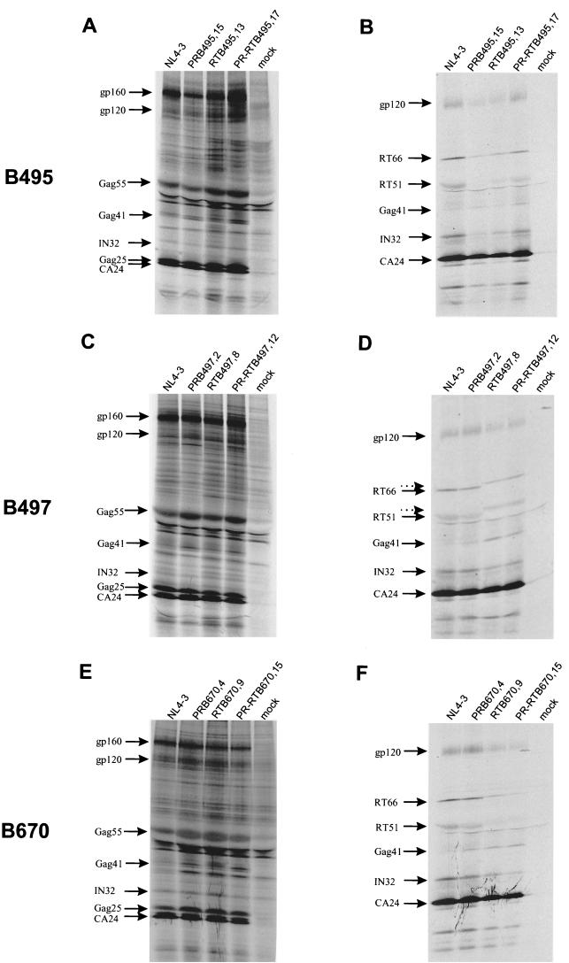
Human immunodeficiency virus type 1 (HIV-1) variants resistant to protease (PR) and reverse transcriptase (RT) inhibitors may display impaired infectivity and replication capacity. The individual contributions of mutated HIV-1 PR and RT to infectivity, replication, RT activity, and protein maturation (herein referred to as "fitness") in recombinant viruses were investigated by separately cloning PR, RT, and PR-RT cassettes from drug-resistant mutant viral isolates into the wild-type NL4-3 background. Both mutant PR and RT contributed to measurable deficits in fitness of viral constructs. In peripheral blood mononuclear cells, replication rates (means +/- standard deviations) of RT recombinants were 72.5% +/- 27.3% and replication rates of PR recombinants were 60.5% +/- 33.6% of the rates of NL4-3. PR mutant deficits were enhanced in CEM T cells, with relative replication rates of PR recombinants decreasing to 15.8% +/- 23.5% of NL4-3 replication rates. Cloning of the cognate RT improved fitness of some PR mutant clones. For a multidrug-resistant virus transmitted through sexual contact, RT constructs displayed a marked infectivity and replication deficit and diminished packaging of Pol proteins (RT content in virions diminished by 56.3% +/- 10.7%, and integrase content diminished by 23.3% +/- 18.4%), a novel mechanism for a decreased-fitness phenotype. Despite the identified impairment of recombinant clones, fitness of two of the three drug-resistant isolates was comparable to that of wild-type, susceptible viruses, suggestive of extensive compensation by genomic regions away from PR and RT. Only limited reversion of mutated positions to wild-type amino acids was observed for the native isolates over 100 viral replication cycles in the absence of drug selective pressure. These data underscore the complex relationship between PR and RT adaptive changes and viral evolution in antiretroviral drug-resistant HIV-1.


Figures

FIG. 1
FIG. 1
Infectivity, replication, and RT activity of clinical isolates. (A) Single-cycle infectivity of wild-type (N1 to N5) and mutant (B495, B497, and B670) viral isolates. The single-cycle titer was determined by FACS analysis of GHOST/CCR5 cell fluorescence as the percent GFP-positive cells. Error bars indicate the ranges of values obtained from triplicate data points. (B) Replication kinetics of wild-type (solid lines) and mutant (dotted lines) viral isolates. PBMCs were infected with p24-normalized amounts of particles, and virus production was monitored by measuring p24 antigen concentration in the culture supernatant. The data are representative of three independent experiments in which comparable results were obtained. Error bars reflect triplicate data points from a single experiment. (C) RT activity of wild-type and mutant viral isolates. Values were normalized to p24 antigen concentration in supernatant. Error bars represent the ranges of values obtained in two independent assays. RT activity was determined in PBMC culture supernatants at the peak of replication.
FIG. 2
FIG. 2
Replication kinetics of B497 and B670 in PBMCs before and after reversion of RT codons 65 from arginine (B497,3) to wild-type lysine (B497,4) and 184 from valine (B670,3) to wild-type methionine (B670,4). PBMCs were infected with p24-normalized amounts of particles, and virus production was monitored by measuring p24 antigen concentration in the culture supernatant. The data are representative of two independent experiments in which comparable results were obtained. Error bars reflect triplicate data points from a single experiment.
FIG. 3
FIG. 3
Single-cycle infectivity of recombinant clones of B495, B497, and B670. The single-cycle titer of each PR (white bars)-, RT (hatched bars)-, and PR-RT (grey bars)-mutated virus was determined by FACS analysis of GHOST/CXCR4 cell fluorescence and expressed as a percentage of the wild-type NL4-3 value (black bars). Error bars indicate the ranges of values obtained from triplicate data points in a single experiment.
FIG. 4
FIG. 4
Replication kinetics of recombinant clones of B495, B497, and B670. PBMCs and CEM cells were infected with p24-normalized amounts of viral particles. Virus production was monitored by measuring p24 antigen concentration in the culture supernatant. The data are from one of two independent experiments in which comparable results were obtained.
FIG. 5
FIG. 5
RT activity of recombinant clones of B495, B497, and B670. RT activity was determined in PBMC culture supernatants at the peak of replication and normalized to p24 antigen concentration. Regression lines show the correlation of viral RT activity with replicative capacity. PR-RTB497,12 is not represented due to limited replication and RT activity outside the assay range. The data are from one of two independent experiments in which comparable results were obtained.
FIG. 6
FIG. 6
Protein processing and maturation profiles in cell lysates (A, C, and E) and supernatant virions (B, D, and F) of recombinant clones of B495, B497, and B670. COS-7 cells were transfected with recombinant clones and metabolically labeled with [35S]methionine-[35S]cysteine. Proteins in cell-associated particles and virions were immunoprecipitated with anti-HIV human immunoglobulin G and analyzed by SDS-PAGE and fluorography. Precursor and processed viral proteins are indicated. Dotted arrows (D) indicate upward shifts in migration of RT66/RT51.

References

    1. Ansari-Lari A M, Donehower L A, Gibbs R A. Analysis of human immunodeficiency virus type 1 integrase mutants. Virology. 1995;211:332–335. - PubMed
    1. Back N K T, Berkhout B. Limiting deoxynucleoside triphosphate concentrations emphasize the processivity defect of lamivudine-resistant variants of human immunodeficiency virus type 1 reverse transcriptase. Antimicrob Agents Chemother. 1997;41:2484–2491. - PMC - PubMed
    1. Back N K T, Nijhuis M, Keulen W, Boucher C A B, Oude Essink B B, van Kuilenburg A B P, van Gennip A H, Berkhout B. Reduced replication of 3TC-resistant HIV-1 variants in primary cells due to a processivity defect of the reverse transcriptase enzyme. EMBO J. 1996;15:4040–4049. - PMC - PubMed
    1. Bahnson A. Centrifugal enhancement of retroviral mediated gene transfer. J Virol Methods. 1995;54:131–134. - PubMed
    1. Bally F, Martinez R, Peters S, Sudre P, Telenti A. Polymorphism of HIV-1 Gag p7/p1 and p1/p6 cleavage sites. Clinical significance and implications for resistance to protease inhibitors. AIDS Res Hum Retrovir. 2000;16:1209–1213. - PubMed

Publication types