. 2001 Apr;75(7):3352-62.
doi: 10.1128/JVI.75.7.3352-3362.2001.

Mitochondrial aconitase binds to the 3' untranslated region of the mouse hepatitis virus genome

Affiliations

Mitochondrial aconitase binds to the 3' untranslated region of the mouse hepatitis virus genome

S K Nanda et al. J Virol. 2001 Apr.

Abstract

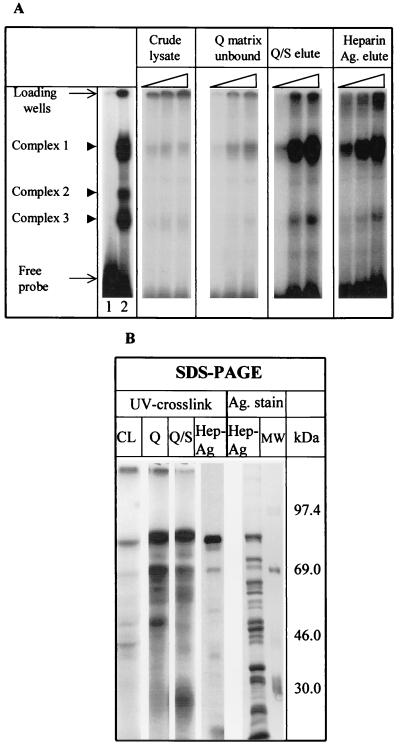
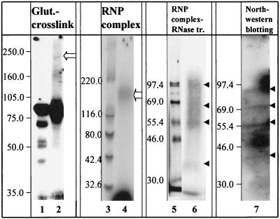
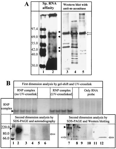
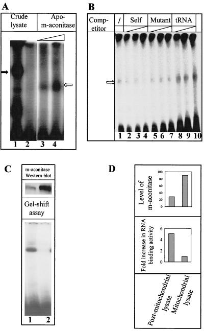
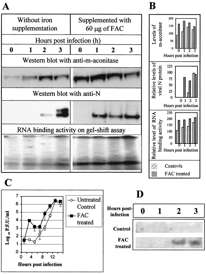
Mouse hepatitis virus (MHV), a member of the Coronaviridae, contains a polyadenylated positive-sense single-stranded genomic RNA which is 31 kb long. MHV replication and transcription take place via the synthesis of negative-strand RNA intermediates from a positive-strand genomic template. A cis-acting element previously identified in the 3' untranslated region binds to trans-acting host factors from mouse fibroblasts and forms at least three RNA-protein complexes. The largest RNA-protein complex formed by the cis-acting element and the lysate from uninfected mouse fibroblasts has a molecular weight of about 200 kDa. The complex observed in gel shift assays has been resolved by second-dimension sodium dodecyl sulfate-polyacrylamide gel electrophoresis into four proteins of approximately 90, 70, 58, and 40 kDa after RNase treatment. Specific RNA affinity chromatography also has revealed the presence of a 90-kDa protein associated with RNA containing the cis-acting element bound to magnetic beads. The 90-kDa protein has been purified from uninfected mouse fibroblast crude lysates. Protein microsequencing identified the 90-kDa protein as mitochondrial aconitase. Antibody raised against purified mitochondrial aconitase recognizes the RNA-protein complex and the 90-kDa protein, which can be released from the complex by RNase digestion. Furthermore, UV cross-linking studies indicate that highly purified mitochondrial aconitase binds specifically to the MHV 3' protein-binding element. Increasing the intracellular level of mitochondrial aconitase by iron supplementation resulted in increased RNA-binding activity in cell extracts and increased virus production as well as viral protein synthesis at early hours of infection. These results are particularly interesting in terms of identification of an RNA target for mitochondrial aconitase, which has a cytoplasmic homolog, cytoplasmic aconitase, also known as iron regulatory protein 1, a well-recognized RNA-binding protein. The binding properties of mitochondrial aconitase and the functional relevance of RNA binding appear to parallel those of cytoplasmic aconitase.


Figures

FIG. 1
FIG. 1
Purification of proteins recognizing the MHV 3′(+)42 protein-binding element. (A) Increasing amounts of 17Cl-1 cytoplasmic extracts (0.5, 1.5, and 2.5 μg) or partially purified fractions were assayed for their ability to bind to a 32P-labeled RNA (nt 16 to 84) by RNase protection-gel mobility shift assays. RNA-protein complexes were resolved by nondenaturing PAGE. The far left panel represents an assay which has been overexposed to clearly demonstrate complexes 2 and 3. Lane 1 represents free probe assayed in the absence of cytoplasmic lysate. Lane 2 represents the probe after incubation with cellular lysate. (B) 32P-labeled RNA (nt 16 to 84) was incubated with 5 μg of either uninfected 17Cl-1 extract (CL), the fraction which failed to bind to High Q matrix (Q), the Q/S tandem eluate (Q/S), or proteins purified through the heparin-agarose step (Hep-Ag), and the RNA-protein complexes formed were irradiated with UV light for 30 min. The samples were digested with RNase A (20 μg/μl) and directly analyzed by SDS-PAGE (10% gel) and autoradiography. A silver stain of the heparin-agarose-eluted material and molecular size markers is also shown (Ag. stain). The sizes of the markers are indicated on the right-hand side (in kilodaltons).
FIG. 2
FIG. 2
Analysis of RNA-protein complex 1. RNA-protein complexes were UV cross-linked in solution and then either treated with glutaraldehyde as described in Materials and Methods (lane 2) or untreated (lane 1). The position of a complex of about 200 kDa is marked by an open arrow. In situ UV cross-linking of RNA-protein (RNP) complexes was performed after native gel electrophoresis as described in Materials and Methods. Complex 1 was then eluted from the native gel and either treated with RNase (lane 6) or untreated (lane 4). The samples were then subjected to SDS-PAGE and autoradiography in parallel with 14C-labeled molecular size markers (lanes 3 and 5). The open arrow in lane 4 indicates the position of a band of approximately 200 kDa. The solid arrowheads in lane 6 indicate the positions of four proteins of approximately 90, 70, 58, and 40 kDa. Lane 7 contains an autoradiograph of a Northwestern blot of 17Cl-1 cytoplasmic extract (10 μg) probed with 32P-labeled MHV 3′(+)42 RNA (2 × 10⁶ cpm). The positions of four proteins of approximately 90, 70, 58, and 40 kDa are indicated by solid arrowheads.
FIG. 3
FIG. 3
Western blot analyses with anti-m-aconitase antibody. (A) Biotinylated RNAs representing nt 42 to 5 at the 3′ end of the MHV genome were immobilized on magnetic beads. Heparin-agarose eluates were incubated with the beads, and the RNA-binding proteins were eluted as described in Materials and Methods, subjected to SDS-PAGE, and stained with the fluorescent dye Sypro-Orange (lane 2) in parallel to molecular size markers (lane 1). The image shown in lanes 1 and 2 is a negative of the image obtained after UV illumination of the gel. The positions of proteins of 90, 70, 50, and 40 kDa are marked by solid arrowheads. We believe that the 50-kDa band corresponds to the approximately 58-kDa band observed in Fig. 1, lane 4, and Fig. 2, lanes 6 and 7. The portion of the gel containing affinity-purified protein (lane 3), crude cytoplasmic lysate (lane 4), and purified m-aconitase (lane 5) was transferred to nitrocellulose and probed with an anti-m-aconitase antibody. The position of m-aconitase is marked with an open arrow. (B) 32P-radiolabeled 3′(+)42 MHV RNA was incubated with a heparin-agarose-eluted protein preparation and analyzed on a 6% nondenaturing polyacrylamide gel. A portion of the gel containing the majority but not all of the lanes was exposed to UV light, and RNA-protein (RNP) complex 1 was visualized by autoradiography (upper panel). Complex 1 was excised from the gel, and the extracted protein (as described in Materials and Methods) was subjected to electrophoresis in an SDS–10% polyacrylamide gel with and without prior RNase treatment. One set of lanes (lanes 1 to 6) was autoradiographed. Lanes 7 to 12 were transferred to nitrocellulose and probed with anti-m-aconitase antibodies. Molecular size markers are shown in lane 1. Lanes 2 and 7 contain complex 1 which was UV cross linked but not treated with RNase. Lanes 3 and 8 represent complex 1 which was UV cross linked and treated with RNase prior to SDS-PAGE. Lanes 4 and 9 represent complex 1 which was not UV cross-linked but was treated with RNase prior to SDS-PAGE. Lanes 5 and 10 represent samples excised from the region of the nondenaturing gel corresponding to complex 1 from binding reactions containing only labeled RNA in the absence of protein. Lanes 6 and 11 represent extracts from a region of the nondenaturing gel containing no RNA-protein complex. Lane 12 represents purified m-aconitase. The position of the 200-kDa complex is indicated by solid arrowheads; the position of m-aconitase is indicated by the open arrows.
FIG. 4
FIG. 4
Interaction of purified apo-m-aconitase with MHV 3′(+)42 RNA is specific. (A) Purified apo-m-aconitase (7 and 14 μg in lanes 3 and 4, respectively) and cytoplasmic lysates (lanes 1 and 2) were incubated with 5 ng of 32P-labeled MHV 3′(+)42 RNA for 20 min at 22°C, exposed to UV light for 30 min, digested with RNase, and analyzed on a nondenaturing polyacrylamide gel. The positions of complex 1 and complex 2 are indicated by solid and open arrows, respectively. (B) Various amounts of unlabeled RNA competitors were added along with 32P-labeled MHV 3′(+)42 RNA prior to the addition of purified apo-m-aconitase. The RNA-protein complexes formed were then analyzed by gel mobility shift assays after UV cross-linking. Reactions displayed in lane 1 contained no competitor; lanes 2 to 4 contained 50, 125, and 250 ng, respectively, of unlabeled specific competitor RNA; lanes 5 to 7 contained 50, 125, and 250 ng, respectively, of mutant (mG1) competitor RNA; lanes 8 to 10 contained increasing amounts (50, 125, and 250 ng, respectively) of unlabeled yeast tRNA. (C) Cytoplasmic extracts of 17Cl-1 cells were separated into mitochondrial (lane 2) and postmitochondrial (lane 1) fractions. Equal amounts of protein from each fraction were used in either RNase protection-gel shift assays or resolved by SDS-PAGE and blotted onto nitrocellulose membranes. (D) Quantitation of the Western blot and RNA-binding assays was performed as described in Materials and Methods.
FIG. 5
FIG. 5
Confocal immunofluorescence of MHV-infected 17Cl-1. At 3 h p.i., MHV-infected 17Cl-1 cells were fixed, labeled with an anti-N monoclonal antibody (green) and with either an anti-Hel (red) polyclonal rabbit antiserum (A) or with rabbit anti-m-aconitase (red) antibody (B). Images were captured by dual laser beam scanning. Colocalization of green (oregon green) and red (rhodamine red X) signals in a single pixel produces yellow, while separated signals remain green or red.
FIG. 6
FIG. 6
Modulation of m-aconitase synthesis and viral replication by iron supplementation. 17Cl-1 cells were either untreated or supplemented with FAC (60 μg/ml) and infected with MHV at an MOI of 1 as described in Materials and Methods. At various times p.i., the cells were harvested for analysis. (A) Equal amounts of protein were electrophoresed, blotted to a nitrocellulose membrane, and probed with either anti-m-aconitase polyclonal rabbit serum or an anti-N monoclonal antibody. RNA-binding activity was assessed by RNase protection-gel mobility shift assay. (B) Quantitation of the Western blot and RNA-binding assays was performed as described in Materials and Methods. (C) Replicate cultures of 17Cl-1 cells were either treated with FAC (60 μg/ml) or untreated and infected with MHV-JHM at an MOI of 0.1. Cultures were harvested at various times p.i., and virus was counted by plaque assay. The data are plotted as virus titer, and each point represents the mean of three experiments, with each experiment containing duplicate or triplicate samples for each time point. Standard errors are indicated by the error bars. (D) Replicate cultures of 17Cl-1 cells were either treated with FAC (60 μg/ml) or untreated and infected with MHV-JHM at an MOI of 0.1. At various times p.i., RNA was extracted and analyzed by Northern blot hybridzation as described in Materials and Methods. The region of the gel containing RNA 7 is shown.

Similar articles

Cited by

References

    1. Beatch M D, Hobman T C. Rubella virus capsid associates with host cell protein p32 and localizes to mitochondria. J Virol. 2000;74:5569–5576. - PMC - PubMed
    1. Beinert H, Kennedy M C. Aconitase, a two-faced protein: enzyme and iron regulatory factor. FASEB J. 1993;7:1442–1449. - PubMed
    1. Berndt P, Hobohm U, Langen H. Reliable automatic protein identification from matrix-assisted laser desorption/ionization mass spectrometric peptide fingerprints. Electrophoresis. 1999;20:3521–3526. - PubMed
    1. Burd C G, Dreyfuss G. Conserved structures and diversity of functions of RNA-binding proteins. Science. 1994;265:615–621. - PubMed
    1. Cavanagh D. Nidovirales: a new order comprising Coronaviridae and Arteriviridae. Arch Virol. 1997;142:629–633. - PubMed

Publication types

MeSH terms