

Single amino acid substitution in the V protein of simian virus 5 differentiates its ability to block interferon signaling in human and murine cells
- PMID: 11238862
- PMCID: PMC114129
- DOI: 10.1128/JVI.75.7.3363-3370.2001
Single amino acid substitution in the V protein of simian virus 5 differentiates its ability to block interferon signaling in human and murine cells
Abstract
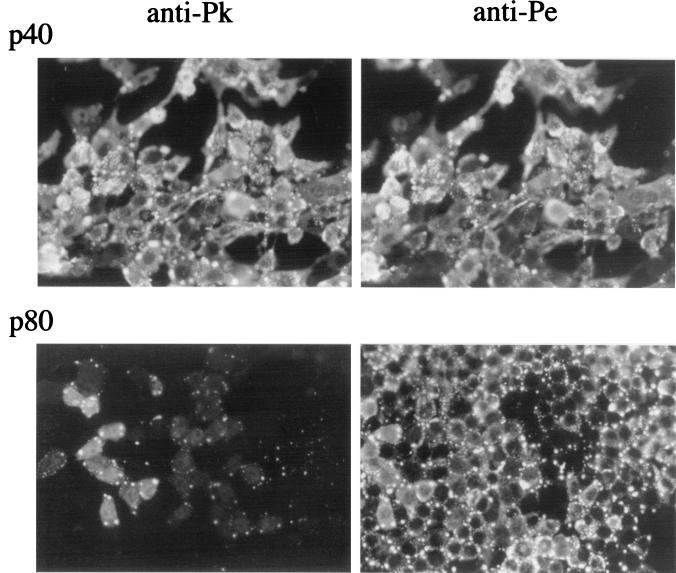
Previous work has demonstrated that the V protein of simian virus 5 (SV5) targets STAT1 for proteasome-mediated degradation (thereby blocking interferon [IFN] signaling) in human but not in murine cells. In murine BF cells, SV5 establishes a low-grade persistent infection in which the virus fluxes between active and repressed states in response to local production of IFN. Upon passage of persistently infected BF cells, virus mutants were selected that were better able to replicate in murine cells than the parental W3 strain of SV5 (wild type [wt]). Viruses with mutations in the Pk region of the N-terminal domain of the V protein came to predominate the population of viruses carried in the persistently infected cell cultures. One of these mutant viruses, termed SV5 mci-2, was isolated. Sequence analysis of the V/P gene of SV5 mci-2 revealed two nucleotide differences compared to wt SV5, only one of which resulted in an amino acid substitution (asparagine [N], residue 100, to aspartic acid [D]) in V. Unlike the protein of wt SV5, the V protein of SV5 mci-2 blocked IFN signaling in murine cells. Since the SV5 mci-2 virus had additional mutations in genes other than the V/P gene, a recombinant virus (termed rSV5-V/P N(100)D) was constructed that contained this substitution alone within the wt SV5 backbone to evaluate what effect the asparagine-to-aspartic-acid substitution in V had on the virus phenotype. In contrast to wt SV5, rSV5-V/P N(100)D blocked IFN signaling in murine cells. Furthermore, rSV5-V/P N(100)D virus protein synthesis in BF cells continued for significantly longer periods than that for wt SV5. However, even in cells infected with rSV5-V/P N(100)D, there was a late, but significant, inhibition in virus protein synthesis. Nevertheless, there was an increase in virus yield from BF cells infected with rSV5-V/P N(100)D compared to wt SV5, demonstrating a clear selective advantage to SV5 in being able to block IFN signaling in these cells.
Figures
Similar articles
-
Naturally occurring substitutions in the P/V gene convert the noncytopathic paramyxovirus simian virus 5 into a virus that induces alpha/beta interferon synthesis and cell death.J Virol. 2002 Oct;76(20):10109-21. doi: 10.1128/jvi.76.20.10109-10121.2002. J Virol. 2002. PMID: 12239285 Free PMC article.
-
Recovery of paramyxovirus simian virus 5 with a V protein lacking the conserved cysteine-rich domain: the multifunctional V protein blocks both interferon-beta induction and interferon signaling.Virology. 2002 Nov 10;303(1):15-32. doi: 10.1006/viro.2002.1738. Virology. 2002. PMID: 12482655
-
A simian virus 5 (SV5) P/V mutant is less cytopathic than wild-type SV5 in human dendritic cells and is a more effective activator of dendritic cell maturation and function.J Virol. 2006 Apr;80(7):3416-27. doi: 10.1128/JVI.80.7.3416-3427.2006. J Virol. 2006. PMID: 16537609 Free PMC article.
-
Growth sensitivity of a recombinant simian virus 5 P/V mutant to type I interferon differs between tumor cell lines and normal primary cells.Virology. 2005 Apr 25;335(1):131-44. doi: 10.1016/j.virol.2005.02.004. Virology. 2005. PMID: 15823612
-
Paramyxovirus accessory proteins as interferon antagonists.Microbiol Immunol. 2001;45(12):787-800. doi: 10.1111/j.1348-0421.2001.tb01315.x. Microbiol Immunol. 2001. PMID: 11838896 Review.
Cited by
-
Paramyxovirus disruption of interferon signal transduction: STATus report.J Interferon Cytokine Res. 2009 Sep;29(9):531-7. doi: 10.1089/jir.2009.0070. J Interferon Cytokine Res. 2009. PMID: 19694544 Free PMC article. Review.
-
The STAT2 activation process is a crucial target of Sendai virus C protein for the blockade of alpha interferon signaling.J Virol. 2003 Mar;77(6):3360-70. doi: 10.1128/jvi.77.6.3360-3370.2003. J Virol. 2003. PMID: 12610111 Free PMC article.
-
Paramyxovirus activation and inhibition of innate immune responses.J Mol Biol. 2013 Dec 13;425(24):4872-92. doi: 10.1016/j.jmb.2013.09.015. Epub 2013 Sep 20. J Mol Biol. 2013. PMID: 24056173 Free PMC article. Review.
-
A single amino acid substitution in the V protein of Nipah virus alters its ability to block interferon signalling in cells from different species.J Gen Virol. 2006 Dec;87(Pt 12):3649-3653. doi: 10.1099/vir.0.82261-0. J Gen Virol. 2006. PMID: 17098981 Free PMC article.
-
Identification of paramyxovirus V protein residues essential for STAT protein degradation and promotion of virus replication.J Virol. 2005 Jul;79(13):8591-601. doi: 10.1128/JVI.79.13.8591-8601.2005. J Virol. 2005. PMID: 15956600 Free PMC article.
References
-
- Choppin P W. Multiplication of a myxovirus (SV5) with minimal cytopathic effects and without interference. Virology. 1964;23:224–233. - PubMed
-
- Dunn C, O'Dowd A M, Randall R E. Fine mapping of the binding sites of monoclonal antibodies raised against the Pk tag. J Immunol Methods. 1999;224:141–150. - PubMed
-
- Fearns R, Young D, Randall R E. The paramyxovirus, simian virus 5, can remain inactive in cytoplasmic inclusion bodies in persistent infections. J Gen Virol. 1994;75:3525–3539. - PubMed
Publication types
MeSH terms
Substances
Grants and funding
LinkOut - more resources
Full Text Sources
Research Materials
Miscellaneous
