. 2001 Apr;75(7):3371-82.
doi: 10.1128/JVI.75.7.3371-3382.2001.

Construction and molecular analysis of gene transfer systems derived from bovine immunodeficiency virus

Affiliations

Construction and molecular analysis of gene transfer systems derived from bovine immunodeficiency virus

R Berkowitz et al. J Virol. 2001 Apr.

Abstract

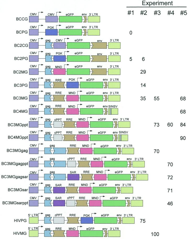
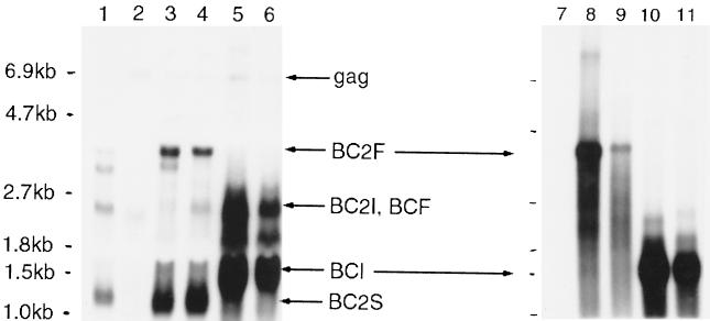
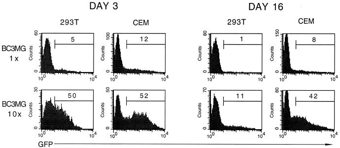
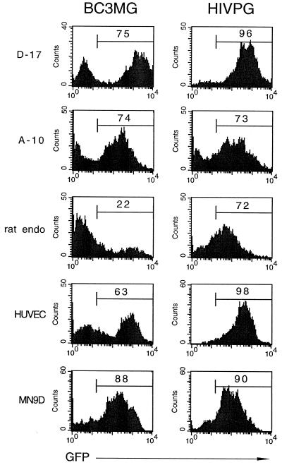
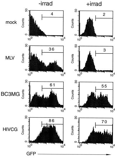
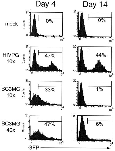
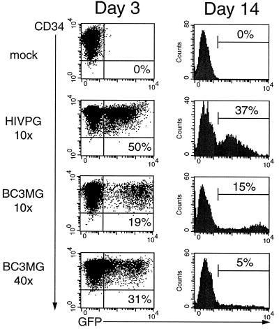
Because lentiviruses are able to infect nondividing cells, these viruses might be utilized in gene therapy applications where the target cell does not divide. However, it has been suggested that the introduction of primate lentivirus sequences, particularly those of human immunodeficiency virus, into human cells may pose a health risk for the patient. To avoid this concern, we have constructed gene transfer systems based on a nonprimate lentivirus, bovine immunodeficiency virus. A panel of vectors and packaging constructs was generated and analyzed in a transient expression system for virion production and maturation, vector expression and encapsidation, and envelope protein pseudotyping. Virion preparations were also analyzed for transduction efficiency in a panel of human and nonhuman primary cells and immortalized cell lines. The virion preparations transduced most of the target cell types, with efficiencies up to 90% and with titers of unconcentrated virus up to 5 x 10(5) infectious doses/ml. In addition, infection of nondividing human cells, including unstimulated hematopoietic stem cells and irradiated endothelial cells, was observed.


Figures

FIG. 1
FIG. 1
(A) Wild-type BIV genome. Viral coding regions are arranged in three horizontal lines representing the different reading frames. The rev and tat genes are composed of two coding regions each. The first coding region of rev and the tmx coding region are in the same reading frame as env. The approximate position of the insertion of CMV-eGFP (in construct BCG) is indicated above the top line. (B) BIV can transduce human cells. VSV-G-pseudotyped construct BCG was produced in 293T cells and exposed to EREp (right column) and fresh 293T (left column) cells; 3 days later, the cells were analyzed for eGFP expression by flow cytometry. The percentage of eGFP+ cells is indicated inside each histogram.
FIG. 1
FIG. 1
(A) Wild-type BIV genome. Viral coding regions are arranged in three horizontal lines representing the different reading frames. The rev and tat genes are composed of two coding regions each. The first coding region of rev and the tmx coding region are in the same reading frame as env. The approximate position of the insertion of CMV-eGFP (in construct BCG) is indicated above the top line. (B) BIV can transduce human cells. VSV-G-pseudotyped construct BCG was produced in 293T cells and exposed to EREp (right column) and fresh 293T (left column) cells; 3 days later, the cells were analyzed for eGFP expression by flow cytometry. The percentage of eGFP+ cells is indicated inside each histogram.
FIG. 2
FIG. 2
BIV packaging constructs and expression in transfected 293T cells. (A) wild-type BIV, CMV-driven BIV, and three packaging constructs (BH1 to -3) are depicted for their gag, pol, and env genes; accessory genes are not shown. Also depicted are the viral major splice donor (MSD), a small chimeric intron (in), the HIV-1 RRE, the puromycin N-acetyltransferase cDNA linked to an IRES from encephalomyocarditis virus (IRES-puro), and the SV40 late polyadenylation signal (SV40 polyA). (B) Northern analysis of the constructs' steady-state cytoplasmic expression levels. Cytoplasmic RNA from transfected 293T cells was probed for gag sequences; the high-molecular weight band in each lane corresponds to the construct's gag mRNA.
FIG. 3
FIG. 3
BIV produces levels of virus similar to HIV-1. BIV packaging construct BH2 and an HIV-1 packaging construct were each modified by insertion of HA tags at the C terminus of Gag and deletion of pol (see text). Parental and modified viruses were produced in 293T cells, collected and subjected to anti-HIV-1 Gag (lanes 1 and 2), anti-BIV Gag (lanes 3 and 4), or anti-HA (lanes 5 and 6) Western blotting. Lane 1, parental HIV-1 packaging construct. Most of the Gag precursor polyprotein (PrGag) has been cleaved. Lane 2, modified HIV-1 packaging construct. The amount of uncleaved Gag polyprotein is similar to the amount of capsid protein (CA) in the parental virus preparation, indicating that the modifications did not alter virus production substantially. Lane 3, parental BIV packaging construct. Lane 4, modified BIV packaging construct. As with the case of the HIV-1 packaging construct, the modification did not alter virus production. Lane 5, modified HIV-1 packaging construct. Lane 6, modified BIV packaging construct, after normalization by RT assay of the parental constructs. The amount of Gag precursor polyprotein is similar to the amount in lane 5, indicating that the RT assay has comparable sensitivity for BIV and HIV-1 virus and hence, that the packaging constructs produce similar levels of virus.
FIG. 4
FIG. 4
BIV vectors and transduction efficiencies. All vectors contain the CMV immediate-early promoter, ending in the TATA box, linked to the BIV 5′ LTR starting immediately after the TATA box. BIV sequences terminate at the gag start codon (BCCG and BCPG) or approximately 510bp into the gag coding region (all others). All vectors contain the eGFP cDNA linked to a heterologous, “internal” promoter: CMV, PGK, or MND (see text). Some vectors contain one or more insertions between the BIV 5′ segment and the internal promoter: these insertions include a potential BIV cPPT, the 3′ segment of the gag gene, the beta interferon SAR, and the putative BIV RRE. Downstream of the eGFP cDNA lies the BIV 3′ LTR and approximately 130 bp (BCCG and BCPG), 1.2 kb (BC2CG, BC2PG, and BC2MG) or 80 bp (all others) of adjacent env sequences. Two vectors (BC4MG and BC4MGppt) contain modified 3′ LTRs in which most of the 3′ LTR U3 region has been replaced by the SV40 late polyadenylation signal enhancer element (SINSV). Transcription start sites and directions are indicated with arrows. At the bottom are depicted two HIV-1 control vectors used in this study. At the right are the transduction efficiencies of the vectors in 293T cells 3 days postinfection, using packaging construct BH2 and VSV-G, in a series of experiments. Infections from experiments 3 to 5 were performed in the presence of an additional buffer to retard pH elevation during spinoculation. As a result, transduction efficiencies were elevated.
FIG. 5
FIG. 5
Northern analysis of BIV RNA in transfected 293T cells (left) and in virus particles shed from the transfected cells (right). Cytoplasmic RNA was probed with BIV sequences at the 3′ end of the genome, to detect all packaging construct and vector RNAs. Viral RNA was probed with eGFP sequences, to detect full-length vector RNA and RNA initiating at the internal promoter. Lane 1, vector BC2PG; lanes 2 and 7, packaging construct BH2; lanes 3 and 8, BH2 and BC2PG; lanes 4 and 9, BH2 and BC2CG; lanes 5 and 10, BH2 and BCPG; lanes 6 and 11, BH2 and BCCG. The positions of certain RNAs are indicated: gag, the gag mRNA encoded by BH2; BC2F, full-length BC2PG and BC2CG vector mRNA; BC2I, internally initiated BC2PG and BC2CG vector mRNA; BCF, full-length BCPG and BCCG vector RNA; BCI, internally initiated BCPG and BCCG vector mRNA; BC2S, spliced BC2PG and BC2CG vector mRNA. Note that this last RNA is only present in the BC2 vectors, since the rev splice acceptor is present in the BC2 vectors but not the BC vectors.
FIG. 6
FIG. 6
Analysis of virus concentration and the duration of transgene expression in transduced human cell lines. A BIV preparation consisting of packaging construct BH2, VSV-G, and vector BC3MG was collected by centrifugation, then suspended in the original volume (1× virus) or in a 10-fold-lower volume (10× virus) and exposed to fresh 293T or CEMSS cells. eGFP expression was measured at 3 and 16 days postinfection by flow cytometry; the percentage of transduced cells is indicated in each histogram.
FIG. 7
FIG. 7
BIV can transduce a variety of cell lines and primary cells. A 1× (D-17 cells) or 10× (all others) BIV preparation consisting of packaging construct BH2, VSV-G, and vector BC3MG was exposed to the indicated cell lines or primary cells (see text for details on each cell type). Two weeks later, the cells were analyzed for eGFP expression by flow cytometry; the percentage of transduced cells is indicated in each histogram. For comparison, the HIVPG histograms indicate the cells exposed to an unconcentrated HIV-1 preparation consisting of a packaging construct, VSV-G, and a vector containing the PGK internal promoter.
FIG. 8
FIG. 8
BIV can transduce nondividing cells. A 10× BIV virus preparation consisting of packaging construct BH2, VSV-G, and vector BC3MG was exposed to HUVEC 2 days after the cells were irradiated to abrograte cell division (right column) or to nonirradiated HUVEC (left column). Two days later, the cells were analyzed for eGFP expression by flow cytometry; the percentage of transduced cells is indicated in the histogram (right column). For comparison, the HIVCG histograms indicate cells exposed to an unconcentrated HIV-1 preparation consisting of a packaging construct, VSV-G, and a vector containing the CMV internal promoter. In addition, the MLV histograms indicate cells exposed to an MLV preparation consisting of a packaging construct, VSV-G, and an LTR-driven eGFP vector.
FIG. 9
FIG. 9
BIV can transduce resting human lymphocytes. Unstimulated human PBLs were infected with 10× and 40× BIV consisting of packaging construct BH2, VSV-G, and vector BC3MG. After infection, the cells were activated and cultured for 4 days (left column) or 2 weeks (right column) in the presence of antibodies specific for CD3 and CD28, after which a portion of the cells were analyzed for eGFP expression by flow cytometry. The percentage of transduced cells is indicated in each histogram. For comparison, the cells were infected with the 10× HIV-1 PGK preparation.
FIG. 10
FIG. 10
BIV can transduce resting HSCs Unstimulated human HSCs were infected with 10x and 40x BIV consisting of packaging construct BH2, VSV-G, and vector BC3MG. After infection, the cells were activated and cultured for 3 days (left column) or 2 weeks (right column) in the presence of tpo mimetic, flt3 ligand, and c-kit ligand, after which a portion of the cells were analyzed for eGFP expression by flow cytometry. The percentage of transduced cells is indicated in each histogram. For comparison, the cells were infected with the 10× HIV-1 PGK preparation.

Similar articles

Cited by

References

    1. Adra C N, Boer P H, McBurney M W. Cloning and expression of the mouse pgk-1 gene and the nucleotide sequence of its promoter. Gene. 1987;60:65–74. - PubMed
    1. Agarwal M, Austin T W, Morel F, Chen J, Böhnlein E, Plavec I. Scaffold attachment region-mediated enhancement of retroviral vector expression in primary T cells. J Virol. 1998;72:3720–3728. - PMC - PubMed
    1. Bender M A, Palmer T D, Gelinas R E, Miller A D. Evidence that the packaging signal of Moloney murine leukemia virus extends into the gag region. J Virol. 1987;61:1639–1646. - PMC - PubMed
    1. Berkowitz R D, Fisher J, Goff S P. RNA packaging. Curr Top Microbiol Immunol. 1996;214:177–218. - PubMed
    1. Berkowitz R D, Ilves H, Plavec I, Veres G. Gene transfer systems derived from Visna virus: analysis of virus production and infectivity. Virology. 2001;279:116–129. - PubMed