. 2001 Apr;75(7):3383-90.
doi: 10.1128/JVI.75.7.3383-3390.2001.

Viral regulation of RANTES expression during human cytomegalovirus infection of endothelial cells

Affiliations

Viral regulation of RANTES expression during human cytomegalovirus infection of endothelial cells

M Billstrom Schroeder et al. J Virol. 2001 Apr.

Abstract

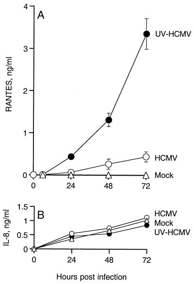
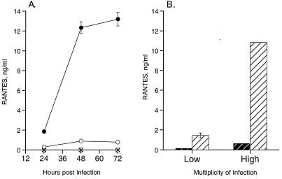
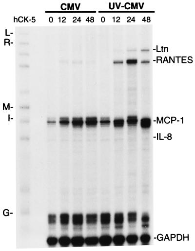
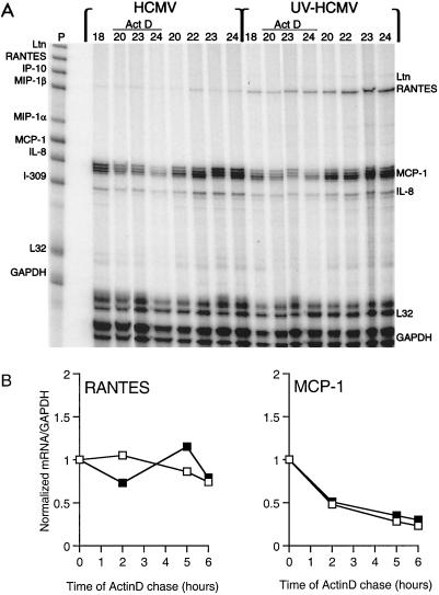
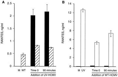
Human cytomegalovirus (HCMV) evades healthy immune responses during infection, and this evasion may allow HCMV to establish latency in the host. The human vasculature has been recognized as a site of HCMV infection and may also be a site of latent HCMV infection. As the interface between circulating cells and underlying parenchymal cells, the vascular endothelium provides signals for local reaction of inflammatory cells. We propose that HCMV down-regulates expression of the proinflammatory chemokine RANTES from the infected endothelium, which may result in reduced recruitment of mononuclear cells to the site of infection. Abortive HCMV infection of primary endothelial cells with the clinical isolate HCMV 4010, under conditions in which viral gene expression could not occur, induced high levels of RANTES expression. Replicative HCMV infection, however, induced cells in parallel cultures to express significantly lower levels of RANTES. Expression of the chemokines interleukin 8 and MCP-1 by endothelial cells was found to be unaffected by replicative HCMV infection and thus may not play an important role during early HCMV infection of the endothelium. HCMV may regulate RANTES expression from endothelial cells as a mechanism to evade the local immune response to infection.


Figures

FIG. 1
FIG. 1
Expression of HCMV IE protein in WT-HCMV-infected endothelial cells but not UV-irradiated HCMV-infected endothelial cells. Subconfluent monolayers of HUVECs were infected with either WT-HCMV (A and B) or UV-HCMV (C and D) for 96 h and then fixed and stained for HCMV IE72 protein (A and C). The bright-field images of the stained WT-HCMV-infected cells (B) and the UV-HCMV-infected cells (D) are presented to confirm cell presence.
FIG. 2
FIG. 2
Expression of chemokines during infection of HUVECs with HCMV. HUVECs were infected with 0.01 PFU of WT-HCMV per cell (○) or 0.01 PFU equivalents of HCMV irradiated by UV light per cell (●). Mock-infected HUVECs (▵) were also assayed for chemokine expression. Expression of RANTES represents cumulative amounts of secreted chemokine over 72 h. Supernatants were collected and assayed for RANTES (A) and IL-8 (B) protein. Error bars represent the standard error of the mean of three experiments.
FIG. 3
FIG. 3
RANTES expression is dependent on particle interaction with the cells. (A) HCMV particle-free inoculum does not induce RANTES expression. Subconfluent monolayers of HUVECs were infected with HCMV inoculum (○), UV-HCMV inoculum (●), and filtrate from HCMV inoculum (□) and filtrate from UV-HCMV inoculum (∗), in which inoculum was passed through a 100-kDa-cutoff membrane to remove virus particles. Supernatants of the cultures were collected at designated time points and assayed for RANTES protein. This assay represents three identical experiments in which the fold differences in RANTES expression between UV-HCMV and WT-HCMV did not differ by more than 10%. (B) RANTES expression is related to PFU equivalents of UV-HCMV. Expression of RANTES was assayed following 72 h of infection at a high multiplicity of infection (1 PFU/cell) and low multiplicity of infection (0.02 PFU/cell). Open hatched bars represent WT-HCMV, and solid hatched bars represent UV-HCMV. Error bars represent the standard error of duplicates within the experiment, and this experiment is representative of three replicative assays.
FIG. 4
FIG. 4
Time course of chemokine mRNA expression during infection of HUVECs with WT-HCMV and UV-HCMV. Total RNA was isolated from infected HUVEC cultures at designated time points (0, 12, 24, and 48 h) postinfection and assayed for specific mRNA expression by RPA with a 32P-labeled multi-RNA probe of human CC chemokines. Unhybridized probe hCK-5 (far left lane) with mRNA of lymphotactin (Ltn [L in the left margin]), RANTES (R), MCP-1 (M), IL-8 (I), and GAPDH (G) is identified. The method is described in detail in Materials and Methods.
FIG. 5
FIG. 5
Stability of mRNA following transcription in HCMV- and UV-HCMV-infected HUVECs. (A) HCMV- and UV-HCMV-infected HUVECs were incubated with actinomycin D (Act D)-supplemented medium following 18 h of infection. At specific time points prior to (18 h) and during the actinomycin D chase (20, 22, 23, and 24 h), the cells were harvested and assayed for steady-state mRNA by RPA (Materials and Methods). Specific chemokine mRNA was detected with a 32P-labeled multi-RNA probe (P), hCK-5 (Pharmingen). Ltn, lymphotactin. (B) Relative degradation of RANTES and MCP-1 mRNA during the actinomycin D (5 μg/ml) chase. The levels of mRNA of RANTES, MCP-1, and GAPDH during the actinomycin D chase depicted on the autoradiogram were quantified by phosphor screen autoradiography. Each value represents the ratio of RANTES or MCP-1 mRNA to levels of control mRNA of GAPDH. □, HCMV-infected HUVECs; ▪, UV-HCMV-infected HUVECs.
FIG. 6
FIG. 6
WT-HCMV inhibits UV-HCMV induction of RANTES expression from endothelial cells. (A) Subconfluent monolayers of HUVECs were mock infected or infected with WT-HCMV at 1.0 PFU/cell. In parallel cultures, 0.2 PFU equivalent of UV-HCMV was added either simultaneously (time zero) or following 90 min of infection with WT-HCMV, at which point the inoculum of WT-HCMV was removed from the cells prior to addition of UV-HCMV. Hatched bars represent cultures infected with WT-HCMV, and solid bars represent mock-infected cultures. (B) HUVECs were mock infected or infected with 1 PFU equivalent of UV-HCMV per cell. In parallel, cultures were infected with 0.2 PFU of WT-HCMV per cell either simultaneously (time zero) or following 90 min of treatment with UV-HCMV, at which point the UV-HCMV inoculum was removed prior to addition of WT-HCMV. Open bars represent cultures infected with UV-HCMV, and solid bars represent mock-infected cultures. Supernatants from all cultures were collected at 72 h postinfection and assayed for RANTES protein. Error bars represent standard errors of duplicates within the experiment, and this experiment is representative of three replicative assays.

References

    1. Alam R, York J, Boyars M, Stafford S, Grant J, Lee J, Forsythe P, Sim T, Ida N. Increased MCP-1, RANTES, and MIP-1α in bronchoalveolar lavage fluid of allergic asthmatic patients. Am J Respir Crit Care Med. 1996;153:1398–1404. - PubMed
    1. Alford C, Britt W. Cytomegalovirus. In: Knipe D, editor. Virology. 2nd ed. New York, N.Y: Raven Press, Ltd.; 1990. pp. 1981–2010.
    1. Arbustini E, Grasso M, Diegoli M, Percivalle E, Grossi P, Bramerio M, Campana C, Goggi C, Gavazzi A, Vigano M. Histophathologic and molecular profile of HCMV infections in patients with heart transplants. Am J Clin Pathol. 1992;98:205–213. - PubMed
    1. Becker S, Reed W, Henderson F, Noah T. RSV infection of human airway epithelial cells causes production of the β-chemokine RANTES. Am J Physiol. 1997;272:L512–L520. - PubMed
    1. Billstrom M A, Johnson G L, Avdi N J, Worthen G S. Intracellular signaling by the chemokine receptor US28 during human cytomegalovirus infection. J Virol. 1998;72:5535–5544. - PMC - PubMed

Publication types

MeSH terms