. 2001 Apr;75(7):3474-9.
doi: 10.1128/JVI.75.7.3474-3479.2001.

Oncolytic activity of vesicular stomatitis virus is effective against tumors exhibiting aberrant p53, Ras, or myc function and involves the induction of apoptosis

Affiliations

Oncolytic activity of vesicular stomatitis virus is effective against tumors exhibiting aberrant p53, Ras, or myc function and involves the induction of apoptosis

S Balachandran et al. J Virol. 2001 Apr.

Abstract

We have recently shown that vesicular stomatitis virus (VSV) exhibits potent oncolytic activity both in vitro and in vivo (S. Balachandran and G. N. Barber, IUBMB Life 50:135-138, 2000). In this study, we further demonstrated, in vivo, the efficacy of VSV antitumor action by showing that tumors that are defective in p53 function or transformed with myc or activated ras are also susceptible to viral cytolysis. The mechanism of viral oncolytic activity involved the induction of multiple caspase-dependent apoptotic pathways was effective in the absence of any significant cytotoxic T-lymphocyte response, and occurred despite normal PKR activity and eIF2alpha phosphorylation. In addition, VSV caused significant inhibition of tumor growth when administered intravenously in immunocompetent hosts. Our data indicate that VSV shows significant promise as an effective oncolytic agent against a wide variety of malignant diseases that harbor a diversity of genetic defects.


Figures

FIG. 1
FIG. 1
VSV replicates and induces apoptosis in C6 cells despite PKR activation and eIF2α phosphorylation. C6 cells were infected with VSV at an MOI of 1 and examined by immunoblotting for total VSV protein synthesis at the indicated time points p.i. using polyclonal antiserum to VSV (A) or analyzed for kinetics of viral replication by following synthesis of VSV G protein at the indicated time points (B). (C) C6 cells were infected with VSV (lane 2) for 4 h in the presence of [32P]orthophosphate and subsequently analyzed by autoradiography or immunoblotting for phosphorylated PKR, total eIF2α, and serine 51-phosphorylated eIF2α. Equivalent levels of tubulin show that approximately equal amounts of protein were loaded in each lane. (D) C6 cells were infected with VSV and subsequently treated with 100 μM concentrations of each of the caspase inhibitors zVAD.fmk (zVAD), zIETD.fmk (zIETD), zLEHD.fmk (zLEHTD), and zDEVD.fmk (zDEVD). Forty-eight hours p.i., cells were assayed for viability by trypan blue exclusion. (E) Cells treated as in panel C were examined for apoptosis using TUNEL followed by fluorescence microscopy (magnification, ×80).
FIG. 2
FIG. 2
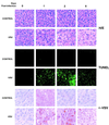
VSV induces apoptosis in C6 tumors in vivo. Athymic nu/nu mice were implanted s.c. with 2 × 10⁶ C6 cells and subsequently infected i.t. with 2 × 10⁷ PFU of VSV/dose (or an equivalent amount of HI VSV as a control) i.t. after palpable tumors had formed. One, two, and six days p.i., tumors were excised, fixed in 4% paraformaldehyde, and sectioned. Paraffin-embedded sections were then stained with hematoxylin and eosin (H/E) and photographed by bright-field microscopy (magnification, ×89) (top panels), assayed for apoptosis using TUNEL, and photographed on a fluorescence microscope (magnification, ×89) (middle panels) or stained for VSV replication using an anti-VSV polyclonal antiserum and photographed by bright-field microscopy (magnification, ×178) (bottom panels).
FIG. 3
FIG. 3
VSV inhibits growth of myc- and ras-transformed tumors in nu/nu mice, can repress tumor growth when administered distally, and inhibits growth of syngeneic tumors in immunocompetent mice. (A) BALB/3T3, BALB/3T3 Myc, and BALB/3T3 Ras cells were treated with or without 1,000 U of alpha/beta murine IFN/ml for 18 h and subsequently infected with VSV at an MOI of 10. Viability was assessed 24 h p.i. by trypan blue exclusion. (B) nu/nu mice with orthotopic s.c. tumors derived from myc-transformed BALB/3T3 cells (n = 5) were injected i.t. with 2 × 10⁷ PFU of VSV/dose. Control tumors (n = 5) received equivalent amounts of HI VSV. Tumor volumes were measured daily for a period of 2 weeks. (C) nu/nu mice with orthotopic s.c. tumors derived from ras-transformed BALB 3T3 cells (n = 5) were injected i.t. with 2 × 10⁷ PFU of VSV/dose. Control tumors (n = 5) received equivalent amounts of HI VSV. Tumor volumes were measured for a period of 10 days, at which time the tumor burden of control-infected animals became excessive. Means ± standard errors of the means (S.E.M.) are given. (D) nu/nu mice were implanted with 2 × 10⁶ C6 cells/flank s.c. into both the right and left rear flanks of each mouse. After palpable tumor formation, the right flank tumor (n = 5) was infected with VSV i.t. (2 × 10⁷ PFU/tumor) or with HI VSV, and all tumor volumes were measured for a period of 15 days. Results are given as means ± S.E.M. (E) nu/nu mice were implanted with 2 × 10⁶ C6 cells s.c. and injected after palpable tumors had formed (n = 5) i.v. with VSV in three serial doses of approximately 2.5 × 10⁷ PFU/dose every 2 days (arrows), and tumor growth was monitored daily. Control tumors (n = 5) received equivalent amounts of HI VSV. Tumor volumes were measured daily for a period of 13 days. (F) C3H mice bearing syngeneic Ag104 sarcoma-derived orthotopic s.c. tumors (n = 5) were injected with three doses of 2.5 × 10⁷ PFU of VSV/dose 3 days apart. Control tumors (n = 5) received equivalent amounts of HI VSV. Tumor volumes were measured for a period of 2 weeks. Results are given as means ± S.E.M.

References

    1. Balachandran S, Barber G N. Vesicular stomatitis virus therapy of tumors. IUBMB Life. 2000;50:135–138. - PubMed
    1. Balachandran S, Roberts P C, Brown L E, Truong H, Pattnaik A K, Archer D R, Barber G N. Essential role for the dsRNA-dependent protein kinase PKR in innate immunity to viral infection. Immunity. 2000;13:129–141. - PubMed
    1. Balachandran S, Roberts P C, Kipperman T, Bhalla K N, Compans R W, Archer D R, Barber G N. Alpha/beta interferons potentiate virus-induced apoptosis through activation of the FADD/caspase 8 death signaling pathway. J Virol. 2000;74:1513–1523. - PMC - PubMed
    1. Benedetti S, et al. Gene therapy of experimental brain tumors using neural progenitor cells. Nat Med. 2000;6:447–450. - PubMed
    1. Bischoff J R, Kirn D H, Williams A, Heise C, Horn S, Muna M, Ng L, Nye J A, Sampson-Johannes A, Fattaey A, McCormick F. An adenovirus mutant that replicates selectively in p53-deficient human tumor cells. Science. 1996;274:373–376. - PubMed

Substances