

doi: 10.1128/JVI.75.7.3480-3482.2001.
Regression of a murine gammaherpesvirus 68-positive b-cell lymphoma mediated by CD4 T lymphocytes
Affiliations
- PMID: 11238875
- PMCID: PMC114142
- DOI: 10.1128/JVI.75.7.3480-3482.2001
Item in Clipboard
Regression of a murine gammaherpesvirus 68-positive b-cell lymphoma mediated by CD4 T lymphocytes
J Virol.
2001 Apr.
Abstract
Murine gammaherpesvirus 68-infected S11 cells were injected subcutaneously into nude mice. Adoptively transferred restimulated lymphocytes consistently elicited the regression of S11 tumors. CD4 T lymphocytes were most effective in preventing tumor formation, and immunohistochemistry highlighted populations of CD4 T cells in regressing tumors.
Figures
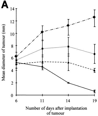
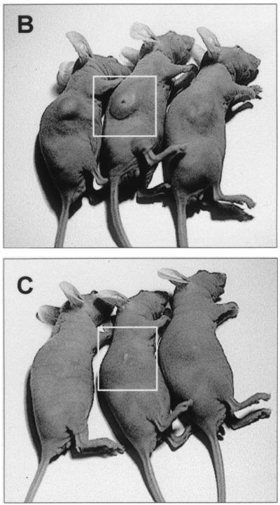
Diameter of subcutaneous S11 tumors in nude mice following the adoptive transfer of splenocyte populations. (A) Nude mice were injected intraperitoneally with 107 restimulated, immune, or normal splenocytes. All mice were then injected subcutaneously with 107 S11 cells on the right flank, and tumor development was assessed by measurement of the tumor diameter at the times indicated. Each point represents the mean diameter of four tumors. Error bars represent the standard error of the mean. ⧫, tumor only; ■, tumor plus normal splenocytes; ▴, tumor plus immune splenocytes; ×, tumor plus S11 restimulated immune splenocytes. (B) S11 tumors in control animals which received no adoptive transfer of cells. (C) A group of animals which received an adoptive transfer of CD8-depleted lymphocytes at the time of tumor implantation. Both photographs were taken at 19 days post-tumor implantation. White squares highlight the subcutaneous tumors.
Diameter of subcutaneous S11 tumors in nude mice following the adoptive transfer of splenocyte populations. (A) Nude mice were injected intraperitoneally with 107 restimulated, immune, or normal splenocytes. All mice were then injected subcutaneously with 107 S11 cells on the right flank, and tumor development was assessed by measurement of the tumor diameter at the times indicated. Each point represents the mean diameter of four tumors. Error bars represent the standard error of the mean. ⧫, tumor only; ■, tumor plus normal splenocytes; ▴, tumor plus immune splenocytes; ×, tumor plus S11 restimulated immune splenocytes. (B) S11 tumors in control animals which received no adoptive transfer of cells. (C) A group of animals which received an adoptive transfer of CD8-depleted lymphocytes at the time of tumor implantation. Both photographs were taken at 19 days post-tumor implantation. White squares highlight the subcutaneous tumors.
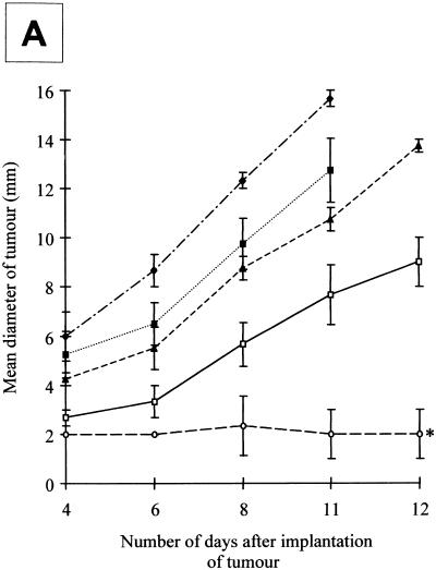
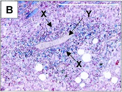
CD4+ T cells elicit S11 tumor regression in nude mice. (A) Nude mice were injected intraperitoneally with 107 splenocytes that were either complete, CD4 depleted, CD8 depleted, or CD4 and CD8 depleted. All mice were then injected subcutaneously with 107 S11 cells on the right flank, and tumor development was assessed by measurement of the tumor diameter at the times indicated. Each point represents the mean diameter of four tumors. Error bars represent the standard error of the mean. ⧫, tumor only; □, complete splenocytes; ▴, CD4-depleted splenocytes; ○, CD8-depleted splenocytes; ■, no CD4 or CD8 lymphocytes. (B) Tumor section immunostained with a rat anti-mouse CD4 monoclonal antibody. CD4+ T cell infiltration (X) was mainly observed in the vicinity of blood vessels (Y) with sporadic staining in other areas of the tumors analyzed. Original magnification, ×200. Bar, 0.2 mm. Arrows show positive staining with relevant antibody.
CD4+ T cells elicit S11 tumor regression in nude mice. (A) Nude mice were injected intraperitoneally with 107 splenocytes that were either complete, CD4 depleted, CD8 depleted, or CD4 and CD8 depleted. All mice were then injected subcutaneously with 107 S11 cells on the right flank, and tumor development was assessed by measurement of the tumor diameter at the times indicated. Each point represents the mean diameter of four tumors. Error bars represent the standard error of the mean. ⧫, tumor only; □, complete splenocytes; ▴, CD4-depleted splenocytes; ○, CD8-depleted splenocytes; ■, no CD4 or CD8 lymphocytes. (B) Tumor section immunostained with a rat anti-mouse CD4 monoclonal antibody. CD4+ T cell infiltration (X) was mainly observed in the vicinity of blood vessels (Y) with sporadic staining in other areas of the tumors analyzed. Original magnification, ×200. Bar, 0.2 mm. Arrows show positive staining with relevant antibody.
References
-
- Biberfeld P, Ensoli B, Sturzl M, Schulz T F. Kaposi sarcoma-associated herpesvirus human herpesvirus 8, cytokines, growth factors and HIV in pathogenesis of Kaposi's sarcoma. Curr Opin Infect Dis. 1998;11:97–105. - PubMed
-
- Brooks L A, Wilson A J, Crook T. Kaposi's sarcoma-associated herpesvirus (KSHV) human herpesvirus 8 (HHV8)—a new human tumour virus. J Pathol. 1997;182:262–265. - PubMed
-
- Greenblatt R M. Kaposi's sarcoma and human herpesvirus-8. Infect Dis Clin N Am. 1998;12:63. - PubMed
-
- Lacerda J F, Ladanyi M, Louie D C, Fernandez J M, Papadopoulos E B, O'Reilly R J. Human Epstein-Barr virus (EBV)-specific cytotoxic T lymphocytes home preferentially to and induce selective regressions of autologous EBV-induced B cell lymphoproliferations in xenografted C.B-17 Scid/Scid mice. J Exp Med. 1996;183:1215–1228. - PMC - PubMed
Publication types
MeSH terms
Substances
LinkOut - more resources
Full Text Sources
Research Materials
