. 2001 Apr;75(7):3483-7.
doi: 10.1128/JVI.75.7.3483-3487.2001.

JC virus-specific cytotoxic T lymphocytes in individuals with progressive multifocal leukoencephalopathy

Affiliations

JC virus-specific cytotoxic T lymphocytes in individuals with progressive multifocal leukoencephalopathy

I J Koralnik et al. J Virol. 2001 Apr.

Abstract

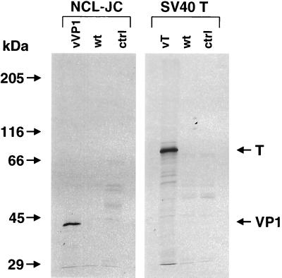
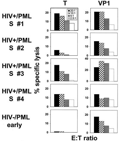
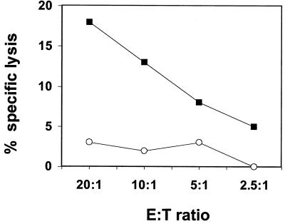
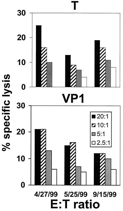
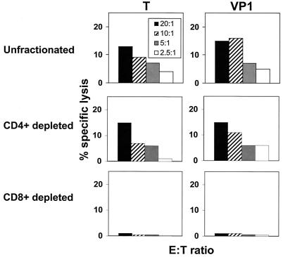
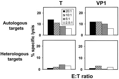
Progressive multifocal leukoencephalopathy (PML) is a demyelinating disease of the central nervous system caused by a reactivation of the polyomavirus JC (JCV) within a setting of immunosuppression. The nature of the immune response that contains replication of this virus is unknown. We have explored JCV-specific cellular immune responses in patients with PML and control subjects. JCV antigen-stimulated peripheral blood mononuclear cells (PBMC) of four human immunodeficiency virus (HIV)-infected patients who were survivors of PML and one HIV-uninfected patient recently diagnosed with PML lysed autologous B-lymphoblastoid cell lines expressing either the JCV T regulatory protein or the VP1 major capsid protein. This lysis was mediated by CD8(+) T lymphocytes and was major histocompatibility complex class I restricted. These cells were therefore cytotoxic T lymphocytes (CTL). JCV-specific CTL could not be detected in PBMC of three HIV-infected PML patients who had progressive neurologic disease and an eventual fatal outcome. These data suggest that the JCV-specific cellular immune response may play a crucial role in the containment of PML. This finding may also prove useful as a favorable prognostic marker in the clinical management of these patients.


Figures

FIG. 1
FIG. 1
Radioimmunoprecipitation of the JCV VP1 and T proteins expressed by recombinant vaccinia viruses. B-LCL from an HIV-negative patient with PML were infected at an MOI of 10 PFU/cell for 16 h with either the recombinant vaccinia virus containing the VP1 (vVP1) or T (vT) genes, or the wild-type vaccinia virus (wt). Uninfected cells served as controls (ctrl). The monoclonal antibodies NCL-JC, specific for the VP1 protein, and SV40 T, which cross-reacts with the T protein of JCV, were used for the immunoprecipitations. Protein products of approximately 43 and 86 kDa were identified, corresponding to the expected size of the JCV VP1 and T proteins, respectively.
FIG. 2
FIG. 2
PBMC from HIV type 1 (HIV-1)-infected survivors of PML (HIV+/PML S # 1 to 4) have cytolytic activity specific for the JCV T and VP1 proteins. T- and VP1-specific effector cells could be detected in three of four and four of four HIV-positive PML survivors, respectively. PBMC from one recently diagnosed HIV-negative PML patient who showed clinical improvement had cytolytic activity against the VP1 protein. PBMC were stimulated with autologous, fixed B-LCL expressing the T or VP1 protein in interleukin-2-containing medium and assessed for effector function using autologous target cells infected with recombinant vaccinia virus-T, -VP1, or the wild-type vaccinia virus. The percent specific lysis indicates the difference in specific 51Cr release between cells infected with the T- or VP1-expressing vaccinia virus and those infected with the wild-type vaccinia virus. E:T ratios are shown in the box in the upper left panel.
FIG. 3
FIG. 3
PBMC of an HIV-negative PML patient stimulated with autologous fixed B-LCL infected with a recombinant vaccinia virus expressing the JCV VP1 protein (filled squares), but not with the wild-type vaccinia virus (open circles), mediated cytolytic activity specific for the JCV VP1 protein.
FIG. 4
FIG. 4
PBMC from an HIV-1-infected survivor of PML mediated cytolytic activity specific for the JCV T and VP1 proteins detectable in three successive assays. The average percentage of specific lysis and standard deviation at an E:T ratio of 20:1 was 19% ± 6% for JCV T protein and 16% ± 4.6% for the VP1 protein.
FIG. 5
FIG. 5
Unfractionated and CD4+ T lymphocyte-depleted but not CD8+ T lymphocyte-depleted PBMC of an HIV-1-infected survivor of PML mediated cytolytic activity specific for the JCV T and VP1 proteins.
FIG. 6
FIG. 6
PBMC of an HIV-1-infected survivor of PML lysed autologous target cells but not fully MHC class I-mismatched target cells infected with recombinant vaccinia viruses expressing the JCV protein T or VP1.

References

    1. Achim C L, Wiley C A. Expression of major histocompatibility complex antigens in the brains of patients with progressive multifocal leukoencephalopathy. J Neuropathol Exp Neurol. 1992;51:257–263. - PubMed
    1. Ammassari A, Cingolani A, Pezzotti P, De Luca D A, Murri R, Giancola M L, Larocca L M, Antinori A. AIDS-related focal brain lesions in the era of highly active antiretroviral therapy. Neurology. 2000;55:1194–1200. - PubMed
    1. Berner B, Krieter D H, Rumpf K W, Grunewald R W, Beuche W, Weber T, Muller G A. Progressive multifocal leukoencephalopathy in a renal transplant patient diagnosed by JCV-specific DNA amplification and an intrathecal humoral immune response to recombinant virus protein 1. Nephrol Dial Transplant. 1999;14:462–465. - PubMed
    1. Coleman D V, Gardner S D, Mulholland C, Fridiksdottir V, Porter A A, Lilford R, Valdimarsson H. Human polyomavirus in pregnancy. A model for the study of defence mechanisms to virus reactivation. Clin Exp Immunol. 1983;53:289–296. - PMC - PubMed
    1. Daniel R, Shah K, Madden D, Stagno S. Serological investigation of the possibility of congenital transmission of papovavirus JC. Infect Immun. 1981;33:319–321. - PMC - PubMed

Publication types

MeSH terms

Substances