. 2001 Mar;21(5):1475-83.
doi: 10.1128/MCB.21.5.1475-1483.2001.

Craniofacial dysmorphogenesis including cleft palate in mice with an insertional mutation in the discs large gene

Affiliations

Craniofacial dysmorphogenesis including cleft palate in mice with an insertional mutation in the discs large gene

G Caruana et al. Mol Cell Biol. 2001 Mar.

Abstract

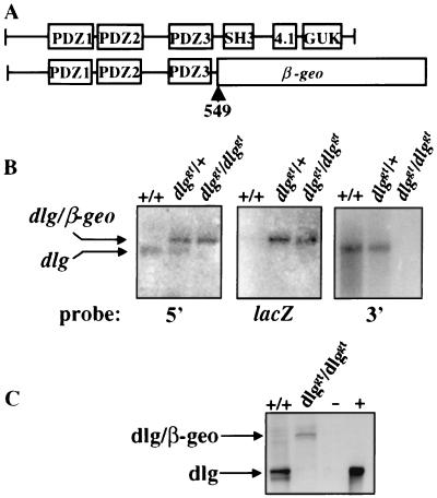
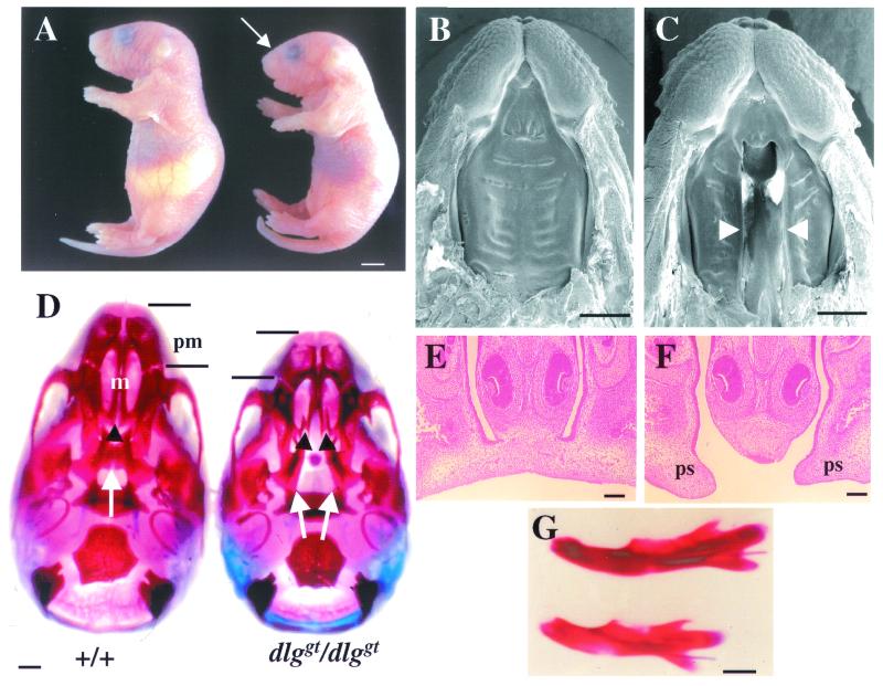
The discs large (Dlg) protein, or synapse-associated protein 97 (SAP97), is a member of the membrane-associated guanylate kinase family of multidomain scaffolding proteins which recruits transmembrane and signaling molecules to localized plasma membrane sites. Murine dlg is the homologue of the Drosophila dlg tumor suppressor gene. The loss of dlg function in Drosophila disrupts cellular growth control, apicobasal polarity, and cell adhesion of imaginal disc epithelial cells, resulting in embryonic lethality. In this study, we isolated a mutational insertion in the murine dlg locus by gene trapping in totipotent embryonic stem cells. This insertion results in a truncated protein product that contains the N-terminal three PSD-95/DLG/ZO-1 domains of Dlg fused to the LacZ reporter and subsequently lacks the src homology 3 (SH3), protein 4.1 binding, and guanylate kinase (GUK)-like domains. The Dlg-LacZ fusion protein is expressed in epithelial, mesenchymal, neuronal, endothelial, and hematopoietic cells during embryogenesis. Mice homozygous for the dlg mutation exhibit growth retardation in utero, have hypoplasia of the premaxilla and mandible, have a cleft secondary palate, and die perinatally. Consistent with this phenotype, Dlg-LacZ is expressed in mesenchymal and epithelial cells throughout palatal development. Our genetic and phenotypic analysis of dlg mutant mice suggests that protein-protein interactions involving the SH3, protein 4.1 binding, and/or GUK-like domains are essential to the normal function of murine Dlg within craniofacial and palatal morphogenesis.


Figures

FIG. 1
FIG. 1
Characterization of the gene trap vector insertion in the dlg locus. (A) Domain structures of wild-type Dlg protein and mutant Dlg protein with the fusion occurring at position 549. Wild-type Dlg contains three PDZ domains and SH3, protein 4.1 binding, and GUK-like domains. (B) Northern blot analysis of brain mRNA extracted from +/+, dlggt/+, and dlggt/dlggt E17.5 embryos for the presence of endogenous dlg and/or dlg-β-geo fusion transcripts. Probes 5′ and 3′ to the vector insertion site were used, as well as a lacZ probe. (C) Western blot analysis of lung lysate from wild-type and dlggt/dlggt mutant newborn pups using antiserum raised against the N terminus of Dlg. A doublet around 120 to 140 kDa was detected in wild-type lysates which was absent in dlggt/dlggt mutant mice; however, the expected Dlg–β-Geo fusion protein of 190 kDa was detected. −, untransfected HEK 293 cells; +, HEK 293 cells transfected with full-length murine dlg cDNA.
FIG. 2
FIG. 2
Expression of Dlg-LacZ during embryonic development in heterozygous dlggt embryos. (A) Expression in E8.5 embryos (dorsal view) was detected in the cephalic neural folds (nf), neural tube (nt), somites (s), and presomitic mesoderm (pm). Bar, 100 μm. Whole-mount LacZ staining in the vasculature of the yolk sac (E8.5) (B) and histological sections (C) demonstrated that the fusion protein was expressed in blood cells (bc), at regions of cell contact in endothelial cells (ec), and at the basal and lateral membranes of the epithelial cells of the endoderm (en). Magnification, ×100; bar, 12 μm. (D) At E8.5, transverse histological sections demonstrated that LacZ was expressed in the neural epithelium of the forebrain neural head folds (fnf), diencephelon (d), and otic pit (op), with the strongest expression detected at the apical surface. In the epithelium of the branchial arch (b1) (D′), foregut diverticulum (fgd) (D"), and surface ectoderm, expression was seen only at the apical face of the cells (arrowheads). Magnifications: panel D, ×20; panels D′ and D", ×40. Bars, 50 μm. (E) Whole-mount Dlg-LacZ expression in E10.5 embryos. (F) Endogenous Dlg expression in E10.5 wild-type embryos detected using Dlg antisera. Bars, 500 μm. (G) Dlg-LacZ expression in the brain (E12.5) was seen in the ventricular zone of the hypothalmus (vzh) and hippocampus (h). In the eye, Dlg-LacZ was expressed in the neural retina (nr), lens (l), retinal pigmented epithelium (rpe), and optic chiasm (oc). Magnification, ×5; bar, 200 μm. (H) Whole-mount Dlg-LacZ expression at E14.5 after only 4 h of X-Gal staining. Bar, 1 mm. (I) In the somites (E9.5), expression was detected in the epithelial dermomyotome (dm) component at the face adjacent to the scleratome (sc) (arrows). Magnification, ×40; bar, 25 μm. (J) Expression was seen predominantly at the apical membrane of epithelial cells (e) (arrows) within the lung (E14.5) but also at all membranes of epithelial cells of the (K) kidney (E14.5) and (L) gut (E14.5). Magnification in panel J, ×20; bar, 50 μm. Magnification in panel K, ×40; bar, 8 μm. Magnification in panel L, ×40; bar, 50 μm. b1 and b2, branchial arches 1 and 2, respectively; cb, cerebellum; ey, eye; g, gut; h, heart; lb, limb bud; l, lung; ma, mandible; m, maxillary component of b1; me, mesenchyme; nc, nasal cavity; np, nasal pit; ne, neural epithelium; n, notochord; ot, otic vesicle; pn, pons; sc, spinal cord; sn, spinal nerve; tb, tailbud; to, tongue; t, telencephalon.
FIG. 3
FIG. 3
Localization of Dlg in colon epithelial cells. Sections of E17.5 colons were stained with an anti-Dlg antibody. (A) Dlg was localized predominantly to the lateral and basal (arrowheads) membranes in wild-type epithelial cells. In dlggt/+ (B) and dlggt/dlggt (C) colon epithelial cells, Dlg-LacZ was expressed at the basal and lateral membranes and also highly expressed at the apical membrane. Arrows depict the apical membrane of epithelial cells. Magnification, ×100; bar, 12.5 μm.
FIG. 4
FIG. 4
Phenotype of dlggt/dlggt embryos and mice. (A) Newborn dlggt/dlggt pups are smaller than their wild-type littermates, lack milk in their stomachs, and have dome-shaped skulls (arrow). Bar, 2.5 mm. SEM of the upper jaws of wild-type (B) and dlggt/dlggt (C) embryos at E16.5 is shown. Arrowheads depict the cleft secondary palate. Bar, 1 mm. (D) Whole-mount alizarin red- and alcian blue-stained skeletal preparations of the skulls of wild-type and dlggt/dlggt mutant E18.5 embryos. In wild-type embryos, the palatine (arrows) and maxillary (m) (arrowheads) shelves are fused. In dlggt/dlggt mutant embryos, both shelves are open. Shortening of the premaxilla (pm) is also indicated. Bar, 1 mm. Coronal sections of E15.5 wild-type (E) and dlggt/dlggt mutant (F) embryos demonstrate partial elevation and lack of contact and fusion of the palatal shelves (ps). Magnification, ×10; bar, 100 μm. (G) Whole-mount alizarin red and alcian blue skeletal staining of the mandibles of wild-type (top) and dlggt/dlggt mutant embryos (bottom). Bar, 1 mm.
FIG. 5
FIG. 5
Histology of palate development and Dlg-LacZ expression in mutant dlggt embryos. (A) Dlg-LacZ was expressed at the apical membrane of the epithelium of the first branchial arch (ba) and the epithelium (e) associated with the olfactory placode in dlggt/+ embryos (E9.5) (arrows). Magnification, ×20; bar, 50 μm. (A′) Higher magnification (×40) of the boxed area in panel A demonstrating the apical expression of Dlg-LacZ in the epithelium of the branchial arch. Bar, 25 μm. Coronal sections of E12.5 wild-type (B) and dlggt/dlggt (C) heads, the latter demonstrating Dlg-LacZ expression in the palatal shelves (ps), toothbud (tb), and facial mesenchyme. Magnification, ×5; bar, 200 μm. (D) Palatal shelves are elevated and fused in dlggt/+ E15.5 embryos. (E) Only one palatal shelf is elevated in dlggt/dlggt embryos. Dlg-LacZ is expressed within the mesenchyme and epithelial cells of the fused palatal shelves (D) and unfused palatal shelves (E). Magnification, ×10; bar, 100 μm. (F) Dlg-LacZ expression in the palatal shelves of dlggt/+ prior to fusion demonstrates low levels of expression in the tips of the shelves (arrows) with higher expression adjacent to this region. Magnification, ×20. Bar, 200 μm. (G) Dlg-LacZ expression was observed in the medial-edge epithelial cells at the time of palatal-shelf fusion (arrows). Magnification, ×100; bar, 100 μm. M, Meckel's cartilage; to, tongue.

References

    1. Adey N B, Huang L, Ormonde P A, Baumgard M L, Pero R, Byreddy D V, Tatigian S V. Threonine phosphorylation of the MMAC/PTEN PDZ binding domain both inhibits and stimulates PDZ binding. Cancer Res. 2000;60:35–37. - PubMed
    1. Alexander C, Stathakis D G, Lin L, Rahman S, Bryant P J, Auburger G, Chishti A H. Fine scale mapping places DLG1, the gene encoding hDlg, telomeric to the OPA1 candidate region. Mamm Genome. 1997;8:795–796. - PubMed
    1. Anderson J M. Cell signalling: MAGUK magic. Curr Biol. 1996;6:382–384. - PubMed
    1. Azim A C, Knoll J H, Marfatia S M, Peel D J, Bryant P J, Chishti A H. DLG1: chromosome location of the closest human homologue of the Drosophila discs large tumor suppressor gene. Genomics. 1995;30:613–616. - PubMed
    1. Dimitratos S D, Woods D F, Stathakis D G, Bryant P J. Signaling pathways are focussed at specialized regions of the plasma membrane by scaffolding proteins of the MAGUK family. Bioessays. 1999;21:912–921. - PubMed

Publication types

MeSH terms