

Dach1 mutant mice bear no gross abnormalities in eye, limb, and brain development and exhibit postnatal lethality
- PMID: 11238885
- PMCID: PMC86694
- DOI: 10.1128/MCB.21.5.1484-1490.2001
Dach1 mutant mice bear no gross abnormalities in eye, limb, and brain development and exhibit postnatal lethality
Abstract
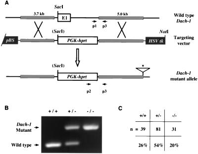
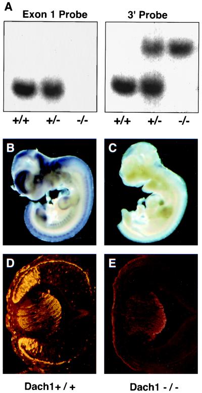
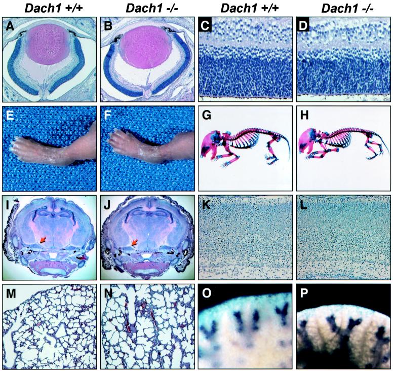
Drosophila dachshund is necessary and sufficient for compound eye development and is required for normal leg and brain development. A mouse homologue of dachshund, Dach1, is expressed in the developing retina and limbs, suggesting functional conservation of this gene. We have generated a loss-of-function mutation in Dach1 that results in the abrogation of the wild-type RNA and protein expression pattern in embryos. Homozygous mutants survive to birth but exhibit postnatal lethality associated with a failure to suckle, cyanosis, and respiratory distress. The heart, lungs, kidneys, liver, and skeleton were examined to identify factors involved in postnatal lethality, but these organs appeared to be normal. In addition, blood chemistry tests failed to reveal differences that might explain the lethal phenotype. Gross examination and histological analyses of newborn eyes, limbs, and brains revealed no detectable abnormalities. Since Dach1 mutants die shortly after birth, it remains possible that Dach1 is required for postnatal development of these structures. Alternatively, an additional Dach homologue may functionally compensate for Dach1 loss of function.
Figures
Similar articles
-
Mouse Dach2 mutants do not exhibit gross defects in eye development or brain function.Genesis. 2006 Feb;44(2):84-92. doi: 10.1002/gene.20188. Genesis. 2006. PMID: 16470613
-
Targeted disruption of mouse Dach1 results in postnatal lethality.Dev Dyn. 2003 Jan;226(1):139-44. doi: 10.1002/dvdy.10210. Dev Dyn. 2003. PMID: 12508235
-
Mouse Dach1 and Dach2 are redundantly required for Müllerian duct development.Genesis. 2008 Apr;46(4):205-13. doi: 10.1002/dvg.20385. Genesis. 2008. PMID: 18395837
-
Dach1, a vertebrate homologue of Drosophila dachshund, is expressed in the developing eye and ear of both chick and mouse and is regulated independently of Pax and Eya genes.Mech Dev. 2002 Feb;111(1-2):75-87. doi: 10.1016/s0925-4773(01)00611-6. Mech Dev. 2002. PMID: 11804780
-
The Dachshund gene in development and hormone-responsive tumorigenesis.Trends Endocrinol Metab. 2010 Jan;21(1):41-9. doi: 10.1016/j.tem.2009.08.002. Epub 2009 Nov 5. Trends Endocrinol Metab. 2010. PMID: 19896866 Free PMC article. Review.
Cited by
-
Comparison of the gene expression profiles from normal and Fgfrl1 deficient mouse kidneys reveals downstream targets of Fgfrl1 signaling.PLoS One. 2012;7(3):e33457. doi: 10.1371/journal.pone.0033457. Epub 2012 Mar 14. PLoS One. 2012. PMID: 22432025 Free PMC article.
-
Global polysome analysis of normal and injured podocytes.Am J Physiol Renal Physiol. 2019 Feb 1;316(2):F241-F252. doi: 10.1152/ajprenal.00115.2018. Epub 2018 Oct 31. Am J Physiol Renal Physiol. 2019. PMID: 30379099 Free PMC article.
-
Genomic analyses in African populations identify novel risk loci for cleft palate.Hum Mol Genet. 2019 Mar 15;28(6):1038-1051. doi: 10.1093/hmg/ddy402. Hum Mol Genet. 2019. PMID: 30452639 Free PMC article.
-
Retinal determination gene networks: from biological functions to therapeutic strategies.Biomark Res. 2023 Feb 8;11(1):18. doi: 10.1186/s40364-023-00459-8. Biomark Res. 2023. PMID: 36750914 Free PMC article. Review.
-
Regulatory Genes in Eyespot Formation and Function of Mytilus coruscus.Mar Biotechnol (NY). 2024 Nov 27;27(1):13. doi: 10.1007/s10126-024-10396-8. Mar Biotechnol (NY). 2024. PMID: 39601898
References
-
- Bonini N M, Bui Q T, Grayboard G L, Warrick J M. The Drosophila eyes absent gene directs ectopic eye formation in a pathway conserved between flies and vertebrates. Development. 1997;124:4819–4826. - PubMed
-
- Bonini N M, Leiserson W M, Benzer S. The eyes absent gene: genetic control of cell survival and differentiation in the developing Drosophila eye. Cell. 1993;72:379–395. - PubMed
-
- Bovolenta P, Mallamaci A, Puelles L, Boncinelli E. Expression pattern of cSix3, a member of the Six/Sine oculis family of transcription factors. Mech Dev. 1998;70:201–203. - PubMed
-
- Caubit X, Thangarajah R, Theil T, Wirth J, Nothwang H G, Ruther U, Krauss S. Mouse Dac, a novel nuclear factor with homology to Drosophila dachshund shows a dynamic expression in the neural crest, the eye, the neocortex, and the limb bud. Dev Dyn. 1999;214:66–80. - PubMed
Publication types
MeSH terms
Substances
Grants and funding
LinkOut - more resources
Full Text Sources
Other Literature Sources
Medical
Molecular Biology Databases