. 2001 Mar;21(5):1662-71.
doi: 10.1128/MCB.21.5.1662-1671.2001.

Zygotic regulation of maternal cyclin A1 and B2 mRNAs

Affiliations

Zygotic regulation of maternal cyclin A1 and B2 mRNAs

Y Audic et al. Mol Cell Biol. 2001 Mar.

Abstract

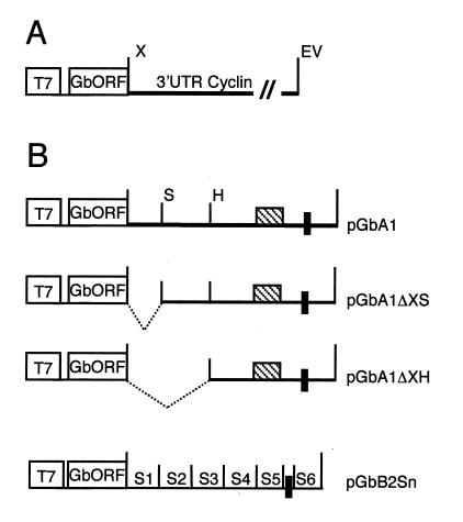
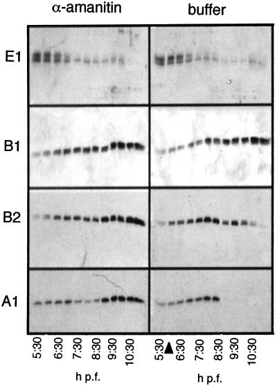
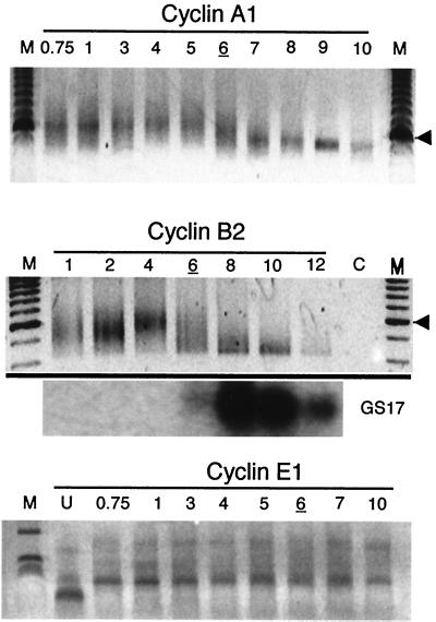
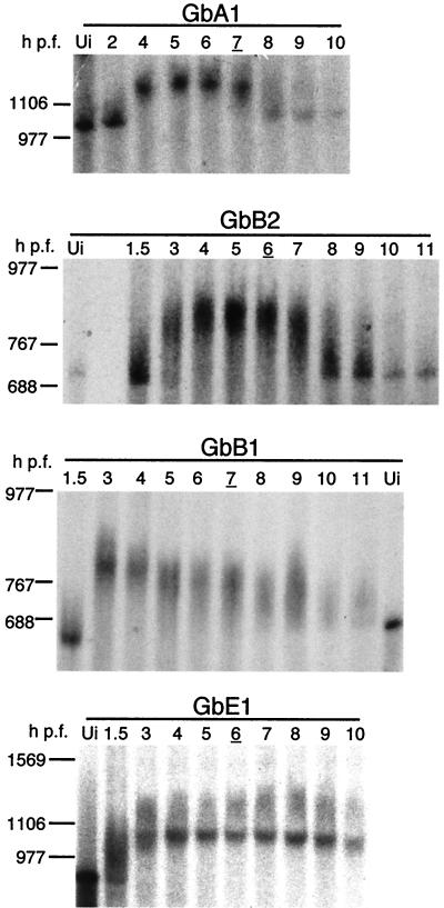
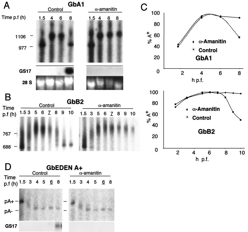
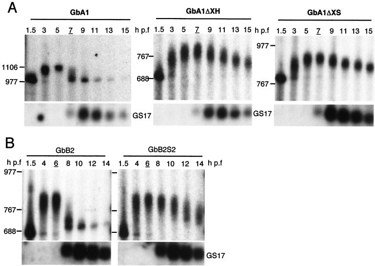
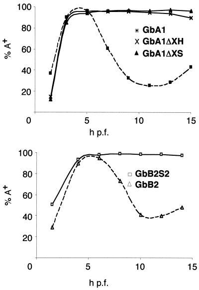
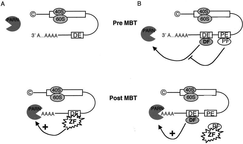
At the midblastula transition, the Xenopus laevis embryonic cell cycle is remodeled from rapid alternations between S and M phases to become the complex adult cell cycle. Cell cycle remodeling occurs after zygotic transcription initiates and is accompanied by terminal downregulation of maternal cyclins A1 and B2. We report here that the disappearance of both cyclin A1 and B2 proteins is preceded by the rapid deadenylation of their mRNAs. A specific mechanism triggers this deadenylation. This mechanism depends upon discrete regions of the 3' untranslated regions and requires zygotic transcription. Together, these results strongly suggest that zygote-dependent deadenylation of cyclin A1 and cyclin B2 mRNAs is responsible for the downregulation of these proteins. These studies also raise the possibility that zygotic control of maternal cyclins plays a role in establishing the adult cell cycle.


Figures

FIG. 1
FIG. 1
Structure of chimeric genes. (A) General structure of the pGb-cyclin 3′-UTR genes. The T7 promoter (T7) and the restriction sites XbaI (X) and EcoRV (EV) are indicated. The β-globin 5′-UTR, open reading frame, and partial globin 3′-UTR sequences are indicated (GbORF). The cyclin 3′ UTRs are complete and contain the hexanucleotide AATAAA. (B) Structure of the mutagenized chimeric genes. The black box represents the nuclear polyadenylation signal, and the striped box corresponds to the CPE. The restriction endonuclease recognition sites used in the deletion analysis of pGbA1 are indicated (S = SpeI; H = HpaI). For pGbB2Sn, mutated sequences S1 to S6 are indicated and have been replaced by the sequence indicated in Materials and Methods.
FIG. 2
FIG. 2
Terminal disappearance of cyclin A1 and B2 proteins during cell cycle remodeling is dependent on zygotic transcription. Western blots for the cyclins indicated depict protein levels during embryogenesis. Embryos were injected with 50 ng of α-amanitin to inhibit the onset of zygotic transcription (left panels), harvested every 30 min at the indicated times after fertilization, and processed for protein extraction. One embryo equivalent was loaded per lane. The same blot was stripped and reprobed for each cyclin. Stage 8 embryos are indicated by an arrowhead.
FIG. 3
FIG. 3
Cyclin A1 and B2 maternal mRNAs are rapidly deadenylated at the MBT. DNA gel analysis of PAT PCR products is shown for cyclins A1, B2, and E1. Total RNA was extracted from embryos at the indicated hours postfertilization and subjected to the PAT. Lanes M, 100-bp DNA ladder. The arrowheads on the right of the top two panels correspond to the 600-bp marker. Northern blot analysis (GS17) of the cyclin B2 mRNA samples shows the expression of the zygotic transcription-specific mRNA GS17. For cyclin E1 PAT, lane M corresponds to a 1-kb ladder. For each analysis, stage 8 embryos are indicated by the underlined hour postfertilization.
FIG. 4
FIG. 4
Cyclin A1 and B2 3′ UTRs trigger rapid deadenylation at the MBT. Representative autoradiograms of deadenylation analysis of chimeric cyclin mRNAs are shown. Radiolabeled, poly(A) GbA1, GbB2, GbB1, or GbE1 mRNAs were injected into two-cell embryos. Total RNA extracted from embryos at the indicated times after fertilization was resolved by denaturing gel electrophoresis. RNA size markers are indicated on the left in bases. The injected mRNA is indicated on the top of each panel. Ui, uninjected poly(A) mRNA. GbA1, n = 8; GbB2, n = 8; GbB1, n = 4; GbE1, n = 2. Stage 8 embryos are indicated by the underlined times postfertilization.
FIG. 5
FIG. 5
Deadenylation triggered by the cyclin A1 and B2 3′ UTRs is dependent on zygotic transcription. Shown are autoradiograms of deadenylation analyses of chimeric GbA1, GbB2, and GbEDEN mRNAs in the presence of α-amanitin. (A) Radiolabeled GbA1 mRNA injected in the presence (α-amanitin) or absence (control) of 50 ng of α-amanitin/embryo. RNA was analyzed as indicated in the legend to Fig. 4. Expression of the zygotic gene GS17 and ethidium bromide-stained 28S rRNA is also shown. (B) Radiolabeled GbB2 mRNA injected in the presence (α-amanitin) or absence (control) of α-amanitin. The positions of RNA size markers are indicated to the left of each panel. GbA1, n = 8; GbA1 plus α-amanitin, n = 4; GbB2, n = 8; GbB2 plus α-amanitin, n = 2. (C) Quantification of the percentage of polyadenylated chimeric mRNA at each time point. (D) Radiolabeled GbORF/mosEDEN poly(A)+ mRNA injected in the presence (α-amanitin) or absence (control) of α-amanitin (n = 2). The positions of A65 (pA+) and A0 (pA) are shown. The expression of the zygotic gene-specific transcript GS17 was monitored by Northern blotting. For each experiment, stage 8 embryos are indicated by underlining of the time postfertilization.
FIG. 6
FIG. 6
Specific regions in the 3′ UTRs of cyclin A1 and B2 mRNAs are required for zygote-dependent deadenylation. (A) Radiolabeled GbA1 mRNA and the deletion mutants GbA1ΔXH and GbA1ΔXS were injected into two-cell embryos. RNAs were analyzed as indicated in the legend to Fig. 4. The expression of the zygotic gene GS17 as determined by Northern blot analysis is shown at the bottom of each panel. (B) Radiolabeled GbB2 mRNA and the substitution mutant GbB2S2 were injected into two-cell embryos and analyzed at the indicated times after fertilization. The positions of RNA size markers are indicated to the left of each panel. GbA1, n = 8; GbA1ΔXH, n = 2; GbA1ΔXS, n = 2; GbB2, n = 8; GbB2S2, n = 2. For each experiment, stage 8 embryos are indicated by the underlined hour postfertilization.
FIG. 7
FIG. 7
Quantification of deadenylation of the GbA1 deletion mutants and the GbB2 substitution mutants. The data collected from the experiments shown in Fig. 6 were quantitated and plotted as the percentage of polyadenylated mRNA for each time point. Upper panel, GbA1 mRNA and the deletion mutants GbA1ΔXH and GbA1ΔXS. Lower panel, GbB2 mRNA and the substitution mutant GbB2S2.
FIG. 8
FIG. 8
Proposed models for zygotically mediated deadenylation of cyclin A1 and B2 mRNAs include a zygotically expressed deadenylation factor (A) or a maternally expressed protective factor (B). © represents the 5′-cap structure. The white box corresponds to the mRNA coding region. DE, deadenylation element; PE, protective element; PF, protective factor; ZF, the unknown zygotic factor that acts either as a deadenylation activator (A) or as an inhibitor of protection mediated by the interaction of the protective factor with the protective element (B).

Similar articles

Cited by

References

    1. Audic Y, Omilli F, Osborne H B. Embryo deadenylation element-dependent deadenylation is enhanced by a cis element containing AUU repeats. Mol Cell Biol. 1998;18:6879–6884. - PMC - PubMed
    1. Audic Y, Omilli F, Osborne H B. Postfertilization deadenylation of mRNAs in Xenopus laevis embryos is sufficient to cause their degradation at the blastula stage. Mol Cell Biol. 1997;17:209–218. - PMC - PubMed
    1. Barnhart K M. Simplified PCR-mediated, linker-scanning mutagenesis. BioTechniques. 1999;26:624–626. - PubMed
    1. Brewer G. Characterization of c-myc 3′ to 5′ mRNA decay activities in an in vitro system. J Biol Chem. 1998;273:34770–34774. - PubMed
    1. Chevalier S, Couturier A, Chartrain I, Le Guellec R, Beckhelling C, Le Guellec K, Philippe M, Ford C C. Xenopus cyclin E, a nuclear phosphoprotein, accumulates when oocytes gain the ability to initiate DNA replication. J Cell Sci. 1996;109:1173–1184. - PubMed

Publication types

LinkOut - more resources