

Actinin-associated LIM protein-deficient mice maintain normal development and structure of skeletal muscle
- PMID: 11238905
- PMCID: PMC86714
- DOI: 10.1128/MCB.21.5.1682-1687.2001
Actinin-associated LIM protein-deficient mice maintain normal development and structure of skeletal muscle
Abstract
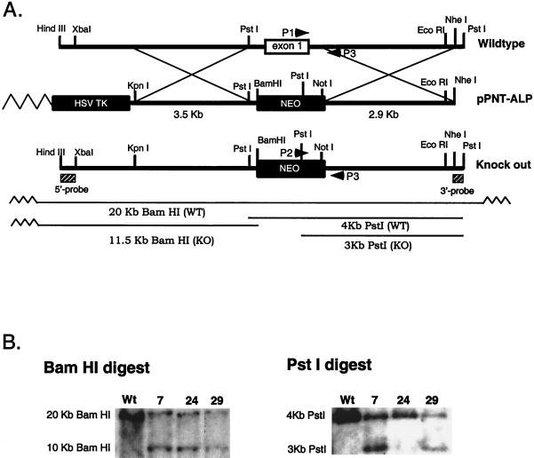
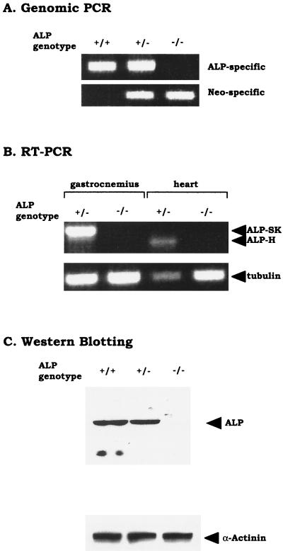
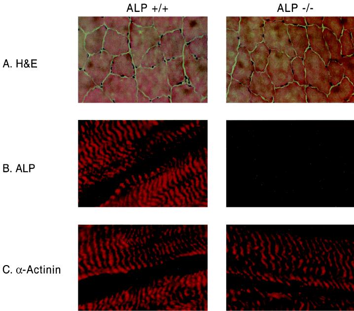
The actinin-associated LIM protein, ALP, is the prototype of a large family of proteins containing an N-terminal PDZ domain and a C-terminal LIM domain. These PDZ-LIM proteins are components of the muscle cytoskeleton and occur along the Z lines owing to interaction of the PDZ domain with the spectrin-like repeats of alpha-actinin. Because PDZ and LIM domains are typically found in proteins that mediate cellular signaling, PDZ-LIM proteins are suspected to participate in muscle development. Interestingly the ALP gene occurs at 4q35 near the heterochromatic region mutated in facioscapulohumeral muscular dystrophy, indicating a possible role for ALP in this disease. Here, we describe the generation and analysis of mice lacking the ALP gene. Surprisingly, the ALP knockout mice show no gross histological abnormalities and maintain sarcolemmal integrity as determined by serum pyruvate kinase assays. The absence of a dystrophic phenotype in these mice suggests that down-regulation of ALP does not participate in facioscapulohumeral muscular dystrophy. These data suggest that ALP does not participate in muscle development or that an alternative PDZ-LIM protein can compensate for the lack of ALP.
Figures
Similar articles
-
Actinin-associated LIM protein: identification of a domain interaction between PDZ and spectrin-like repeat motifs.J Cell Biol. 1997 Oct 20;139(2):507-15. doi: 10.1083/jcb.139.2.507. J Cell Biol. 1997. PMID: 9334352 Free PMC article.
-
Exclusion of muscle specific actinin-associated LIM protein (ALP) gene from 4q35 facioscapulohumeral muscular dystrophy (FSHD) candidate genes.Neuromuscul Disord. 1999 Jan;9(1):3-10. doi: 10.1016/s0960-8966(98)00087-x. Neuromuscul Disord. 1999. PMID: 10063829
-
The ZASP-like motif in actinin-associated LIM protein is required for interaction with the alpha-actinin rod and for targeting to the muscle Z-line.J Biol Chem. 2004 Jun 18;279(25):26402-10. doi: 10.1074/jbc.M401871200. Epub 2004 Apr 14. J Biol Chem. 2004. PMID: 15084604
-
ALP/Enigma PDZ-LIM domain proteins in the heart.J Mol Cell Biol. 2010 Apr;2(2):96-102. doi: 10.1093/jmcb/mjp038. Epub 2009 Dec 30. J Mol Cell Biol. 2010. PMID: 20042479 Free PMC article. Review.
-
PDZ and LIM domain-encoding genes: molecular interactions and their role in development.ScientificWorldJournal. 2007 Sep 1;7:1470-92. doi: 10.1100/tsw.2007.232. ScientificWorldJournal. 2007. PMID: 17767364 Free PMC article. Review.
Cited by
-
Actuated tissue engineered muscle grafts restore functional mobility after volumetric muscle loss.Biomaterials. 2023 Nov;302:122317. doi: 10.1016/j.biomaterials.2023.122317. Epub 2023 Sep 8. Biomaterials. 2023. PMID: 37717406 Free PMC article.
-
Ablation of Cypher, a PDZ-LIM domain Z-line protein, causes a severe form of congenital myopathy.J Cell Biol. 2001 Nov 12;155(4):605-12. doi: 10.1083/jcb.200107092. Epub 2001 Nov 5. J Cell Biol. 2001. PMID: 11696561 Free PMC article.
-
Goodpasture antigen-binding protein (GPBP) directs myofibril formation: identification of intracellular downstream effector 130-kDa GPBP-interacting protein (GIP130).J Biol Chem. 2011 Oct 7;286(40):35030-43. doi: 10.1074/jbc.M111.249458. Epub 2011 Aug 9. J Biol Chem. 2011. PMID: 21832087 Free PMC article.
-
Zasp52, a Core Z-disc Protein in Drosophila Indirect Flight Muscles, Interacts with α-Actinin via an Extended PDZ Domain.PLoS Genet. 2016 Oct 26;12(10):e1006400. doi: 10.1371/journal.pgen.1006400. eCollection 2016 Oct. PLoS Genet. 2016. PMID: 27783625 Free PMC article.
-
The sarcomeric Z-disc: a nodal point in signalling and disease.J Mol Med (Berl). 2006 Jun;84(6):446-68. doi: 10.1007/s00109-005-0033-1. Epub 2006 Jan 17. J Mol Med (Berl). 2006. PMID: 16416311 Review.
References
-
- Altherr M R, Bengtsson U, Markovich R P, Winokur S T. Efforts toward understanding the molecular basis of facioscapulohumeral muscular dystrophy. Muscle Nerve. 1995;2:S32–S38. - PubMed
-
- Arber S, Halder G, Caroni P. Muscle LIM protein, a novel essential regulator of myogenesis, promotes myogenic differentiation. Cell. 1994;79:221–231. - PubMed
-
- Arber S, Hunter J J, Ross J, Jr, Hongo M, Sansig G, Borg J, Perriard J C, Chien K R, Caroni P. MLP-deficient mice exhibit a disruption of cardiac cytoarchitectural organization, dilated cardiomyopathy, and heart failure. Cell. 1997;88:393–403. - PubMed
-
- Bouju S, Piétu G, Le Cunff M, Cros N, Malzac P, Pellissier J F, Pons F, Léger J J, Auffray C, Dechesne C A. Exclusion of muscle specific actinin-associated LIM protein (ALP) gene from 4q35 facioscapulohumeral muscular dystrophy (FSHD) candidate genes. Neuromuscul Disord. 1999;9:3–10. - PubMed
-
- Brenman J E, Chao D S, Xia H, Aldape K, Bredt D S. Nitric oxide synthase complexed with dystrophin and absent from skeletal muscle sarcolemma in Duchenne muscular dystrophy. Cell. 1995;82:743–752. - PubMed
Publication types
MeSH terms
Substances
Grants and funding
LinkOut - more resources
Full Text Sources
Molecular Biology Databases