. 2001 Mar;21(6):1973-85.
doi: 10.1128/MCB.21.6.1973-1985.2001.

Antisense promoter of human L1 retrotransposon drives transcription of adjacent cellular genes

Affiliations

Antisense promoter of human L1 retrotransposon drives transcription of adjacent cellular genes

M Speek. Mol Cell Biol. 2001 Mar.

Abstract

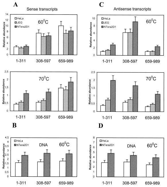
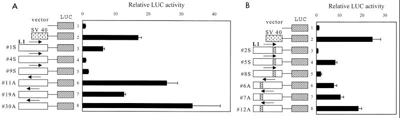
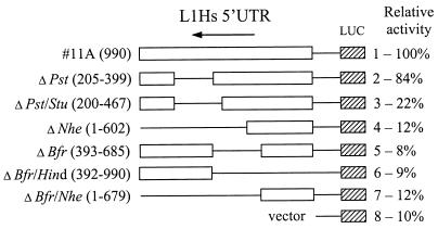
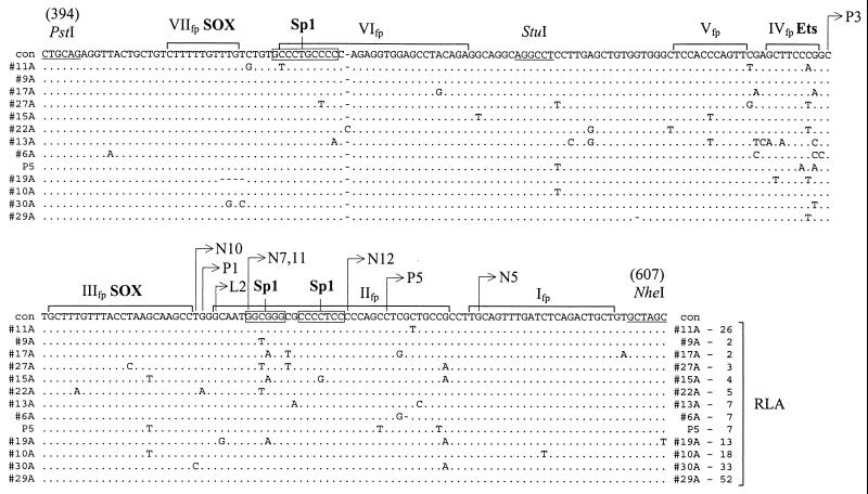
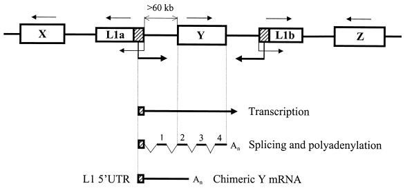
In the human genome, retrotranspositionally competent long interspersed nuclear elements (L1Hs) are involved in the generation of processed pseudogenes and mobilization of unrelated sequences into existing genes. Transcription of each L1Hs is initiated from its internal promoter but may also be driven from the promoters of adjacent cellular genes. Here I show that a hitherto unknown L1Hs antisense promoter (ASP) drives the transcription of adjacent genes. The ASP is located in the L1Hs 5' untranslated region (5'UTR) and works in the opposite direction. Fifteen cDNAs, isolated from a human NTera2D1 cDNA library by a differential screening method, contained L1Hs 5'UTRs spliced to the sequences of known genes or non-proteincoding sequences. Four of these chimeric transcripts, selected for detailed analysis, were detected in total RNA of different cell lines. Their abundance accounted for roughly 1 to 500% of the transcripts of four known genes, suggesting a large variation in the efficiency of L1Hs ASP-driven transcription. ASP-directed transcription was also revealed from expressed sequence tag sequences and confirmed by using an RNA dot blot analysis. Nine of the 15 randomly selected genomic L1Hs 5'UTRs had ASP activities about 7- to 50-fold higher than background in transient transfection assays. ASP was assigned to the L1Hs 5'UTR between nucleotides 400 to 600 by deletion and mutation analysis. These results indicate that many L1Hs contain active ASPs which are capable of interfering with normal gene expression, and this type of transcriptional control may be widespread.


Figures

FIG. 1
FIG. 1
Strategy for differential screening of the L1Hs cDNAs. L1Hs transcripts with homogeneous 3′ ends and heterogeneous 5′ ends, shown at the top, are aligned to the general L1 retrotransposon structure. Below this structure, positions of DNA probes corresponding to the L1Hs promoter (5′UTR) and ORF1 are indicated. A search for an unknown gene or mRNA with an L1-like internal promoter by screening NTera2D1 cDNA library for promoter-positive and ORF1-negative cDNAs (marked by + and − signs, respectively) yielded novel cDNAs (marked by ?) containing the L1Hs 5′UTR linked to known and unknown sequences located upstream of the L1Hs (bottom structure). Vertical bars mark the 5′ and 3′ ends of the 5′UTR.
FIG. 2
FIG. 2
Mapping of the cDNA 5′-terminal sequences to the L1Hs 5′UTR. (A) cDNA sequences with the mRNA sequence polarity (5′ to 3′) indicated by arrows were mapped to the L1Hs 5′UTR. Numbering of the top L1Hs strand starts with +1 (34), and numbering of the bottom strand starts from nt 86 of ORF1. Splicing of the N4 and P3 cDNAs within the 5′UTR is indicated by thin lines. Two cDNAs (N9 and P2) showed opposite polarity. (B) Six cDNA sequences (N4, N5, N7, N12, P1, and P3) compared to the corresponding genomic sequences (Table 1) were aligned to show their exon and intron structures, indicated by upper- and lowercase letters, respectively. The conserved 5′ and 3′ intronic dinucleotide sequences (GT and AG) are underlined. Two cDNAs (N4 and P3) are spliced within the L1Hs 5′UTR, and their identical exon II sequences compared to the corresponding L1 sequence are boxed. The others are spliced to known or unknown sequences located further upstream of the L1Hs. Numbering is according to the L1Hs 5′UTR bottom strand. L1.2, accession no. M80343 (8). Identical nucleotides are marked below sequences by asterisks; sequences not shown are indicated by dots.
FIG. 3
FIG. 3
Graphical representation and detection in different cell lines of chimeric transcripts generated from the L1Hs ASP. Diagrams show alignments of N12 and SPT3 mRNA (A), N4 and GAP-43 mRNA (B), N10 and CHRM3 gene (C), and P5 and OATP mRNA (D). Chimeric cDNAs and mRNAs are indicated by lines, the CHRM3 gene (mRNA sequence unknown) is shown by an open box, and L1Hs 5′UTR sequences transcribed from the L1Hs ASP and linked to the known mRNA and gene sequences are indicated by hatched boxes. Identical regions between mRNA (gene) and chimeric cDNAs are shown by vertical lines. Introns, determined from the alignment of genomic and cDNA sequences (Table 1), are shown by arrowheads below cDNA structures only for aligned portions and 5′ regions of chimeric cDNAs. Initiator AUG/ATG and stop codons are shown above the lines; polyadenylation signals (not found for the structures of GAP-43 mRNA and CHRM3 gene depicted) are marked by oval arrows. Additional sequences were found at the 5′ end of N4 and 3′ end of N10 cDNAs. Their origin is unclear. Detection of the four chimeric transcripts in different cell lines by using RNase protection (27) is shown at the right. All protected RNAs were analyzed on a 5% denaturing polyacrylamide gel by using the following 32P-labeled probes. (A) A 465-nt riboprobe encompassing 131 nt of the L1Hs 5′UTR, three exons of SPT3 mRNA (85, 87, and 94 nt), and 68 nt of vector sequences was generated from N12, cut with NheI, and transcribed with T7 RNA polymerase. (B) A 829-nt riboprobe encompassing two exons of the L1 5′UTR (150 and 202 nt), 409 nt of 5′ region of the GAP-43 mRNA, and 59 nt of vector sequence was generated from a HindIII deletion subclone of N4, cut with EcoRI, and transcribed with T3 RNA polymerase. (C) A 934-nt riboprobe encompassing 450 nt of the L1 5′UTR plus 16 nt of non-L1 sequence, three exons (63, 103, and 127 nt) and 5′ coding region of the CHMR3 gene (111 nt), and 61 nt of vector sequences was generated from a SmaI deletion subclone of N10, cut with EcoRI, and transcribed with T7 RNA polymerase. (D) A 1,009-nt riboprobe encompassing 429 nt of L1 5′UTR plus 103 nt of non-L1 sequence, 5′ region of OATP mRNA (three exons, 382 nt), and 94 nt of vector sequences was generated from a HindIII deletion subclone of P5, cut with XhoI, and transcribed with T7 RNA polymerase. Restriction enzymes used to make riboprobes and deletion subclones are shown below cDNA structures. Chimeric and alternative mRNAs detected are shown on the right of each panel. For these mRNAs, the number of exons (xex) and their sizes in nucleotides are indicated in parentheses. Protected fragments of N10 and P5 predict three and one additional exons for the 5′ ends of CHMR3 and OATP mRNAs, respectively. Protections with the L1Hs 5′UTR of N10 (C) generate several fragments of <466 nt representing highly homologous transcripts derived other genomic L1Hs. Similar protections for N12 and N4 (fragments <200 and <300, respectively) are not shown (A and B). Traces of the undigested probe (B) are shown by the asterisk. A 32P-labeled 100-bp ladder (BRL) was used as a molecular weight marker. P, uncut probe; t, tRNA. In each experiment, 5 μg of total RNAs from NTera2D1 (N), JEG 3 (J), and HeLa cells (H) were used. Alt sp, possible alternatively spliced products; ?, unknown product.
FIG. 4
FIG. 4
Graphical representation of EST sequences similar to the L1Hs 5′UTR-ORF1 region. Sense (A) and antisense (B) strands of a 996-bp L1.2 5′UTR-ORF1 sequence (accession. no. M80343) were searched for homologous sequences in EST databank using the WU-BLAST2 program. Only the 60 first sequences with the highest scores are shown. Numbering of the L1Hs sense strand starts from +1 (34), and numbering of the antisense strand starts from nt 86 of ORF1. A possible transcriptional initiation region from nt 500 to 700 and termination site around nt 300 are shaded by wide and narrow zones, respectively (see the text for explanation).
FIG. 5
FIG. 5
Distribution of L1Hs transcripts in total RNAs of different cell lines. Hybridization to the transcripts and total DNA (HeLa and NTera2D1) encompassing different L1Hs 5′UTR-ORF1 regions (shown below the columns) was carried out with L1Hs antisense (A and B) and sense (C and D) riboprobes as described in Materials and Methods. Probes 1–311, 308–597, and 659–989, encompassing 5′, central, and 3′ portions of the L1 5′UTR (sense-strand numbering), were used. Columns show relative abundance of transcripts or genomic DNA regions normalized with respect to the hybridization signals obtained from synthetic sense and antisense control RNAs and control DNA (SFL1). Upper and lower parts of panels A and C represent the same blot washed at 60 and 70°C, respectively. Data for DNA blots washed at 60°C are shown in panels B and D.
FIG. 6
FIG. 6
L1Hs 5′UTR promoter activity test. Twelve randomly selected genomic L1Hs 5′UTR-ORF1 fragments cloned in the luciferase (LUC) expression vector were tested for sense and ASP activities in transfected HeLa cells. RLAs were measured for the sense (1S, 4S and 9S) and antisense (11A, 19A and 30A) L1Hs 5′UTR-ORF1 constructs (A) and the sense (2S, 5S, and 8S) and antisense (6A, 7A, and 12A) L1Hs 5′UTR-ORF1 constructs containing an insertion of 110 to 130 bp around nt 780 (B). Luciferase activity was normalized to β-galactosidase activity for each construct in three separate experiments. Vector, promotorless pGL3-basic plasmid; SV 40, pGL3-promoter plasmid.
FIG. 7
FIG. 7
Definition of the L1Hs ASP region by deletion analysis. Various deletion constructs were prepared from 990-bp genomic clone 11A containing L1Hs 5′UTR-ORF1 ligated to the luciferase (LUC) expression vector in antisense orientation. Deleted regions are indicated in parentheses (except for 11A) and marked with thin lines. Numbering of the antisense strand starts at nt 86 of the ORF1 (accession no. M80343). The restriction enzymes used to make these deletions are shown at the left. Each construct was cotransfected into HeLa cells with β-galactosidase vector, and both luciferase and galactosidase activities were measured. RLA, normalized to β-galactosidase activity, is shown at the right. Activity of construct 11A was set to 100%.
FIG. 8
FIG. 8
Multiple sequence alignment and L1Hs ASP activity of the L1Hs genomic clones. Sequences of the 12 randomly selected genomic clones and the P5 genomic clone encompassing a putative L1Hs ASP region (nt 394 to 607; numbering of the antisense strand starting from nt 86 of ORF1 [accession no. M80343]) were aligned and compared to the RLA data (shown at the end of the alignment) measured for each genomic clone as described in Materials and Methods. For the alignment, only sequence differences are shown. Identical nucleotides are marked by dots; dashes indicate deletions. Con, consensus sequence. DNase footprints, marked above the sequences by brackets and roman numerals I to VII, and binding sites for SOX and Ets associated with these footprints are from data of others (36, 40). 5′-terminal nucleotides of different cDNAs are mapped to the sequence positions by arrows. Possible binding sites for Sp1 in footprints II and VI are boxed. RLA for the promotorless vector pGL3 was set to 1.
FIG. 9
FIG. 9
Transcriptional regulation by the L1Hs ASP. A fragment of the human genome containing two randomly positioned full-length L1 retrotransposons (L1a and L1b) and genes X, Y, and Z is shown. Direction of transcription for each gene is indicated by a thin arrow above the gene. Transcriptions driven by the L1a and L1b internal promoters (34) and ASPs, driving in opposite direction, are indicated by thin and thick arrows, respectively. Note that gene Y has the same direction of transcription as the L1a ASP but is located >60 kb further downstream. Genes X and Z do not match the L1a ASP orientation. Transcription of gene Y from the L1a ASP can generate a long precursor mRNA which upon splicing and polyadenylation according to the gene Y structure yields a chimeric Y mRNA. This mRNA contains the 5′-terminal L1a 5′UTR in antisense orientation spliced to the exon 1 derived from the intergenic region and exons 2 to 4 of gene Y. The latter structure is depicted at the bottom.

References

    1. Adams J W, Kaufman R E, Kretschmer P J, Harrison M, Nienhuis A W. A family of long reiterated DNA sequences, one copy of which is next to the human beta globin gene. Nucleic Acids Res. 1980;8:6113–6128. - PMC - PubMed
    1. Altschul S F, Madden T L, Schaffer A A, Zhang J, Zhang Z, Miller W, Lipman D J. Gapped BLAST and PSI-BLAST: a new generation of protein database search programs. Nucleic Acids Res. 1997;25:3389–3402. - PMC - PubMed
    1. Becker K G, Swergold G D, Ozato K, Thayer R E. Binding of the ubiquitous nuclear transcription factor YY1 to a cis regulatory sequence in the human LINE-1 transposable element. Hum Mol Genet. 1993;2:1697–702. - PubMed
    1. Birnboim H C, Doly J. A rapid alkaline extraction procedure for screening recombinant plasmid DNA. Nucleic Acids Res. 1979;7:1513–1523. - PMC - PubMed
    1. Boeke J D. LINEs and Alus—the polyA connection. Nat Genet. 1997;16:6–7. - PubMed

Publication types

LinkOut - more resources