. 2001 Mar;21(6):2203-12.
doi: 10.1128/MCB.21.6.2203-2212.2001.

Activation of Akt (protein kinase B) in mammary epithelium provides a critical cell survival signal required for tumor progression

Affiliations

Activation of Akt (protein kinase B) in mammary epithelium provides a critical cell survival signal required for tumor progression

J Hutchinson et al. Mol Cell Biol. 2001 Mar.

Abstract

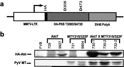
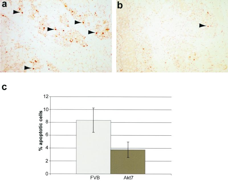
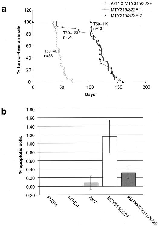
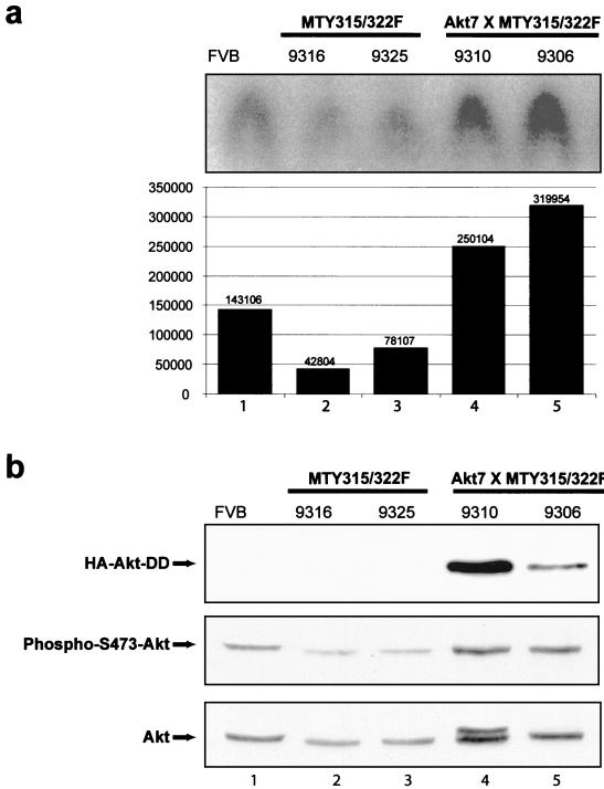
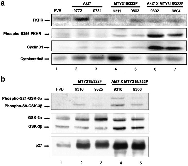
Activation of Akt by the phosphatidylinositol 3'-OH kinase (PI3K) results in the inhibition of proapoptotic signals and the promotion of survival signals (L. P. Kane et al., Curr. Biol. 9:601-604, 1999; G. J. Kops et al., Nature 398:630-634, 1999). Evidence supporting the importance of the PI3K/Akt signaling pathway in tumorigenesis stems from experiments with transgenic mice bearing polyomavirus middle T antigen under the control of the mouse mammary tumor virus long terminal repeat promoter. Mammary epithelium-specific expression of polyomavirus middle T antigen results in the rapid development of multifocal metastatic mammary tumors, whereas transgenic mice expressing a mutant middle T antigen decoupled from the phosphatidylinositol 3'-OH kinase (MTY315/322F) develop extensive mammary gland hyperplasias that are highly apoptotic. To directly assess the role of Akt in mammary epithelial development and tumorigenesis, we generated transgenic mice expressing constitutively active Akt (HAPKB308D473D or Akt-DD). Although expression of Akt-DD interferes with normal mammary gland involution, tumors were not observed in these strains. However, coexpression of Akt-DD with MTY315/322F resulted in a dramatic acceleration of mammary tumorigenesis correlated with reduced apoptotic cell death. Furthermore, coexpression of Akt-DD with MTY315/322F resulted in phosphorylation of the FKHR forkhead transcription factor and translational upregulation of cyclin D1 levels. Importantly, we did not observe an associated restoration of wild-type metastasis levels in the bitransgenic strain. Taken together these observations indicate that activation of Akt can contribute to tumor progression by providing an important cell survival signal but does not promote metastatic progression.


Figures

FIG. 1
FIG. 1
Activated Akt transgene expression. (a) Structure of the MMTV/Akt transgene. The Bluescript vector backbone is represented by a thin line on either side of the expression cassette, with the white region corresponding to the MMTV-LTR derived from plasmid pAp, the black portion corresponding to the hemagglutinin tag, the dark grey region corresponding to the Akt (HAPKBT308D/S473D) cDNA with aspartate substitutions at amino acid positions 308 and 473, and the mid-grey region corresponding to the transcriptional processing sequences derived from the SV40 early transcription unit. The transcription start site is indicated by an arrow. (b) Immunoblot analysis of expression of HAPKB and PyV mT in bitransgenic Akt7 × MTY315/322F strains. Note that Akt7 × MTY315/322F tumor samples coexpress both Akt and PyV mT proteins. The numbers above each lane indicate individual mouse identification numbers.
FIG. 2
FIG. 2
Coexpression of Akt and mutant PyV mT oncogene results in the induction of multifocal mammary tumors. These digital images illustrate the histological patterns observed in the Akt7 (a and b), MTY315/322F (c and d), and Akt7 × MTY315/322F (e and f) bigenic mice. Note that the whole-mount preparations (a, c, and e) demonstrate that the Akt strains have a relatively normal mammary tree (a) compared to the cystic hyperplasias seen in the MTY315/322F strains at the same age (c) (8 weeks) (scale bar = 1 mm). In contrast, the bigenic mammary gland does not fill the fat pad (e) and is a solid mass at this age (f). The histological patterns seen at high magnification (scale bar = 0.01 mm) demonstrate that the Akt7 strain has a normal epithelium (b), while the MTY315/322F strain has a cystic hyperplasia of the ducts and glands without significant atypia (d). In contrast, the Akt7 × MTY315/322F cross has acinar or lobular hyperplasia with low-grade atypia at 8 weeks (f). Normal mammary gland morphologies for the FVB strain can be viewed at the following website: http://ccm.ucdavis.edu/tgmouse/wmtable.htm.
FIG. 3
FIG. 3
Mammary epithelial expression of Akt results in defect in mammary gland involution. Digital images of the involution patterns in wild-type (a, c, e, and g) and Akt7 (b, d, f, and h) mammary glands. The images compare whole-mount preparations (a, e, b, and f) of the mammary gland (scale bar = 1 mm) with the histological pattern (c, d, g, and h) (scale bar = 0.1 mm) on days 1 (a to d) and 3 (e to h) of involution. Note the delayed involution in the Akt7 mouse mammary gland (b to h).
FIG. 4
FIG. 4
Mammary epithelial expression of Akt results in decreased mammary gland apoptosis during involution. (a and b) TUNEL analysis of involuting mammary glands from FVB/n (a) and Akt7 (b) at 3 days postparturition. Arrows indicate representative apoptotic cells. (c) Mammary apoptotic indices of FVB/n and Akt7 at 3 days postparturition. Values shown represent the percentage of total cells stained positive for apoptosis by TUNEL assay in age-matched singly parous female mice at 15 weeks of age.
FIG. 5
FIG. 5
Mammary tumor kinetics and apoptotic indices in transgenic strains. (a) Mammary tumor kinetics of MTY315/322F and Akt7 × MTY315/322F strains. Two different kinetics curves are shown for the MTY315/322F strain, from the original published data (MTY315/322F-1) and confirmatory data us (MTY315/322F-2), to account for possible differences in palpation technique between researchers. The age indicated is that at which a mammary tumor is first palpable in each transgenic strain. The number of animals analyzed for each strain (n) and the median age at which tumors were palpable are also shown. (b) Mammary apoptotic indices of FVB/n, MT634 (wild-type mT), Akt7, MTY315/322F, and Akt7 × MTY315/322F strains. Values shown represent the percentage of total cells stained positive for apoptosis by TUNEL assay in virgin female mice at 10 to 12 weeks of age.
FIG. 6
FIG. 6
Akt kinase activity in transgenic strains. (a) Total Akt kinase activity analysis in 8- to 10-week-old virgin females from FVB/n (lane 1), MTY135/322F (lanes 2 and 3), and bitransgenic Akt7 × MTY315/322F (lanes 4 and 5) strains. Assays were conducted using the cross-tide peptide as an Akt kinase substrate. Kinase activities were quantified by phosphorimager analysis and are represented here both graphically and numerically. (b) Immunoblot analysis of expression of HA-Akt-DD, phospho-S473-Akt, and Akt in 8- to 10-week-old virgin females from FVB/n (lane 1), MTY135/322F (lanes 2 and 3), and bitransgenic Akt7 × MTY315/322F (lanes 4 and 5) strains. All tissues were derived from 8- to 10-week-old virgin mammary glands. The arrows indicate the migration of transgenic HA-Akt-DD (upper panel), phospho-S473-Akt (middle panel), and total Akt (bottom panel). The numbers above each lane indicate individual mouse identification numbers.
FIG. 7
FIG. 7
Coexpression of activated Akt and MTY315/322F results in FKHR phosphorylation at serine 256 and increased cyclin D1 levels but does not affect GSK-3 phosphorylation or p27 levels. (a) Immunoblot analysis of expression of FKHR, phospho-FKHR (Ser256), cyclin D1, and cytokeratin in 8- to 10-week-old virgin females from FVB/n (lane 1), Akt7 (lanes 2 and 3), MTY135/322F (lanes 4 and 5), and bitransgenic Akt7 × MTY315/322F (lanes 6 and 7) strains. All tissues were derived from 8- to 10-week virgin mammary glands. The arrows indicate the migration of FKHR (upper panel), phospho-FKHR (Ser256) (second panel), cyclin D1 (third panel), and cytokeratin proteins (lower panel). The numbers above each lane indicate individual mouse identification numbers. (b) Immunoblot analysis of expression of phospho-S21-GSK-3α, phospho-S9-GSK-3β, GSK-3α/β, and p27 in 8- to 10-week-old virgin females from FVB/n (lane 1), MTY135/322F (lanes 2 and 3), and bitransgenic Akt7 × MTY315/322F (lanes 4 and 5) strains. All tissues were derived from 8- to 10-week virgin mammary glands. The arrows indicate the migration of phospho-S21-GSK-3α and phospho-S9-GSK-3β (upper panel), GSK-3α/β (middle panel), and p27 (lower panel). The numbers above each lane indicate individual mouse identification numbers.

References

    1. Alessi D R, Andjelkovic M, Caudwell B, Cron P, Morrice N, Cohen P, Hemmings B A. Mechanism of activation of protein kinase B by insulin and IGF-1. EMBO J. 1996;15:6541–6551. - PMC - PubMed
    1. Alessi D R, James S R, Downes C P, Holmes A B, Gaffney P R, Reese C B, Cohen P. Characterization of a 3-phosphoinositide-dependent protein kinase which phosphorylates and activates protein kinase Bα. Curr Biol. 1997;7:261–269. - PubMed
    1. Brunet A, Bonni A, Zigmond M J, Lin M Z, Juo P, Hu L S, Anderson M J, Arden K C, Blenis J, Greenberg M E. Akt promotes cell survival by phosphorylating and inhibiting a Forkhead transcription factor. Cell. 1999;96:857–868. - PubMed
    1. Cardone M H, Roy N, Stennicke H R, Salvesen G S, Franke T F, Stanbridge E, Frisch S, Reed J C. Regulation of cell death protease caspase-9 by phosphorylation. Science. 1998;282:1318–1321. - PubMed
    1. Chang H W, Aoki M, Fruman D, Auger K R, Bellacosa A, Tsichlis P N, Cantley L C, Roberts T M, Vogt P K. Transformation of chicken cells by the gene encoding the catalytic subunit of PI 3-kinase. Science. 1997;276:1848–1850. - PubMed

Publication types

MeSH terms