. 2001 Mar;21(6):2213-20.
doi: 10.1128/MCB.21.6.2213-2220.2001.

Disruption of the mouse mu-calpain gene reveals an essential role in platelet function

Affiliations

Disruption of the mouse mu-calpain gene reveals an essential role in platelet function

M Azam et al. Mol Cell Biol. 2001 Mar.

Abstract

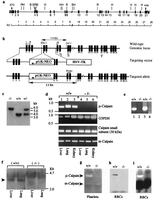
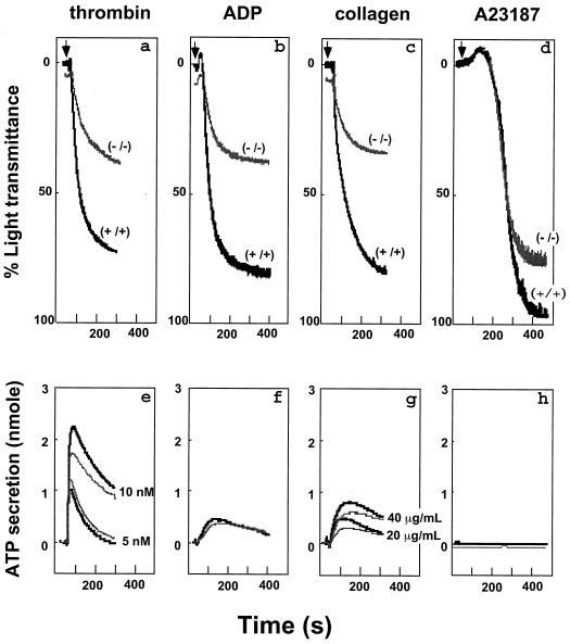
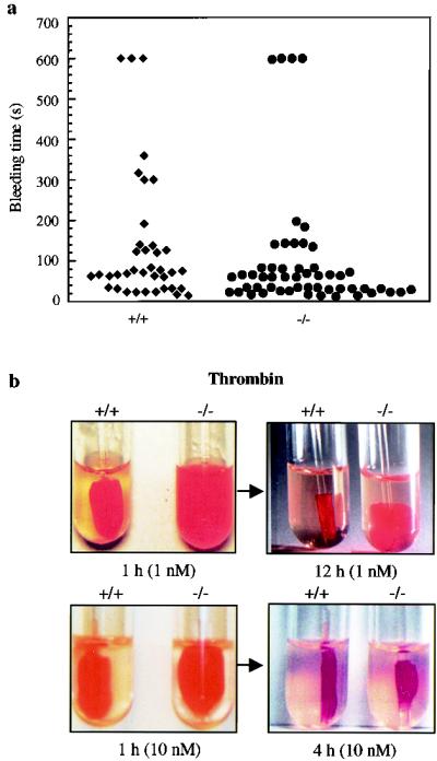
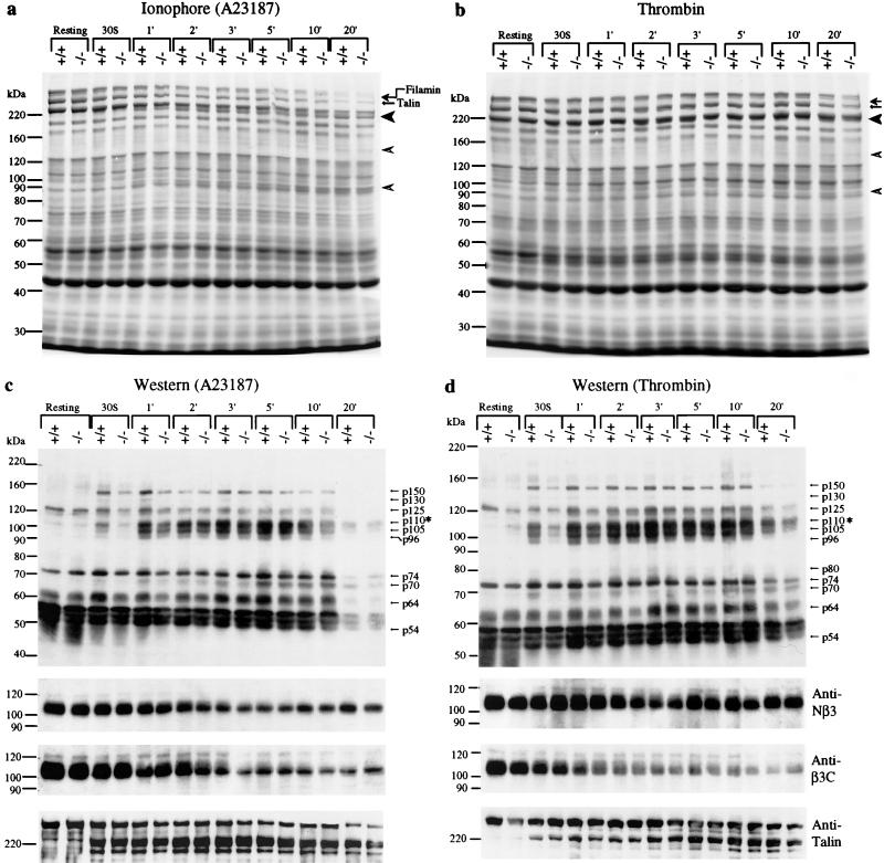
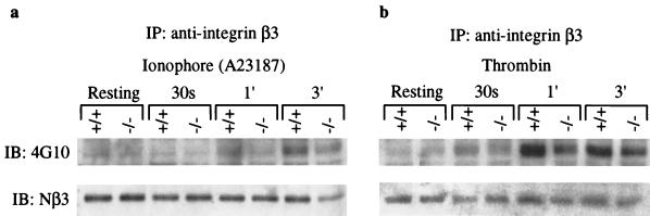
Conventional calpains are ubiquitous calcium-regulated cysteine proteases that have been implicated in cytoskeletal organization, cell proliferation, apoptosis, cell motility, and hemostasis. There are two forms of conventional calpains: the mu-calpain, or calpain I, which requires micromolar calcium for half-maximal activation, and the m-calpain, or calpain II, which functions at millimolar calcium concentrations. We evaluated the functional role of the 80-kDa catalytic subunit of mu-calpain by genetic inactivation using homologous recombination in embryonic stem cells. The mu-calpain-deficient mice are viable and fertile. The complete deficiency of mu-calpain causes significant reduction in platelet aggregation and clot retraction but surprisingly the mutant mice display normal bleeding times. No detectable differences were observed in the cleavage pattern and kinetics of calpain substrates such as the beta3 subunit of alphaIIbbeta3 integrin, talin, and ABP-280 (filamin). However, mu-calpain null platelets exhibit impaired tyrosine phosphorylation of several proteins including the beta3 subunit of alphaIIbbeta3 integrin, correlating with the agonist-induced reduction in platelet aggregation. These results provide the first direct evidence that mu-calpain is essential for normal platelet function, not by affecting the cleavage of cytoskeletal proteins but by potentially regulating the state of tyrosine phosphorylation of the platelet proteins.


Figures

FIG. 1
FIG. 1
Targeted disruption of the Capn1 locus and generation of mutant mice. (a) Schematic representation of the genomic locus of mouse μ-calpain drawn to scale. Sequence information for the intron-exon boundaries is shown in Table 1. The restriction sites shown on the map are as follows: BamHI (B), HindIII (H), and SalI (S). (b) The Capn1 locus (wild type; top line), the targeting vector (middle line), and the disrupted Capn1 locus (bottom line). Shown is the expected size of an XbaI and SalI fragment that hybridized with the probe for the wild-type locus (top) and the mutated allele (bottom). Small arrows indicate the positions of calpain primers that were used to conduct the RT-PCR analysis. (c) Southern blot analysis of genomic DNA isolated from the F2 generation of Capn1+/− mice was digested with XbaI and SalI and was blotted and hybridized with the probe shown in panel b. (d) RT-PCR analysis of mouse μ-calpain using primers Cal 1 and Cal 2. Glyceraldehyde 3-phosphate dehydrogenase (G3PDH) was used as a control to normalize the samples. The calpain regulatory 30-kDa subunit and the m-calpain catalytic subunit were amplified from the total RNA of liver, lung, and kidney of wild-type and Capn1−/− mice using gene-specific primers as described in Materials and Methods. (e) To rule out the possibility that alternate splicing may have occurred and produced low levels of truncated transcripts, RT-PCR analysis was conducted using total RNA pooled from liver, lung, and kidney. Lanes 1 and 2 show the results obtained using primer pair Cal 10 and Cal 19 (upstream transcript), and lanes 3 and 4 show the results obtained with primer pair Cal 18 and Cal 6 (downstream transcript). (f) Northern blot analysis of total RNA isolated from liver and lung of Capn1+/+ and Capn1−/− mice. The blot was hybridized with a cDNA probe (1.5 kb; exons 2 to 16) of murine μ-calpain. (g) Casein zymogram showing the distribution of μ-calpain and m-calpain in the platelet extract of Capn1+/+ and Capn1−/− mice. Note that only the μ-calpain activity was lost in the Capn1−/− mice. (h) Western blot analysis of μ-calpain in the whole red blood cell lysate of Capn1+/+ (left lane) and Capn1−/− (right lane) mice. (i) Casein zymogram of calpain activity in the red blood cell lysate of Capn1+/+ (left lane) and Capn1−/− (right lane) mice. The dark band represents the position of hemoglobin in the red cell lysate.
FIG. 2
FIG. 2
Effects of μ-calpain deficiency on platelet aggregation and granule secretion. Aggregation (top panel) and granule secretion (bottom panel) responses of washed platelets (1.5 × 10⁸/ml) from Capn1+/+ (black) and Capn1−/− (gray) mice are shown. (a) Thrombin, 10 nM. (b) ADP, 20 μM. (c) Collagen, 20 μg/ml. (d) Calcium ionophore A23187, 1 μM. In separate experiments, calcium ionophore at 0.1 μM was also used and produced an aggregation response consistent with other agonists. All measurements of platelet aggregation and granule secretion (19) were performed on the apyrase-treated (5 U/ml) platelets. Data are representative of four experiments.
FIG. 3
FIG. 3
Effects of μ-calpain deficiency on hemostasis and clot retraction. (a) Bleeding time was measured as described in Materials and Methods. Each symbol represents bleeding time measurement on a single Capn1+/+ mouse (left) and Capn1−/− mouse (right). (b) Photographs show the extent of in vitro clot retraction using PRP from wild-type and calpain null mice. Samples were treated with either 1.0 or 10 nM thrombin. As mentioned in Materials and Methods, 5 μl of red blood cells was added to enhance the color contrast for photography. The defective clot retraction in the μ-calpain null platelets was not influenced by the addition of red blood cells. Each photograph is representative of three independent experiments.
FIG. 4
FIG. 4
Proteolysis and tyrosine phosphorylation of platelet proteins. Washed platelets were activated by either calcium ionophore A23187 (1 μM) or thrombin (10 nM) at 37°C with stirring, and samples were taken out at indicated times for gel electrophoresis. (a) SDS-PAGE and Coomassie blue-stained 6% gel of total platelet lysate (40 μl). Calcium ionophore A23187 was used as an agonist. Arrows indicate the positions of intact filamin and talin. The 190-kDa fragment of talin (solid arrowhead) and the 130- and 93-kDa cleavage products of filamin (open arrowheads) are shown. (b) SDS-PAGE and Coomassie blue-stained 6% gel of total platelet lysate (40 μl). Thrombin was used as an agonist. Other symbols are the same as shown in the previous panel. (c and d) Western blot analysis of total platelet protein samples shown in panels a and b. (c) Calcium ionophore A23187 treatment. (d) Thrombin-treated platelets. An equal amount of platelet lysate, normalized by protein concentration, was analyzed by SDS-PAGE (7% gel), transferred to nitrocellulose, and probed with an antiphosphotyrosine monoclonal antibody (4G10). Note that the asterisk at p110 indicates the position of the β3 subunit of αIIbβ3 integrin. The same blots were stripped and reprobed to examine the proteolysis of β3 integrin and talin using defined antibodies. The bottom three panels show the results of Western blots using antibodies against β3 integrin and talin. The anti-Nβ3 antibody is specific for the N-terminal region of β3 integrin and was used to determine the total amount of β3 integrin in each lane. The anti-β3C antibody is cleavage sensitive and detects only the intact C terminus of β3 integrin (10). The anti-talin 8d4 antibody (Sigma) recognized the intact talin and cleaved the 190-kDa fragment but not the 50-kDa fragment. The same results were obtained in four independent experiments.
FIG. 5
FIG. 5
Immunoprecipitation and tyrosine phosphorylation of the β3 subunit of αIIbβ3 integrin. The β3 integrin was immunoprecipitated using biotin-conjugated anti-mouse antibody against the integrin β3 chain. Immunoprecipitates (IP) were analyzed by SDS-PAGE (8% gel), transferred to a nitrocellulose membrane, and blotted (IB) an antiphosphotyrosine antibody, 4G10 (upper panel). The same blot was stripped and blotted with an anti-Nβ3 antibody to normalize the amount of β3 integrin in each lane (lower panel).

Similar articles

Cited by

References

    1. Arthur J S C, Elce J S, Hegadorn C, Williams K, Greer P A. Disruption of the murine calpain small subunit gene, Capn4: calpain is essential for embryonic development but not for cell growth and division. Mol Cell Biol. 2000;20:4474–4481. - PMC - PubMed
    1. Baier L J, Permana P A, Yang X, Pratley R E, Hanson R L, Shen G Q, Mott D, Knowler W C, Cox N J, Horikawa Y, Oda N, Bell G I, Bogardus C. A calpain-10 gene polymorphism is associated with reduced muscle mRNA levels and insulin resistance. J Clin Investig. 2000;106:R69–R73. - PMC - PubMed
    1. Calderwood D A, Zent R, Grant R, Rees D J, Hynes R O, Ginsberg M H. The Talin head domain binds to integrin beta subunit cytoplasmic tails and regulates integrin activation. J Biol Chem. 1999;274:28071–28074. - PubMed
    1. Cichowski K, McCormick F, Brugge J S. p21rasGAP association with Fyn, Lyn, and Yes in thrombin-activated platelets. J Biol Chem. 1992;267:5025–5028. - PubMed
    1. Clark E A, Brugge J S. Redistribution of activated pp60c-src to integrin-dependent cytoskeletal complexes in thrombin-stimulated platelets. Mol Cell Biol. 1993;13:1863–1871. - PMC - PubMed

Publication types

MeSH terms