Review
. 2001 Mar;65(1):1-43.
doi: 10.1128/MMBR.65.1.1-43.2001.

Hyperthermophilic enzymes: sources, uses, and molecular mechanisms for thermostability

Affiliations
Review

Hyperthermophilic enzymes: sources, uses, and molecular mechanisms for thermostability

C Vieille et al. Microbiol Mol Biol Rev. 2001 Mar.

Abstract

Enzymes synthesized by hyperthermophiles (bacteria and archaea with optimal growth temperatures of > 80 degrees C), also called hyperthermophilic enzymes, are typically thermostable (i.e., resistant to irreversible inactivation at high temperatures) and are optimally active at high temperatures. These enzymes share the same catalytic mechanisms with their mesophilic counterparts. When cloned and expressed in mesophilic hosts, hyperthermophilic enzymes usually retain their thermal properties, indicating that these properties are genetically encoded. Sequence alignments, amino acid content comparisons, crystal structure comparisons, and mutagenesis experiments indicate that hyperthermophilic enzymes are, indeed, very similar to their mesophilic homologues. No single mechanism is responsible for the remarkable stability of hyperthermophilic enzymes. Increased thermostability must be found, instead, in a small number of highly specific alterations that often do not obey any obvious traffic rules. After briefly discussing the diversity of hyperthermophilic organisms, this review concentrates on the remarkable thermostability of their enzymes. The biochemical and molecular properties of hyperthermophilic enzymes are described. Mechanisms responsible for protein inactivation are reviewed. The molecular mechanisms involved in protein thermostabilization are discussed, including ion pairs, hydrogen bonds, hydrophobic interactions, disulfide bridges, packing, decrease of the entropy of unfolding, and intersubunit interactions. Finally, current uses and potential applications of thermophilic and hyperthermophilic enzymes as research reagents and as catalysts for industrial processes are described.


Figures

FIG. 1
FIG. 1
Hydrogen-deuterium exchange recorded in S. acidocaldarius and porcine muscle cytosol adenylate kinases during a temperature gradient experiment. Fractions of unexchanged protons as a function of temperature were calculated from the normalized amide II intensities at 1,546 cm−1 (S. acidocaldarius enzyme) and 1,542 cm−1 (porcine enzyme). The exchange was completed at 56 and 97°C for the porcine and S. acidocaldarius enzymes, respectively. Reprinted from reference with permission of the publisher. (Note that the two enzymes are not directly comparable because the pig enzyme is a monomer whereas the Sulfolobus enzyme is a trimer.)
FIG. 2
FIG. 2
Comparison of theoretical ΔGstab-versus-T curves for mesophilic and hyperthermophilic proteins. M, theoretical ΔGstab-versus-T curve for a mesophilic protein. (a), (b), and (c), theoretical ΔGstab-versus-T curves for a hyperthermophilic protein. In curve (a), the hyperthermophilic protein has the same temperature of maximal stability (Ts) as the mesophilic protein, and the ΔGstab-versus-T curve of the hyperthermophilic protein is shifted upward to higher ΔGstab values. In curve (b), hyperthermophilic and mesophilic protein have same Ts values and the same ΔGstab values at Ts. The ΔGstab-versus-T curve of the hyperthermophilic protein is flatter. In curve (c), hyperthermophilic and mesophilic proteins have different Ts values but have the same ΔGstab at their respective Ts. The ΔGstab-versus-T curve of the hyperthermophilic protein is shifted toward higher temperatures.
FIG. 3
FIG. 3
Mechanisms of protein degradation involving Asn residues.
FIG. 4
FIG. 4
Stereo view of the ion pair between Arg19 and Asp111 in S. solfataricus indole-3-glycerol phosphate synthase. The Arg19 guanidinium group also forms a cation-π interaction with the Tyr93 π system and two H bonds with Thr84. Reprinted from reference with permission of the publisher.
FIG. 5
FIG. 5
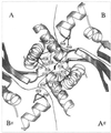
Schematic representation of the ion-pair network that stabilizes the intersubunit interactions in the hexameric P. furiosus GDH. The twofold axis of symmetry between the dimers is indicated by the dyad symbol. Dotted lines represent ion pair interactions. Reprinted from reference with permission of the publisher.
FIG. 6
FIG. 6
Comparison of the Phe59 loop structures in Thermoanaerobacterium thermosulfurigenes (left) and Thermotoga neapolitana xylose isomerases (right). Thin lines represent the structures of the wild-type enzymes; thick lines represent the model of the T. thermosulfurigenes xylose isomerase Gln58Pro mutant derivative.
FIG. 7
FIG. 7
Stabilization of the N-terminal β-strand by H bonding in T. maritima ferredoxin. β-Strand Lys2-Val5 forms a two-stranded β-sheet with β-strand Ile56-Glu59. The N-terminal β-strand is fixed to the protein core by three H bonds (gray dotted lines) to turn D: two bonds between Met1 and Thr39, and one bond between Val3 and Pro37. Reprinted from reference with permission of the publisher.
FIG. 8
FIG. 8
Activity-temperature profiles of wild-type subtilisin E (○), subtilisin E variant 5-3H5 (●), and thermitase (◊). Variant 5-3H5 was obtained by directed evolution. It is the subtilisin E eightfold mutant derivative Pro14Leu-Asn76Asp-Asn118Ser-Ser161Cys-Gly166Arg-Asn181Asp-Ser194Pro-Asn218Ser. Reprinted from reference with permission of the publisher.

References

    1. Achenbach-Richter L, Gupta R, Stetter K-O, Woese C R. Were the original eubacteria thermophiles? Syst Appl Microbiol. 1987;9:34–39. - PubMed
    1. Adams M W W. Enzymes and proteins from organisms that grow near and above 100°C. Annu Rev Microbiol. 1993;47:627–658. - PubMed
    1. Adams M W W, Kelly R M. Enzymes from microorganisms from extreme environments. C & E News. 1995;73:32–42.
    1. Adams M W W, Perler F B, Kelly R M. Extremozymes: expanding the limits of biocatalysis. Bio/Technology. 1995;13:662–668. - PubMed
    1. Aguilar C F, Sanderson I, Moracci M, Ciaramella M, Nucci R, Rossi M, Pearl L H. Crystal structure of the β-glycosidase from the hyperthermophilic archeon Sulfolobus solfataricus: resilience as a key factor in thermostability. J Mol Biol. 1997;271:789–802. - PubMed

Publication types

LinkOut - more resources