. 2001 Mar 15;29(6):1341-51.
doi: 10.1093/nar/29.6.1341.

Chromatin-bound PCNA complex formation triggered by DNA damage occurs independent of the ATM gene product in human cells

Affiliations

Chromatin-bound PCNA complex formation triggered by DNA damage occurs independent of the ATM gene product in human cells

A S Balajee et al. Nucleic Acids Res. .

Abstract

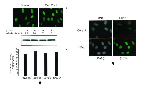
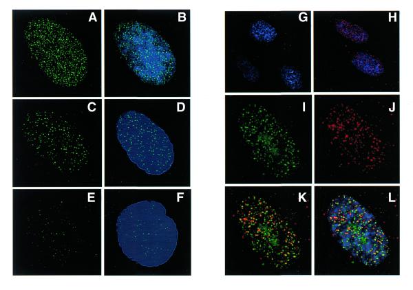
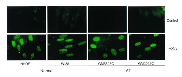
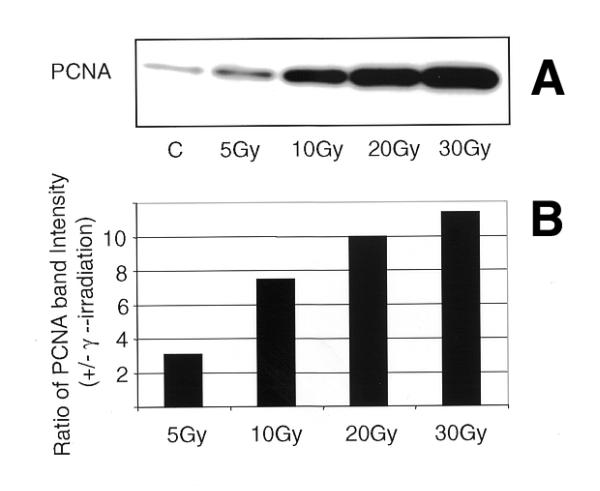
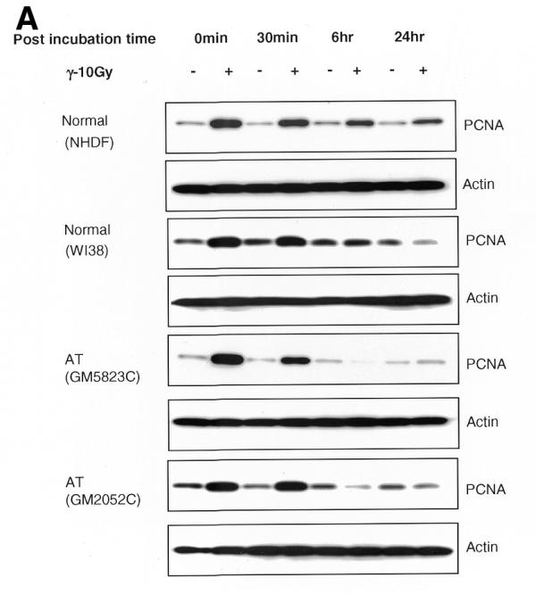
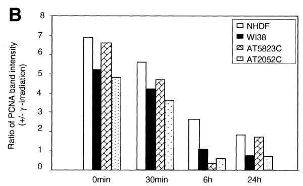
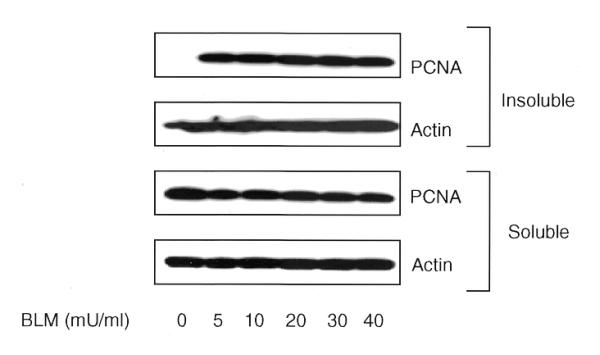
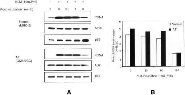
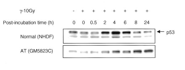
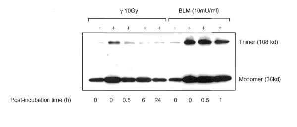
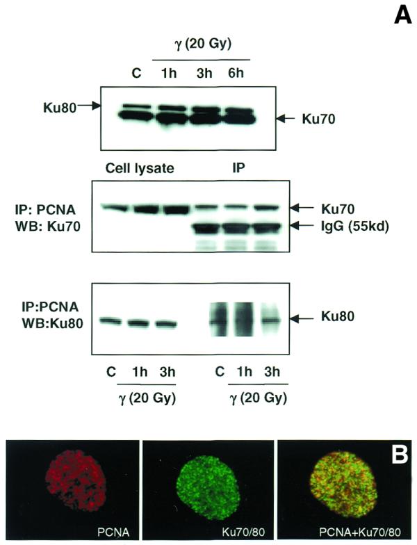
Proliferating cell nuclear antigen (PCNA), a processivity factor for DNA polymerases delta and epsilon, is involved in DNA replication as well as in diverse DNA repair pathways. In quiescent cells, UV light-induced bulky DNA damage triggers the transition of PCNA from a soluble to an insoluble chromatin-bound form, which is intimately associated with the repair synthesis by polymerases delta and epsilon. In this study, we investigated the efficiency of PCNA complex formation in response to ionizing radiation-induced DNA strand breaks in normal and radiation-sensitive Ataxia telangiectasia (AT) cells by immunofluorescence and western blot techniques. Exposure of normal cells to gamma-rays rapidly triggered the formation of PCNA foci in a dose-dependent manner in the nuclei and the PCNA foci (40-45%) co-localized with sites of repair synthesis detected by bromodeoxyuridine labeling. The chromatin-bound PCNA gradually declined with increasing post-irradiation times and almost reached the level of unirradiated cells by 6 h. The PCNA foci formed after gamma-irradiation was resistant to high salt extraction and the chromatin association of PCNA was lost after DNase I digestion. Interestingly, two radiosensitive primary fibroblast cell lines, derived from AT patients harboring homozygous mutations in the ATM gene, displayed an efficient PCNA redistribution after gamma-irradiation. We also analyzed the PCNA complex induced by a radiomimetic agent, Bleomycin (BLM), which produces predominantly single- and double-strand DNA breaks. The efficiency and the time course of PCNA complex induced by BLM were identical in both normal and AT cells. Our study demonstrates for the first time that the ATM gene product is not required for PCNA complex assembly in response to DNA strand breaks. Additionally, we observed an increased interaction of PCNA with the Ku70 and Ku80 heterodimer after DNA damage, suggestive of a role for PCNA in the non-homologous end-joining repair pathway of DNA strand breaks.


Figures

Figure 1
Figure 1
Immunofluorescence analysis of PCNA complex triggered by γ-ray irradiation in NHDF cells. Cells synchronized at G1 stage were irradiated (5 Gy), untreated or treated with buffer I and fixed after 30 min in a mixture of acetone:methanol (1:1). The cells were indirectly immunostained with a primary mouse monoclonal antibody to PCNA and a FITC-conjugated secondary antibody to mouse IgG2a. In order to determine whether or not the total cellular PCNA level is elevated by IR, the cells were immediately fixed in acetone:methanol without the extraction of soluble proteins (A). Immunofluorescence of PCNA in control and γ-treated cells without buffer I extraction (a) and the western blot detection of PCNA in the total cellular proteins isolated by RIPA buffer (b) show similar levels of PCNA. A histogram shows the intensity of PCNA band in control and irradiated NHDF cells (c). NDHF cells were extracted with buffer to reveal the chromatin-bound PCNA formation (B). The cells stained with DAPI are given in gray scale.
Figure 2
Figure 2
Time-dependent distribution of PCNA foci induced by γ- ray irradiation in the interphase nuclei of WI38 cells (AF). WI38 cells in G1 phase were irradiated with 10 Gy and the cells were incubated for different post-incubation times. The cells were extracted with buffer I and immunostained for PCNA with FITC conjugated antibody. The pattern of PCNA foci (green, FITC) induced by γ-rays at 0 min (A), 3 h (C) and 6 h (E) after irradiation is shown in the left panel and the combination of DNA and PCNA staining of the same cell (blue, DAPI; green, FITC) is shown in the right panel (B, D and F). MRC5 cells in G1 phase were irradiated with 20 Gy of γ-rays and the cells were incubated in complete medium containing 100 µM BrdU for 3 h. BrdU sites were detected with Avidin-Texas red (red) and the PCNA foci (green) with FITC. Control cells did not show any BrdU labeling (G), while irradiated cells show a punctuated pattern of repair foci detected by BrdU antibody (H). The cells were counterstained with DAPI to show both DNA (blue) and BrdU labeling (red). A single representative nucleus of an irradiated cell under high magnification shows the spatial distribution of PCNA foci (I), repair foci (J) and a combination of repair and PCNA foci (K). The merging of all the three colors (DNA, blue; PCNA, green; BrdU, red) is shown in (L). The co-localization of PCNA and repair foci appears as yellow spots due to the merging of green and red colors (K and L).
Figure 3
Figure 3
Immunofluorescence analysis of IR-induced PCNA complexes in the interphase nuclei of normal and AT cells. The normal (NHDF and WI38) and AT (GM5823C and GM2052C) cells in G1 phase were exposed to 5 Gy of γ-rays and incubated for 30 min. The cells were extracted with hypotonic buffer I and fixed in acetone:methanol. The cells were indirectly immunolabeled for insoluble PCNA as described before.
Figure 4
Figure 4
Western blot analysis of insoluble proteins isolated from NHDF cells exposed to increasing doses of γ-rays (A). NHDF cells in G1 phase were irradiated (0, 5,10, 20 and 30 Gy) and the insoluble proteins were extracted after 30 min. The proteins were size fractionated on 4–20% SDS–PAGE, transferred to PVDF membrane and probed with antibody to PCNA. The signal was detected by the ECL method. (B) Histogram showing the PCNA band intensity in the insoluble proteins.
Figure 5
Figure 5
(A) Time course kinetics of PCNA complex formation induced by IR in normal and AT cell lines. The normal and AT cells in G1 phase were exposed to γ-rays (10 Gy) and incubated for different recovery times. The insoluble proteins were extracted, size fractionated and transferred to PVDF membrane. The membranes were immunoreacted with PCNA antibody and the signal was detected by the ECL method. The 0 time point represents an irradiation time of 10 min only. The PCNA complex analyzed at 15 min post-irradiation was essentially similar to 0 min while that analyzed at 3 h showed a decline in PCNA band intensity (data not shown) consistent with the immunofluorescence results (Fig. 2C and D). In order to verify equal loading of proteins, the same membranes were stripped off and reprobed with actin antibody. (B) Histogram showing the intensity of PCNA in normal and AT cell lines at different post-incubation times.
Figure 5
Figure 5
(A) Time course kinetics of PCNA complex formation induced by IR in normal and AT cell lines. The normal and AT cells in G1 phase were exposed to γ-rays (10 Gy) and incubated for different recovery times. The insoluble proteins were extracted, size fractionated and transferred to PVDF membrane. The membranes were immunoreacted with PCNA antibody and the signal was detected by the ECL method. The 0 time point represents an irradiation time of 10 min only. The PCNA complex analyzed at 15 min post-irradiation was essentially similar to 0 min while that analyzed at 3 h showed a decline in PCNA band intensity (data not shown) consistent with the immunofluorescence results (Fig. 2C and D). In order to verify equal loading of proteins, the same membranes were stripped off and reprobed with actin antibody. (B) Histogram showing the intensity of PCNA in normal and AT cell lines at different post-incubation times.
Figure 6
Figure 6
Immunoblot analysis of soluble and insoluble proteins isolated from NHDF cells after treatment with different concentrations of BLM for 30 min at 37°C. The proteins were size fractionated on 4–20% SDS–PAGE, transferred to PVDF membrane and probed with antibody to PCNA. The signal was detected by the ECL method. Equal loading of proteins was verified by actin antibody.
Figure 7
Figure 7
(A) Time course kinetics of PCNA complex formation induced by BLM treatment in normal and AT cells. The cells were treated with 10 mU/ml of BLM for 30 min and incubated for various recovery times. The insoluble proteins isolated from control and treated cells were electrophoresed and transferred to PVDF membrane. The membrane was reacted with antibodies to PCNA, p53 and actin. (B) Histogram showing the time course kinetics of PCNA complex in normal and AT cells.
Figure 8
Figure 8
Time course kinetics of p53 protein induction by IR in normal and AT cells. The cells in G1 phase were exposed to 10 Gy of γ-rays and incubated for various recovery times. The insoluble proteins were isolated, size fractionated and transferred to PVDF membrane. The membranes were probed for p53 and the signal was detected by the ECL method.
Figure 9
Figure 9
Analysis of PCNA trimeric structures induced by IR and BLM in NHDF cells. The G1 cells were exposed to either 10 Gy of γ-rays or 10 mU/ml of BLM. Prior to electrophoresis, the PCNA trimeric structures in the pool of insoluble proteins were preserved by crosslinking with 0.1% glutaraldehyde for 10 min on ice. The reaction was terminated by addition of glycine to a final concentration of 0.1 M. The samples were heated at 80°C for 5 min, electrophoresed, transferred to PVDF membrane and immunoreacted with PCNA. The film was exposed longer to reveal the trimeric structures induced by γ-rays, which has resulted in the darkening of the PCNA band at the monomer position.
Figure 10
Figure 10
(A) Immunoblot analysis of the association of PCNA complex with Ku70/80 heterodimer by immunoprecipitation. Total cellular proteins were isolated from control and γ-irradiated (20 Gy) WI38 cells. Western blot analysis of Ku70 and Ku80 proteins in control and irradiated cells is shown in the top panel. Protein (500 µg) was immunoprecipitated using agarose-conjugated PCNA antibody (see Materials and Methods). The immunoprecipitated complex was size fractionated by 4–20% SDS–PAGE and transferred to PVDF membrane. The membranes were immunoreacted with mouse monoclonal antibodies to Ku70 (middle panel) and Ku80 (lower panel) and the signal was detected using the ECL kit. Ku70 antibody detected a cross-reacting immunoglobulin band of 55 kDa (middle panel). (B) Immunological co-localization of PCNA and Ku70/80 heterodimer in the interphase nucleus after γ-irradiation. WI38 cells in G1 phase were irradiated with 10 Gy of γ-rays, post-incubated for 3 h and fixed in acetone:methanol (1:1) after extraction with hypotonic buffer. The cells were sequentially immunostained for PCNA (detected by Texas red-conjugated secondary antibody to mouse IgG) and Ku70/80 heterodimer (detected by FITC-conjugated secondary antibody to mouse IgG). The immunostaining of PCNA (red), Ku70/80 heterodimer (green) and the merging of both PCNA and Ku70/80 heterodimer (yellow) are shown.

References

    1. Bravo R., Frank R., Blundell,P.A. and Macdonald-Bravo,H. (1987) Cyclin/PCNA is the auxiliary protein of DNA polymerase-δ. Nature, 326, 515–517. - PubMed
    1. Burgers P.M. (1991) Saccharomyces cerevisiae replication factor C. II. Formation and activity of complexes with the proliferating cell nuclear antigen and with DNA polymerases δ and ɛ. J. Biol. Chem., 266, 22698–22706. - PubMed
    1. Krishna T.S., Kong,X.P., Gary,S., Burgers,P.M. and Kuriyan,J. (1994) Crystal structure of the eukaryotic DNA polymerase processivity factor PCNA. Cell, 79, 1233–1243. - PubMed
    1. Xiong Y., Zhang,H. and Beach,D. (1992) D type cyclins associate with multiple protein kinases and the DNA replication and repair factor PCNA. Cell, 71, 505–514. - PubMed
    1. Zhang H., Xiong,Y. and Beach,D. (1993) Proliferating cell nuclear antigen and p21 are components of multiple cell cycle kinase complexes. Mol. Biol. Cell, 4, 897–906. - PMC - PubMed

Publication types

MeSH terms