. 2001 Mar 15;29(6):1389-98.
doi: 10.1093/nar/29.6.1389.

Topological testing of the mechanism of homology search promoted by RecA protein

Affiliations

Topological testing of the mechanism of homology search promoted by RecA protein

L Cai et al. Nucleic Acids Res. .

Abstract

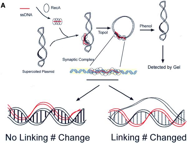
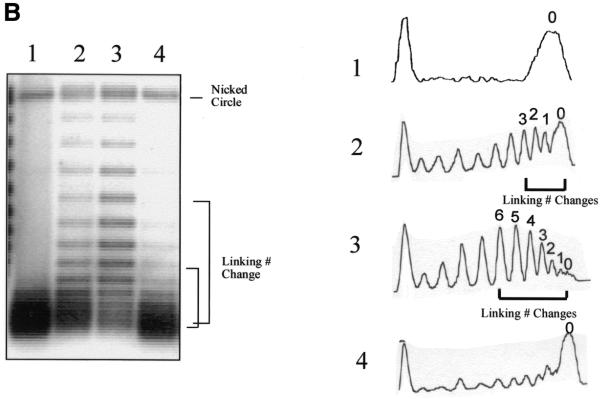
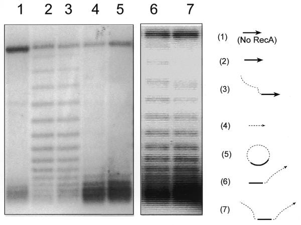
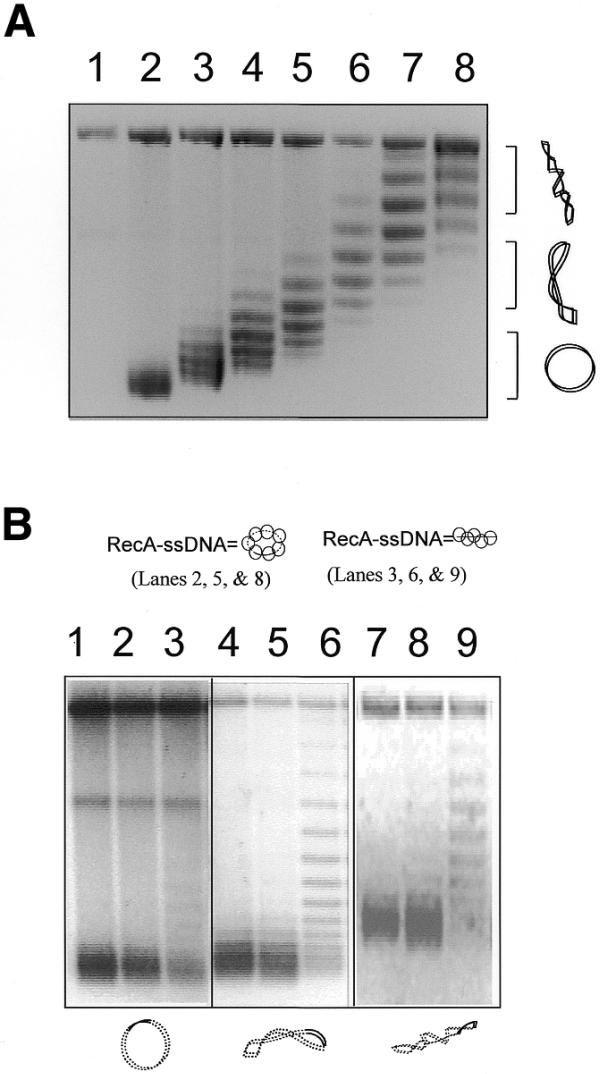
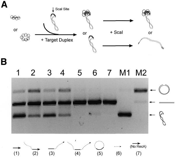
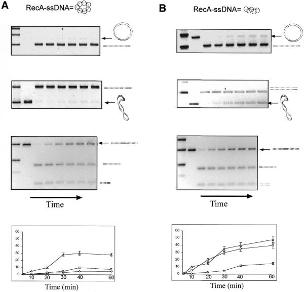
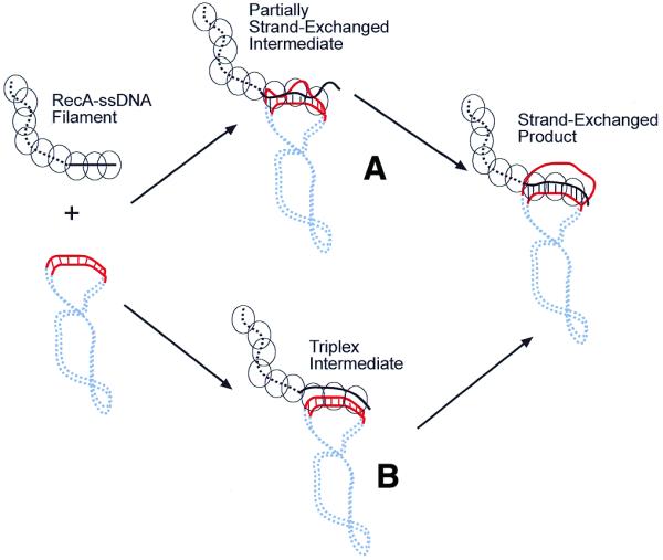
To initiate homologous recombination, sequence similarity between two DNA molecules must be searched for and homology recognized. How the search for and recognition of homology occurs remains unproven. We have examined the influences of DNA topology and the polarity of RecA-single-stranded (ss)DNA filaments on the formation of synaptic complexes promoted by RecA. Using two complementary methods and various ssDNA and duplex DNA molecules as substrates, we demonstrate that topological constraints on a small circular RecA-ssDNA filament prevent it from interwinding with its duplex DNA target at the homologous region. We were unable to detect homologous pairing between a circular RecA-ssDNA filament and its relaxed or supercoiled circular duplex DNA targets. However, the formation of synaptic complexes between an invading linear RecA-ssDNA filament and covalently closed circular duplex DNAs is promoted by supercoiling of the duplex DNA. The results imply that a triplex structure formed by non-Watson-Crick hydrogen bonding is unlikely to be an intermediate in homology searching promoted by RecA. Rather, a model in which RecA-mediated homology searching requires unwinding of the duplex DNA coupled with local strand exchange is the likely mechanism. Furthermore, we show that polarity of the invading RecA-ssDNA does not affect its ability to pair and interwind with its circular target duplex DNA.


Figures

Figure 1
Figure 1
Unwinding of a target duplex DNA by a RecA–ssDNA filament. (A) A linear (or circular in the experiments shown in Fig. 2) RecA–ssDNA filament was used to search for its homologous sequence (120 bp) on a circular duplex plasmid DNA (pBluescript, 3 kb). The synaptic complex was then treated with Topo I to relax the target plasmid. If the invading RecA–ssDNA filament had interwound with its complementary strand in the duplex (as shown lower right), after dissociating the protein with phenol the duplex plasmid DNA would become negatively supercoiled. The degree of supercoiling reflects the extent of unwinding induced by the RecA–ssDNA filament. The change in linking number could then be determined by running the final products in agarose gels with chloroquine. If the invading RecA–ssDNA filament just wound itself around the duplex, then no linking number change should be detected (as shown lower left). Solid lines represent homologous regions. Red represents the invading ssDNA. (B) (Left) An agarose gel containing 2 mg/ml chloroquine; (right) densitometric scans of each lane of the gel. Lane 3 demonstrates that a linear 120mer RecA–ssDNA filament can cause a target supercoiled DNA (pBluescript, 3 kb) with 120 bp of homology to unwind on average about six helical turns. Lane 1 is the same experiment as in lane 3, but no RecA has been used. With a 60mer homologous RecA–ssDNA as the searching entity, the linking number change is on average ∼3 (lane 2). When a RecA-coated heterologous 124mer ssDNA is used, no linking number change can be detected (lane 4).
Figure 1
Figure 1
Unwinding of a target duplex DNA by a RecA–ssDNA filament. (A) A linear (or circular in the experiments shown in Fig. 2) RecA–ssDNA filament was used to search for its homologous sequence (120 bp) on a circular duplex plasmid DNA (pBluescript, 3 kb). The synaptic complex was then treated with Topo I to relax the target plasmid. If the invading RecA–ssDNA filament had interwound with its complementary strand in the duplex (as shown lower right), after dissociating the protein with phenol the duplex plasmid DNA would become negatively supercoiled. The degree of supercoiling reflects the extent of unwinding induced by the RecA–ssDNA filament. The change in linking number could then be determined by running the final products in agarose gels with chloroquine. If the invading RecA–ssDNA filament just wound itself around the duplex, then no linking number change should be detected (as shown lower left). Solid lines represent homologous regions. Red represents the invading ssDNA. (B) (Left) An agarose gel containing 2 mg/ml chloroquine; (right) densitometric scans of each lane of the gel. Lane 3 demonstrates that a linear 120mer RecA–ssDNA filament can cause a target supercoiled DNA (pBluescript, 3 kb) with 120 bp of homology to unwind on average about six helical turns. Lane 1 is the same experiment as in lane 3, but no RecA has been used. With a 60mer homologous RecA–ssDNA as the searching entity, the linking number change is on average ∼3 (lane 2). When a RecA-coated heterologous 124mer ssDNA is used, no linking number change can be detected (lane 4).
Figure 2
Figure 2
A circular RecA–ssDNA filament cannot unwind its supercoiled target DNA. Using the assay described in Figure 1A, shown here is an agarose gel with 2 mg/ml chloroquine. The diagrams to the right of the gel indicate the ssDNA substrates used in each reaction; the target duplex DNA used in all reactions was the 3 kb supercoiled pBluescript plasmid. The solid line indicates the homologous region (120 nt) and the dashed line indicates the heterologous region (400 nt). Lane 5 shows a reaction in which circular RecA–ssDNA filament (C-520, 520 nt long with 120 nt homologous to the target duplex DNA) was used as the searching entity. Lane 3 shows the same reaction but linear 520mer ssDNA (with the 3′-end 120 nt homologous to the target duplex) was used as the searching entity. Lane 2 is the reaction in which a 120mer homologous ssDNA (H-120) was used. Lanes 1 and 4 are control reactions either without RecA protein or with heterologous 124mer ssDNA (Het-124). Lanes 6 and 7 show two reactions with linear 520mer ssDNA with 120 nt homologous to the target duplex located at the 5′-end (lane 6) or at the center (lane 7).
Figure 3
Figure 3
Unwinding of target DNAs with different degrees of supercoiling by a linear RecA–ssDNA filament. (A) An agarose gel with chloroquine (2 mg/ml) shows the covalently closed circular duplex DNA (pBluescript, 3 kb with 120 bp homologous to the ssDNA used) with high to zero supercoiling that was used in the reactions shown in (B). In the chloroquine gel the relaxed duplex DNA will become positively supercoiled. Therefore, it runs faster than the negatively supercoiled DNA that has become relaxed under these conditions. Lane 1 is a nicked circle as the marker for relaxed plasmid. Lanes 2–8 are the covalently closed plasmids (pBluescript, 3 kb) with increasing negative superhelicity. (B) An agarose gel with chloroquine (2 mg/ml) showing unwinding of the circular target DNA caused by either linear or circular homologous RecA–ssDNA. The diagrams at the top and bottom of the gel indicate the RecA–ssDNA and the duplex target DNA used in the reactions; solid lines represent homology between the two molecules. Lanes 1–3 are reactions where relaxed duplex was used as the target. Lanes 4–6 are reactions where medium supercoiled duplexes (superhelical density ∼–0.016) were used as target. Lanes 7–9 are reactions where highly supercoiled duplex (superhelical density ∼–0.033) were used as the target. Lane 3, 6 and 9 show reactions in which a homologous linear 120mer RecA–ssDNA filament (H-120) was used as the searching molecule. Lanes 1, 4 and 7 are reactions in which 124mer heterologous RecA–ssDNA filament (Het-124) was used as the searching molecule. Lanes 2, 5 and 8 show a reaction in which circular RecA–ssDNA filament (C-520, 520 nt long with 120 nt homologous to the target duplex DNA) was used as the searching molecule.
Figure 4
Figure 4
Restriction enzyme protection assay for homologous pairing. (A) In this assay a RecA–ssDNA filament (linear or circular) is used to search for its homologous sequence located on a plasmid DNA (pBluescript, linear, supercoiled or relaxed circular). Once the RecA–ssDNA filament finds its homologous target, it forms a synaptic complex with the duplex DNA that inhibits digestion of the duplex DNA by a restriction enzyme (ScaI) that recognizes the center of the homologous region. The increasing inability of ScaI to cleave the duplex DNA provides a direct measurement of synaptic complex formation. Agarose gel electrophoresis allows us to separate the unprotected (digested) product from the protected substrate. Here, only a linear RecA–ssDNA filament and supercoiled duplex target are shown as an example. (B) This agarose gel shows homologous pairing, assayed by the restriction enzyme protection method, between various RecA–ssDNA filaments (indicated at the bottom of the gel) and supercoiled duplex DNA as target. The solid lines represent the 120 bp of homology between the two molecules. The dashed lines represent 400 nt heterologous sequences in the ssDNA (except for the substrate used in lane 6, which is a 124mer heterologous ssDNA). Lanes M1 and M2 are markers for supercoiled, linear and nicked forms of the duplex substrates.
Figure 5
Figure 5
Restriction enzyme protection assay for homologous pairing between linear or circular RecA–ssDNA with relaxed, supercoiled or linear duplex DNA with 120 bp of homology. (A) (Top) An agarose gel that shows homologous pairing between a 520mer circular RecA–ssDNA (C-520, with 120 nt homologous to the duplex) filament and relaxed circular duplex target. (Upper middle) A reaction in which supercoiled duplex DNA was used as the target; (lower middle) the same reaction with linear duplex DNA as the target. (Bottom) Quantification of the three reactions (diamonds represent the reaction with relaxed duplex target; squares represent the reaction with supercoiled duplex target; triangles represent the reaction with linear duplex target). The reaction times were 5, 10, 20, 30, 40 and 60 min after formation of synaptic complexes and prior to 8 min of restriction digestion (see Materials and Methods for details). (B) Here, instead of 520mer circular RecA–ssDNA a 120mer homologous linear RecA–ssDNA (H-120) was used as the searching molecule. Top to bottom the panels show the formation of synaptic complexes with all three types of target duplex DNA as in (A). Solid lines indicate homology between the two molecules. Arrows to the right of each gel indicate the initial substrate used in that reaction.
Figure 6
Figure 6
Proposed model for RecA-mediated homology recognition. (A) Homology recognition is likely coupled to local strand exchanges between the two molecules. These locally unwound regions on the target duplex DNA could either be induced through contacts with the RecA–ssDNA filament or simply be the result of thermal fluctuation of the duplex DNA. Thus, the searching RecA–ssDNA filament tests for homology by base pairing with its complementary strand in the target duplex DNA. If homology is found through these short strand exchanges, then branch migration can extend the exchanged region to a greater distance, eventually resulting in full strand exchange. (B) In this model the RecA–ssDNA forms a triplex intermediate with the target duplex DNA prior to strand exchange. The results presented in this paper do not support this model, where in the triplex intermediate the RecA–ssDNA (which has been extended to ∼18 bp/turn) needs to be aligned in register with its target duplex DNA (10 bp/turn) on the homologous region, yet does not unwind the target duplex DNA.

Similar articles

Cited by

References

    1. Cox M.M. (2000) Recombinational DNA repair in bacteria and the RecA protein. Prog. Nucleic Acid Res. Mol. Biol., 63, 311–366. - PubMed
    1. Kowalczykowski S.C. and Eggleston,A.K. (1994) Homologous pairing and DNA strand exchange proteins. Annu. Rev. Biochem., 63, 991–1043. - PubMed
    1. Rao B.J.,Chiu,S.K., Bazemore,L.R., Reddy,G. and Radding,C.M. (1995) How specific is the first recognition step of homologous recombination? Trends Biochem. Sci., 20, 109–113. - PubMed
    1. West S.C. (1992) Enzymes and molecular mechanisms of genetic recombination. Annu. Rev. Biochem., 61, 603–640. - PubMed
    1. West S.C. (1994) The processing of recombination intermediates: mechanistic insights from studies of bacterial proteins. Cell, 76, 9–15. - PubMed

Publication types

MeSH terms