. 2001 Apr;12(4):219-31.
doi: 10.1002/1097-0193(200104)12:4<219::aid-hbm1017>3.0.co;2-l.

Neural basis of novel and well-learned recognition memory in schizophrenia: a positron emission tomography study

Affiliations

Neural basis of novel and well-learned recognition memory in schizophrenia: a positron emission tomography study

B Crespo-Facorro et al. Hum Brain Mapp. 2001 Apr.

Abstract

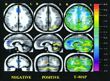
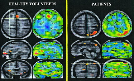
The level of familiarity of a given stimulus plays an important role in memory processing. Indeed, the novelty/familiarity of learned material has been proven to affect the pattern of activations during recognition memory tasks. We used visually presented words to investigate the neural basis of recognition memory for relatively novel and familiar stimuli in schizophrenia. Subjects were 34 healthy volunteers and 19 schizophrenia spectrum patients. Two experimental cognitive conditions were used: 1 week and again 1 day prior to the PET imaging subjects had to thoroughly learn a list of 18 words (well-learned memory). Subjects were also asked to learn another set of 18 words presented 1 min before the PET experiment (novel memory). During the PET session, subjects had to recognize the list of 18 words among 22 new (distractor) words. Subjects also performed a control task (reading words). A nonparametric randomization test and a statistical t-mapping method were used to determine between- and within-group differences. In patients the recognition of novel material produced relatively less flow in several frontal areas, superior temporal gyrus, insular cortex, and parahippocampal areas, and relatively higher activity in parietal areas, visual cortex, and cerebellum, compared to controls. No significant differences in flow were seen when comparing well-learned memory activations between groups. These results suggest that different neural pathways are engaged during novel recognition memory in patients with schizophrenia compared to healthy individuals. During recognition of novel material, patients failed to activate frontal/limbic regions, recruiting a set of posterior perceptual brain regions instead.


Figures

Figure 1
Figure 1
Randomization analysis of novel recognition minus well‐learned subtraction in schizophrenia patients compared to healthy volunteers: Negative activations (left column), positive activations (middle column), and statistical t‐map of activations (right column). Three orthogonal views are shown: transaxial, sagittal, and coronal. Crosshairs are used to show the location of the slice. Statistical (randomization) maps of the PET data are superimposed on a composite magnetic resonance (MR) image derived by averaging the MR scans from the subjects. The “t‐map” (right column of the image) represents the value of t for all voxels in the image and shows the general geography of the activations. Color bar on the right shows the t statistic values. The “peak map” (left and middle columns of the image) provides a descriptive picture of areas where all contiguous voxels exceed negatively (left) and positively (middle), the predefined threshold for statistical significance. Orange and red colors portray a relative increase in blood flow; blue and purple colors portray a relative decrease in blood flow. On the left column, the slices were chosen to show several frontal areas (e.g., superior frontal gyrus, anterior cingulate, orbitofrontal cortex, and straight gyrus) with a relative decrease in flow during the novel memory. The crosshairs point to the right anterior cingulate. On the middle column, the view of the three planes indicates that blood flow in the parietal cortex, occipital cortex, and cerebellum was significantly higher in patients.
Figure 2
Figure 2
Positive activations in healthy volunteers and patients with schizophrenia during novel memory activations (novel minus well‐learned increases in rCBF). (a) This figure shows the subtraction of novel minus well‐learned conditions in healthy volunteers. Regions in yellow/red tones indicate higher flow during the novel condition (novel memory activations). Novel activations encompass bilateral insular cortex, the orbitofrontal cortex, and the rostral anterior cingulate. (b) This figure shows the subtraction of novel minus well‐learned conditions in patients. Yellow/red tones indicate a relative increase in flow in patients during the novel condition (novel memory activations). The sagittal and coronal planes indicate a relative increase in flow in the SMA, left superior cerebellum, the right lingual gyrus, and the left parietal cortex.
Figure 3
Figure 3
Positive activations during the novel condition (left) and during the well‐learned condition (right) compared to a reading task in patients with schizophrenia. Brain regions activated by patients with schizophrenia during the novel condition are shown on the left figure. Patients showed an increase in flow in the superior cerebellum, the occipital and the parietal cortex when novel and reading words conditions are subtracted. Interestingly, patients display a similar subset of brain activations when well‐learned and reading words are subtracted. However, smaller activation sizes were observed in the well‐learned minus reading subtraction compared to those found in the novel‐reading subtraction.

References

    1. Abdullaev YG, Posner MI (1998): Event‐related brain potential imaging of semantic encoding during processing single words. Neuroimage 7: 1–13. - PubMed
    1. Andreasen NC (1983): Scale for the assessment of negative symptoms (SANS). Iowa City, University of Iowa.
    1. Andreasen NC (1984): Scale for the assessment of positive symptoms (SAPS). Iowa City, University of Iowa.
    1. Andreasen NC, Flaum M, Arndt S (1992a): The Comprehensive Assessment of Symptoms and History (CASH). An instrument for assessing diagnosis and psychopathology. Arch Gen Psychiatry 49: 615–623. - PubMed
    1. Andreasen NC, Cohen G, Harris G, Cizadlo T, Parkkinen J, Rezai K, Swayze VW (1992b): Image processing for the study of brain structure and function: problems and programs. J Neuropsychiatry Clin Neurosci 4: 125–133. - PubMed

Publication types